AUCTORES
Globalize your Research

Review Article | DOI: https://doi.org/10.31579/2640-1045/122
1 Department of Research, Veteran Administration University of California Irvine Medical Center, Long Beach, CA 90822.
2 Analytical Pharmacology Core Facility, City of Hope, Duarte, CA 91010.
3 Department of Obstetrics and Gynecology, University of California Irvine, Irvine, CA 92697.
*Corresponding Author: Sing-Yung Wu, M.D., Ph.D., Radiology and Research Service (151), VA-UCI Medical Center, Long Beach, CA 90822, USA.
Citation: Sing-Yung Wu, Haibo Zhao, Bi-Xin Xi, Dong-Bao Chen, and Maria E. Fucito. (2022). W-Compound can be used as a Biomarker for Fetal Thyroid Function and a Potential Tool for Screening Congenital Hypothyroidism. J. Endocrinology and Disorders. 6(3): DOI:10.31579/2640-1045/122
Copyright: © 2022 Sing-Yung Wu, This is an open-access article distributed under the terms of the Creative Commons Attribution License, which permits unrestricted use, distribution, and reproduction in any medium, provided the original author and source are credited.
Received: 16 June 2022 | Accepted: 24 June 2022 | Published: 30 June 2022
Keywords: congenital hypothyroidism; fetal thyroid function; W-Compound; biomarker; neonatal screening
Sulfoconjugation is the major pathway for thyroid hormone (TH) metabolism, converting T4 to inactive metabolites, T4S, rT3S, and T3S in fetus, via sulfotransferases (SULT) and type 3 deiodinase in gestation. Consistent with high production rate of T4S and rT3S, there are high serum sulfated iodothyronine analogs, including T4S, T3S, rT3S, and 3,3’-T2S (T2S), in ovine and human fetal and preterm infants. Fetal TH metabolic pathways predict T2S as the major TH metabolite in the fetus. Since maternal T2S appears to be quantitatively derived from fetal T3 (the active TH), the amount of T2S in the maternal compartment correlates with fetal thyroid function in sheep. In humans, maternal serum contains high levels of radioimmunoassayable T2S; however, it displays as a peak adjacent to but unidentical to synthetic T2S on HLPC and we named it the W-Compound. Levels of W-Compound increase during pregnancy and peak as high as 20-fold to that of nonpregnant women. Maternal serum levels of W-Compound significantly correlate with fetal T4 and W-compound concentrations but not maternal serum T4 in euthyroid or hyperthyroid women, showing a distinct difference between fetal and maternal in TH metabolism. Fetal T2S is actively transferred to the mother via placenta and the quantity of T2S or its metabolite (W-Compound) in maternal compartment reflects fetal thyroid function. Thus, maternal serum W-Compound may be a biomarker for monitoring fetal thyroid function in utero, although more investigations are needed to determine if it can be used as an alternative strategy for screening/managing congenital hypothyroidism due to dysregulated thyroid hormone metabolism.
The current screening program for congenital hypothyroidism (CHT) has allowed early treatment of this disorder and clearly improving long term outcomes [1-4]. However, despite the systematic screening and treatment of CHT, mild brain damages do occur [5, 6]. Since thyroid hormone (TH) is involved early first trimester fetal brain development including the neuronal cells [7-9], it is expected that developmental neuronal defects cannot be totally reversed postnatally. In developing mammals including humans, a deficiency or excess of TH in the developing brain during the fetal and neonatal periods can lead to morphological and functional abnormalities. Cretin is a serious form of congenital hypothyroidism (CHT), deficiency in TH in the newborn. These neonates suffer from not only impaired neurological function, but also stunted growth and physical deformities. This condition may occur in babies with a hypofunctioning thyroid gland. An estimated 15 to 20% of cases of CHT are inherited including gene mutations [1]. Many inherited cases are autosomal recessive but those with a mutation in the PAX8 gene or certain thyroid stimulating hormone receptor (TSHR) gene mutations have an autosomal dominant pattern of inheritance [3]. Other possible cause of fetal hypothyroidism is anti-thyroid medication treatment for maternal hyperthyroidism and lack of iodine during pregnancy. The incidence of babies born with CHT is 1 in 2,000 - 4,000 live birth in developed countries [3], representing a significant public health problem; this calls for more attention in better perinatal and neonatal care that needs new screening tools for fetal thyroid function and CHT. Our work in the sulfation pathway in mammalian fetal TH metabolism has obtained data that suggest the fetal-to-maternal transferred 3,3’-diiodothyronine sulfate (T2S)-like metabolite (W-Compound) can be used as novel marker for fetal thyroid function (10-12). The measurement of this compound in maternal serum and urine may serve as new marker for fetal thyroid function during in utero development.
Current neonatal screening of thyroid function and CHT
Neonatal screening programs began detecting neonates with CHT over 45 years ago. At present, 38 million births yearly worldwide undergo screening for this disorder [3]. The screening program for CHT has allowed early treatment of this disorder and clearly improving long term outcomes [2-4]. However, despite the systematic screening and treatment of CHT, mild brain damages do occur [5, 6]. Since thyroid hormone (TH) is involved early first trimester fetal brain development including the proliferation, migration, and differentiation of neuronal cells [7-9], it is expected that developmental neuronal defects cannot be totally reversed postnatally. These irreversible changes can impact on child IQ, cognitive and motor measures [2, 5, 13-16]. Children affected may present reduced socio-educational achievement [17, 18], greater risk of autistic trait [14], and more ADHD (attention-deficit/hyperactivity disorder) symptoms [19]. Recently, it has been found that higher preconception maternal iodine intakes are associated with higher child IQ [20], indicating intervention before or during pregnancy may help the future outcome of children.
Unfortunately, the incidence of CHT in the United States showed a trend of increasing from ~ 1:4100 in 1987 to ~ 1:2400 in 2002 [21]. Similar increases (Table 1) were also observed in Australia [22], Italy [23], and Ireland [24]. Furthermore, some infants display a delayed thyroid stimulating hormone (TSH) rise that missed by neonatal screening [25]. Recent studies suggest that delayed TSH rise may be more common and more severe than previously recognized [26].

In addition, despite the U.S. being iodine sufficient for the general population, the U.S. dietary iodine intakes have decreased drastically since the 1970s, with deficiency reemerging in vulnerable groups such as women of reproductive age [26]. All these findings indicate that there is room for improvement in the current strategy with neonatal CHT screening. Further study of fetal thyroid hormone metabolism and function is warranted as these studies may provide alternative strategies for managing CHT to avert unwanted sequelae.
What are the differences in thyroid hormone metabolism between fetus and adult?
Our lab at University of California (Irvine) - Long Beach VA Medical Center, in collaboration with Professor D. A. Fisher at UCLA-Harbor General Medical Center, has found in mammalian fetuses that sulfo-conjugation is the major pathway for TH metabolism (Figure. 1) [10, 27, 28].
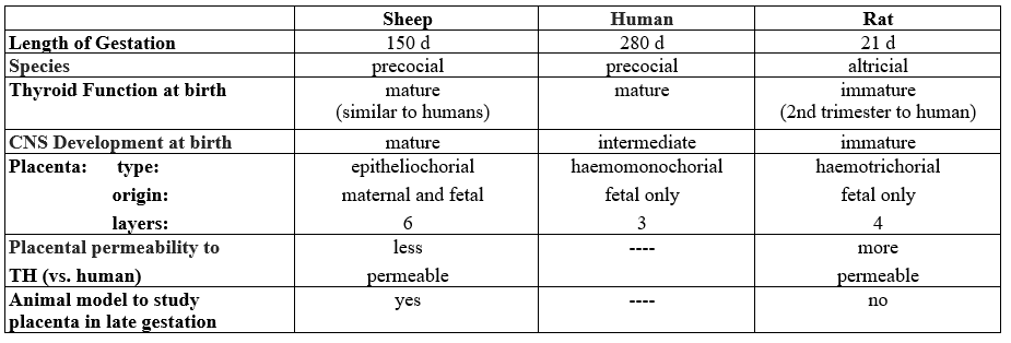
Before the onset of active synthesis and release of TH, iodothyronines detected in the fetus clearly are maternal origin [15, 29]. This period is approximately the first 17 gestational days (d) in rats, 50d in sheep, and 90d in humans (Table 2 and Figure. 1, the upper horizontal light dotted line). The proposed scheme for ovine fetal iodothyronine metabolism in late gestation (near term) depicts the production rates for sulfoconjugated TH analogs (shown as numbers in parentheses along the thick arrows in Figure. 1).

A kinetic study using the steady state constant infusion method in sheep showed that the major pathways of TH metabolism in the fetus convert T4 to inactive metabolites, rT3, T4S, rT3S, and T3S, via sulfotransferase and D3 enzyme systems in late gestation [10, 27, 28]. The high production rate (µg/kg/d) of T4 sulfate (T4S) (Figure. 1) reflects the active activity of the sulfation pathway in the fetus [27,28]. The rT3S production rate likely represents both sulfation of rT3 and inner-ring deiodination of T4S.
Consistent with the high production rate of T4S and rT3S, we have shown high serum concentrations of sulfated iodothyronine analogs in ovine and human fetal and preterm infant sera. These include T4S, T3S, rT3S, and 3,3’-T2S (T2S) [27, 28, 31-40]. Elevated iodothyronine sulfoconjugates are also detectable in amphibians during metamorphosis [41].
Thus, in developing mammals, sulfoconjugation of iodothyronine is an important pathway, in particular, during late gestation when the hypophyseal-pituitary-thyroid system becomes more mature in precocial species including sheep and humans. As term approaches, fetal thyroid gland secretion increases progressively while the effects of TH in many peripheral tissues must be delayed to the postpartum period. D3 and SULTs may serve to moderate the circulating THs before parturition. In addition, the shunting of iodothyronine metabolites of fetal origin into maternal circulation, the fetal to maternal transfer, is also an important mechanism in keeping an optimal active TH level in the developmental fetus.
The most common maternal circulating iodothyronine metabolite of fetal origin (-- the fetal to maternal transfer).
Thyroid hormone (TH) plays an important role in early fetal neurological maturation. Iodothyronines detected in the fetus before the onset of fetal thyroid function is of maternal origin. The maternal-fetal transfer of TH and their metabolites are apparently a two-way street. The high gradient between fetal and maternal serum concentrations of iodothyronine sulfates raises the possibility of significant fetal to maternal transfer of iodothyronine sulfoconjugates.
Sack et al. [42] reported that umbilical cord cutting, thus removing the lamb from placental D3 and transfer, triggers hypertriiodothyroninemia in the newborn lamb and that the postnatal T3 peak can be delayed until well after the TSH peak by delaying umbilical cord cutting. Santini et al. [43] reported that the placenta plays an important role in maintaining the low serum T3 in fetuses late in gestation. These findings suggest an important role of the placenta in fetal T3 metabolism, (Fig. 1, the blue line); it is possible that fetal-to-maternal transfer of the sulfated iodothyronines (via placenta) is one mechanism responsible for reducing serum T3 concentrations in the fetus. Increasing fetal-to-maternal transfer of iodothyronines occurs in late gestation.
The scheme shown in Fig. 1 also predicts 3,3’-T2S is the major thyroid hormone metabolite in the fetus. Intravenous infusion of radioiodine labeled T3 and T4 into near-term fetuses, demonstrated a rapid clearance of labeled T3 from fetal serum (disappearance T1/2 of 0.7 hours). Labeled T2S was identified as the major fetal iodothyronine metabolite in maternal urine [34]. Fetal T3 undergoes rapid inner-ring monodeiodination to 3,3’-T2 which is an excellent substrate for all known mammalian iodothyronine sulfotransferases [10]. The rapid sulfoconjugation of the hydroxyl group in the outer-ring of 3,3’-T2 forms a hydrophilic sulfated T2 (T2S) with enhanced permeability through placental membranes, facilitating the transfer of THs to maternal compartments. The T2S of fetal origin appears to be rapidly cleared from the maternal circulation via excretion in urine [44]. Fetal T4, on the other hand, disappears from the fetal circulation at a slower rate; a fast phase (T1/2=2.4 hours) in the first 3 hours followed by a slow phase (T1/2 = 17.5 hours). The major metabolites in fetal circulation after infusion of 125I-T4 were rT3 andT3 as well as their sulfates, T4S, rT3S and 3, 3’-T2S. Negligible amounts of T3S, roughly 0.7 – 1.2%, were also detected [44].
Similar to fetal T3 infusion, the most abundant metabolite found in maternal urine following radioactive T4 infusion is T2S. The T4 infusion study also confirms previous data in ovine fetuses [34, 35], indicating that the production of active thyroid hormone (T3) is less than the production of inactive products, rT3, T2S, rT3S and T3S [44].
T3 derived from T4 formed in the fetal circulation is converted to T2S, which is then transferred to the maternal compartment for deiodination/excretion. Recently, we have found sulfated [125I]-T2S was readily detected in the maternal compartment as the major metabolite of T3 following the perfusion of placenta with [125I]-T3 in guinea pig (12), suggesting that placental deiodinase and sulfotransferase may play an important role in fetal T3 homeostasis and in the fetal to maternal transfer of sulfated iodothyronine metabolites. This process would contribute to the low circulating T3 levels in the fetus. Since T2S appears to be quantitatively derived from circulating T3 (the active TH in the fetus), a significant increase or decrease in T2S in the maternal circulation would suggest hyper- or hypothyroidism in the fetus. In thyroidectomized sheep model, we found that 3,3’- T2S excretion in maternal urine reflects fetal thyroid function [45]. These data indicate clearly that maternal-fetal transfer of TH and its metabolites is a two-way street despite ovine placenta is less permeable as compared to rat and/or human (Table 2).
Furthermore, studies in rats have shown that 3,3’-T2 stimulates mitochondrial respiration in various tissues [46]. It is possible that a tight regulation of T2 concentration by sulfation and fetal-to-maternal transfer would have physiological value. Enhancing fetal-to-maternal transfer may protect the fetus from excessive mitochondrial thermogenesis stimulated by high fetal concentrations of T2. Another T2, i.e. 3,5-T2, was also shown to stimulate mitochondrial thermogenesis [46, 47]; however, its production rate is much lower in the fetus due to the inactive D1 (Figure 1).
W-Compound, a T2S-immuno-crossreactive compound, ought to be considered as a fetal thyroid function marker.
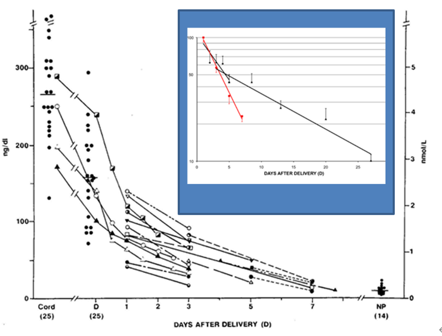
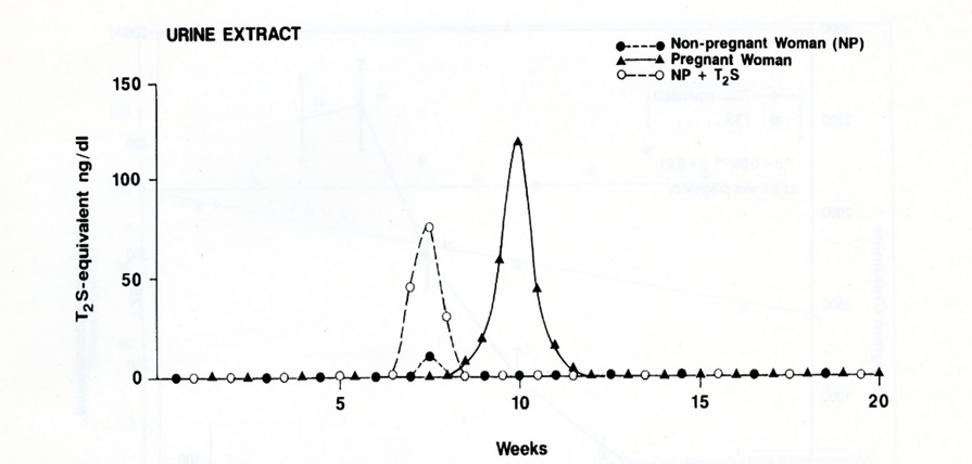
In humans, we have found high levels of radioimmunoassayable T2S in maternal serum [37, 39]; its levels increase with gestational age and peaked just prior to parturition. At delivery, a 20-fold increase in serum “T2S” is present compared to nonpregnant women (Figure 2) and “T2S” levels return to nonpregnant values in 7 to 10 days after delivery (Figure.3). Serum levels were measured by a T2S-specific radioimmunoassay (RIA) in 60 serum samples from newborns with hyperbilirubinemia, age 1 to 30 days. It is found that radioimmunoassayable T2S is cleared at similar rates in newborn as in postpartum maternal sera. This is consistent with the hypothesis that this “T2S” is produced in the placenta [46] (Figure 3).

On closer examination, the radioimmunoassayable “T2S” did not cochromatography with synthetic T2S by HPLC [39], (Figure 4). Over 40 known synthetic thyroid hormone analogs that were examined, none was found to be identical to the serum T2S-like material in pregnant women [49]. Thus, the name W-Compound was given. It is postulated that W-Compound is a side-chain modification of T2S, which cross-reacts with T2S antibody but is slightly more hydrophobic than T2S. Consistent with being an analogue of iodothyronine, we found high level of iodine content in highly purified W-Compound preparation analyzed by a Triple Quadrupole ICP-MS (Inductively Coupled Plasma Mass Spectrum) [50].
In normal pregnancy, both maternal and fetal W-Compound levels increase progressively with a significant direct correlation (p less than 0.001, in both mothers and fetuses) [51], (Figure 5). In addition, in 436 paired cord and maternal sera obtained from women at delivery, there is a highly significant correlation between the concentrations of Compound W in newborn cord and maternal sera (p less than 0.01) [49], (Figure 6).


A significant positive correlation is also observed between fetal serum concentrations of W-Compound and fetal T4 (p less than 0.003) and between maternal and fetal W-Compound concentrtions (p less than 0.0001) [51], (Figure. 7). However, no significant correlations were observed between maternal serum W-Compound and maternal serum T4 in euthyroid or hyperthyroid women. These data strongly suggest the fetal origin of W-Compound.

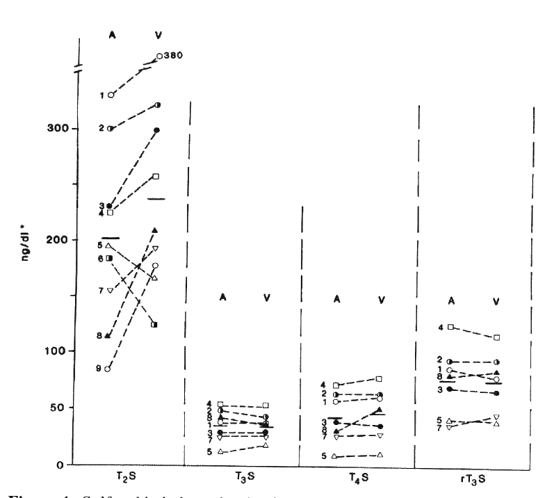
To further explore the possible origin of W-Compound, the serum concentrations of sulfated iodothyronines from cord arterial and venous blood samples were compared [49]. There were no significant differences between the mean T3S, T4S, or reverse-T3S concentrations of arterial and venous serum samples. However, the venous concentration of the T2S-equivalent material was higher than that in arterial blood in seven of the paired samples and lower in two. The mean “corrected” concentration of W-Compound in nine pairs of cord sera was found to be significantly higher in venous than arterial blood samples suggesting the fetal origin of W [49]. In addition, the mean of the maternal serum concentrations of T2S-reactive material was significantly lower than that of the paired cord serum concentrations. The rapid disappearance of W-Compound from maternal blood immediately after delivery supports this hypothesis [39], (Figure 3). A similar disappearance slope of serum W-Compound was also found in newborn infants [48], (Figure. 3, insert). These findings support the postulation that W-Compound is produced in placenta with iodothyronine precursor of fetal origin.
The Measurement of W-Compound: a technical consideration.
The original method for the measurement of W-Compound involves the use of RIA which was developed by Wu et al. [39]. Radioimmunoassay, in general, is not convenient to most clinical laboratories due to the involvement of using a radioisotope I125.
In a recent study, we have applied a highly sensitive and rapid homogeneous time-resolved fluorescence immunoassay to establish an indirect competitive W-Compound quantitative detection method called AlphaLisa (ICW-AlphaLisa), to measure the levels of W-Compound in maternal serum during pregnancy [52]. We developed specific polyclonal antibodies against W-Compound [a 3,3′-diiodothyronine sulfate (T2S) immuno-crossreactive material] and established an ICW quantitative detection method using AlphaLISA. In this method, photosensitive particles (donor beads) were coated with purified W-Compound or T2S and rabbit anti- W-Compound antibody, followed by incubation with biotinylated goat anti-rabbit antibody. This constitutes a detection system with streptavidin-coated acceptor particle. We have optimized the test conditions and evaluated the detection performance. The sensitivity of the method was 5 pg/ml in a detection range of 5-10,000 pg/ml. The intra-assay coefficient of variation averages less than 10 percentage with stable reproducibility. The ICW-AlphaLISA shows good stability and high sensitivity and can measure a wide range of W-compound levels in extracts of maternal serum samples. This may have clinical application to screen congenital hypothyroidism in utero [52].
Brominated flame retardants (BFRs) have been recently shown to disrupt TH homeostasis through multiple mechanisms (53), including inhibition of enzymes that regulate intracellular levels of THs, such as sulfotransferases (SULTs). As discussed in the present review, the placenta plays a critical role in expressing D3 and SULTs to prevent the developing fetuses from exposure to high level of active thyroid hormone T3, which are needed immediately after birth. The adverse effect of BFRs is concerning, given that disruption of TH regulation within the placenta could potentially harm the developing fetus [28, 29]. Iodothyronines and their sulfoconjugates in these studies were measured by liquid chromatography-tandem mass spectrometry (LC/MS-MS) [54, 55]. Even though the claim was made that this method was comparable to RIA, however, the sensitivities to detect for 3,3’-T2 and T2S were difficult to judge. Nevertheless, the lowest concentrations of standards used to optimize and calibrate the LC/MS-MS varied between 1-10 ng/ml that was much higher than the serum levels of 3,3’-T2 and T2S in physiological states [37, 53- 56].
Sulfoconjugation is a major metabolic pathway for thyroid hormone in developing mammals. The significant rise of sulfated iodothyronines in fetal compartments raises the possibilities that remarkable fetal to maternal transfer of the TH sulfoconjugates may occur throughout the second and third trimester in humans. This transfer may be a novel mechanism to maintain low T3 states or regulate serum 3,3’-T2, a thermogenic hormone, that is important for normal tissue maturity. The possibility that the transferred iodothyronine sulfate, especially 3,3’-T2S and its metabolite, may serve as a biomarker of fetal thyroid function needs to be further explored. Because the placenta plays a critical role in expressing D3 and SULTs to prevent the developing fetuses from exposure to high level of active thyroid hormone T3, which is needs immediately after birth. To this end, the non-isotopic method we developed [49] provides a very valuable means to facilitate future studies on W-Compound as a fetal thyroid function biomarker. Because disruption of TH regulation within the placenta could potentially harm the developing fetus [28], further studies are warranted to explore the possibility of the maternal serum or urine levels of W-Compound as a biomarker for BFR toxicity.
ADHD : Attention Deficit/Hyperactivity Disorder
BFR :Brominated flame retardants
CHT : Congenital Hypothyroidism
D1, D2, and D3: Type I, Type II, And Type III Iodothyronine Deiodinase
DiacS :Sulfated 3,3’-Diiodothyroacetic Acid
LAO/AT : L-Amino Acid Oxidase/Aminotransferase
SULT or ST : Sulfotransferases
T1, T2 and T3 : Mono-, Di-, and Tri-iodothyronine
T4 : Thyroxine
T4S, T3S, rT3S, T2S and T1S: Sulfated T4, T3, rT3, T2 and T1
TH : Thyroid Hormone
TriacS : Sulfated 3,3’,5-Triiodothyroacetic Acid
TSH : Thyroid Stimulating Hormone
TSHR : TSH Receptor
This work was supported in part by the Department of Veterans Affairs to S-y W and the National Institutes of Health (NIH) grants 1RO1 AR073298 and RO178843 to H-b Z, RO1 HL70562 and NIH R21 HD097498 to D-b C. The content is solely the responsibility of the authors and does not necessarily the official views of DVA and NIH.
None
Clearly Auctoresonline and particularly Psychology and Mental Health Care Journal is dedicated to improving health care services for individuals and populations. The editorial boards' ability to efficiently recognize and share the global importance of health literacy with a variety of stakeholders. Auctoresonline publishing platform can be used to facilitate of optimal client-based services and should be added to health care professionals' repertoire of evidence-based health care resources.

Journal of Clinical Cardiology and Cardiovascular Intervention The submission and review process was adequate. However I think that the publication total value should have been enlightened in early fases. Thank you for all.

Journal of Women Health Care and Issues By the present mail, I want to say thank to you and tour colleagues for facilitating my published article. Specially thank you for the peer review process, support from the editorial office. I appreciate positively the quality of your journal.

Journal of Clinical Research and Reports I would be very delighted to submit my testimonial regarding the reviewer board and the editorial office. The reviewer board were accurate and helpful regarding any modifications for my manuscript. And the editorial office were very helpful and supportive in contacting and monitoring with any update and offering help. It was my pleasure to contribute with your promising Journal and I am looking forward for more collaboration.

We would like to thank the Journal of Thoracic Disease and Cardiothoracic Surgery because of the services they provided us for our articles. The peer-review process was done in a very excellent time manner, and the opinions of the reviewers helped us to improve our manuscript further. The editorial office had an outstanding correspondence with us and guided us in many ways. During a hard time of the pandemic that is affecting every one of us tremendously, the editorial office helped us make everything easier for publishing scientific work. Hope for a more scientific relationship with your Journal.

The peer-review process which consisted high quality queries on the paper. I did answer six reviewers’ questions and comments before the paper was accepted. The support from the editorial office is excellent.

Journal of Neuroscience and Neurological Surgery. I had the experience of publishing a research article recently. The whole process was simple from submission to publication. The reviewers made specific and valuable recommendations and corrections that improved the quality of my publication. I strongly recommend this Journal.

Dr. Katarzyna Byczkowska My testimonial covering: "The peer review process is quick and effective. The support from the editorial office is very professional and friendly. Quality of the Clinical Cardiology and Cardiovascular Interventions is scientific and publishes ground-breaking research on cardiology that is useful for other professionals in the field.

Thank you most sincerely, with regard to the support you have given in relation to the reviewing process and the processing of my article entitled "Large Cell Neuroendocrine Carcinoma of The Prostate Gland: A Review and Update" for publication in your esteemed Journal, Journal of Cancer Research and Cellular Therapeutics". The editorial team has been very supportive.

Testimony of Journal of Clinical Otorhinolaryngology: work with your Reviews has been a educational and constructive experience. The editorial office were very helpful and supportive. It was a pleasure to contribute to your Journal.

Dr. Bernard Terkimbi Utoo, I am happy to publish my scientific work in Journal of Women Health Care and Issues (JWHCI). The manuscript submission was seamless and peer review process was top notch. I was amazed that 4 reviewers worked on the manuscript which made it a highly technical, standard and excellent quality paper. I appreciate the format and consideration for the APC as well as the speed of publication. It is my pleasure to continue with this scientific relationship with the esteem JWHCI.

This is an acknowledgment for peer reviewers, editorial board of Journal of Clinical Research and Reports. They show a lot of consideration for us as publishers for our research article “Evaluation of the different factors associated with side effects of COVID-19 vaccination on medical students, Mutah university, Al-Karak, Jordan”, in a very professional and easy way. This journal is one of outstanding medical journal.

Dear Hao Jiang, to Journal of Nutrition and Food Processing We greatly appreciate the efficient, professional and rapid processing of our paper by your team. If there is anything else we should do, please do not hesitate to let us know. On behalf of my co-authors, we would like to express our great appreciation to editor and reviewers.

As an author who has recently published in the journal "Brain and Neurological Disorders". I am delighted to provide a testimonial on the peer review process, editorial office support, and the overall quality of the journal. The peer review process at Brain and Neurological Disorders is rigorous and meticulous, ensuring that only high-quality, evidence-based research is published. The reviewers are experts in their fields, and their comments and suggestions were constructive and helped improve the quality of my manuscript. The review process was timely and efficient, with clear communication from the editorial office at each stage. The support from the editorial office was exceptional throughout the entire process. The editorial staff was responsive, professional, and always willing to help. They provided valuable guidance on formatting, structure, and ethical considerations, making the submission process seamless. Moreover, they kept me informed about the status of my manuscript and provided timely updates, which made the process less stressful. The journal Brain and Neurological Disorders is of the highest quality, with a strong focus on publishing cutting-edge research in the field of neurology. The articles published in this journal are well-researched, rigorously peer-reviewed, and written by experts in the field. The journal maintains high standards, ensuring that readers are provided with the most up-to-date and reliable information on brain and neurological disorders. In conclusion, I had a wonderful experience publishing in Brain and Neurological Disorders. The peer review process was thorough, the editorial office provided exceptional support, and the journal's quality is second to none. I would highly recommend this journal to any researcher working in the field of neurology and brain disorders.
Dear Agrippa Hilda, Journal of Neuroscience and Neurological Surgery, Editorial Coordinator, I trust this message finds you well. I want to extend my appreciation for considering my article for publication in your esteemed journal. I am pleased to provide a testimonial regarding the peer review process and the support received from your editorial office. The peer review process for my paper was carried out in a highly professional and thorough manner. The feedback and comments provided by the authors were constructive and very useful in improving the quality of the manuscript. This rigorous assessment process undoubtedly contributes to the high standards maintained by your journal.

International Journal of Clinical Case Reports and Reviews. I strongly recommend to consider submitting your work to this high-quality journal. The support and availability of the Editorial staff is outstanding and the review process was both efficient and rigorous.

Thank you very much for publishing my Research Article titled “Comparing Treatment Outcome Of Allergic Rhinitis Patients After Using Fluticasone Nasal Spray And Nasal Douching" in the Journal of Clinical Otorhinolaryngology. As Medical Professionals we are immensely benefited from study of various informative Articles and Papers published in this high quality Journal. I look forward to enriching my knowledge by regular study of the Journal and contribute my future work in the field of ENT through the Journal for use by the medical fraternity. The support from the Editorial office was excellent and very prompt. I also welcome the comments received from the readers of my Research Article.

Dear Erica Kelsey, Editorial Coordinator of Cancer Research and Cellular Therapeutics Our team is very satisfied with the processing of our paper by your journal. That was fast, efficient, rigorous, but without unnecessary complications. We appreciated the very short time between the submission of the paper and its publication on line on your site.

I am very glad to say that the peer review process is very successful and fast and support from the Editorial Office. Therefore, I would like to continue our scientific relationship for a long time. And I especially thank you for your kindly attention towards my article. Have a good day!

"We recently published an article entitled “Influence of beta-Cyclodextrins upon the Degradation of Carbofuran Derivatives under Alkaline Conditions" in the Journal of “Pesticides and Biofertilizers” to show that the cyclodextrins protect the carbamates increasing their half-life time in the presence of basic conditions This will be very helpful to understand carbofuran behaviour in the analytical, agro-environmental and food areas. We greatly appreciated the interaction with the editor and the editorial team; we were particularly well accompanied during the course of the revision process, since all various steps towards publication were short and without delay".

I would like to express my gratitude towards you process of article review and submission. I found this to be very fair and expedient. Your follow up has been excellent. I have many publications in national and international journal and your process has been one of the best so far. Keep up the great work.

We are grateful for this opportunity to provide a glowing recommendation to the Journal of Psychiatry and Psychotherapy. We found that the editorial team were very supportive, helpful, kept us abreast of timelines and over all very professional in nature. The peer review process was rigorous, efficient and constructive that really enhanced our article submission. The experience with this journal remains one of our best ever and we look forward to providing future submissions in the near future.

I am very pleased to serve as EBM of the journal, I hope many years of my experience in stem cells can help the journal from one way or another. As we know, stem cells hold great potential for regenerative medicine, which are mostly used to promote the repair response of diseased, dysfunctional or injured tissue using stem cells or their derivatives. I think Stem Cell Research and Therapeutics International is a great platform to publish and share the understanding towards the biology and translational or clinical application of stem cells.

I would like to give my testimony in the support I have got by the peer review process and to support the editorial office where they were of asset to support young author like me to be encouraged to publish their work in your respected journal and globalize and share knowledge across the globe. I really give my great gratitude to your journal and the peer review including the editorial office.

I am delighted to publish our manuscript entitled "A Perspective on Cocaine Induced Stroke - Its Mechanisms and Management" in the Journal of Neuroscience and Neurological Surgery. The peer review process, support from the editorial office, and quality of the journal are excellent. The manuscripts published are of high quality and of excellent scientific value. I recommend this journal very much to colleagues.

Dr.Tania Muñoz, My experience as researcher and author of a review article in The Journal Clinical Cardiology and Interventions has been very enriching and stimulating. The editorial team is excellent, performs its work with absolute responsibility and delivery. They are proactive, dynamic and receptive to all proposals. Supporting at all times the vast universe of authors who choose them as an option for publication. The team of review specialists, members of the editorial board, are brilliant professionals, with remarkable performance in medical research and scientific methodology. Together they form a frontline team that consolidates the JCCI as a magnificent option for the publication and review of high-level medical articles and broad collective interest. I am honored to be able to share my review article and open to receive all your comments.

“The peer review process of JPMHC is quick and effective. Authors are benefited by good and professional reviewers with huge experience in the field of psychology and mental health. The support from the editorial office is very professional. People to contact to are friendly and happy to help and assist any query authors might have. Quality of the Journal is scientific and publishes ground-breaking research on mental health that is useful for other professionals in the field”.

Dear editorial department: On behalf of our team, I hereby certify the reliability and superiority of the International Journal of Clinical Case Reports and Reviews in the peer review process, editorial support, and journal quality. Firstly, the peer review process of the International Journal of Clinical Case Reports and Reviews is rigorous, fair, transparent, fast, and of high quality. The editorial department invites experts from relevant fields as anonymous reviewers to review all submitted manuscripts. These experts have rich academic backgrounds and experience, and can accurately evaluate the academic quality, originality, and suitability of manuscripts. The editorial department is committed to ensuring the rigor of the peer review process, while also making every effort to ensure a fast review cycle to meet the needs of authors and the academic community. Secondly, the editorial team of the International Journal of Clinical Case Reports and Reviews is composed of a group of senior scholars and professionals with rich experience and professional knowledge in related fields. The editorial department is committed to assisting authors in improving their manuscripts, ensuring their academic accuracy, clarity, and completeness. Editors actively collaborate with authors, providing useful suggestions and feedback to promote the improvement and development of the manuscript. We believe that the support of the editorial department is one of the key factors in ensuring the quality of the journal. Finally, the International Journal of Clinical Case Reports and Reviews is renowned for its high- quality articles and strict academic standards. The editorial department is committed to publishing innovative and academically valuable research results to promote the development and progress of related fields. The International Journal of Clinical Case Reports and Reviews is reasonably priced and ensures excellent service and quality ratio, allowing authors to obtain high-level academic publishing opportunities in an affordable manner. I hereby solemnly declare that the International Journal of Clinical Case Reports and Reviews has a high level of credibility and superiority in terms of peer review process, editorial support, reasonable fees, and journal quality. Sincerely, Rui Tao.

Clinical Cardiology and Cardiovascular Interventions I testity the covering of the peer review process, support from the editorial office, and quality of the journal.

Clinical Cardiology and Cardiovascular Interventions, we deeply appreciate the interest shown in our work and its publication. It has been a true pleasure to collaborate with you. The peer review process, as well as the support provided by the editorial office, have been exceptional, and the quality of the journal is very high, which was a determining factor in our decision to publish with you.

The peer reviewers process is quick and effective, the supports from editorial office is excellent, the quality of journal is high. I would like to collabroate with Internatioanl journal of Clinical Case Reports and Reviews journal clinically in the future time.

Clinical Cardiology and Cardiovascular Interventions, I would like to express my sincerest gratitude for the trust placed in our team for the publication in your journal. It has been a true pleasure to collaborate with you on this project. I am pleased to inform you that both the peer review process and the attention from the editorial coordination have been excellent. Your team has worked with dedication and professionalism to ensure that your publication meets the highest standards of quality. We are confident that this collaboration will result in mutual success, and we are eager to see the fruits of this shared effort.

Dear Dr. Jessica Magne, Editorial Coordinator 0f Clinical Cardiology and Cardiovascular Interventions, I hope this message finds you well. I want to express my utmost gratitude for your excellent work and for the dedication and speed in the publication process of my article titled "Navigating Innovation: Qualitative Insights on Using Technology for Health Education in Acute Coronary Syndrome Patients." I am very satisfied with the peer review process, the support from the editorial office, and the quality of the journal. I hope we can maintain our scientific relationship in the long term.

Dear Monica Gissare, - Editorial Coordinator of Nutrition and Food Processing. ¨My testimony with you is truly professional, with a positive response regarding the follow-up of the article and its review, you took into account my qualities and the importance of the topic¨.

Dear Dr. Jessica Magne, Editorial Coordinator 0f Clinical Cardiology and Cardiovascular Interventions, The review process for the article “The Handling of Anti-aggregants and Anticoagulants in the Oncologic Heart Patient Submitted to Surgery” was extremely rigorous and detailed. From the initial submission to the final acceptance, the editorial team at the “Journal of Clinical Cardiology and Cardiovascular Interventions” demonstrated a high level of professionalism and dedication. The reviewers provided constructive and detailed feedback, which was essential for improving the quality of our work. Communication was always clear and efficient, ensuring that all our questions were promptly addressed. The quality of the “Journal of Clinical Cardiology and Cardiovascular Interventions” is undeniable. It is a peer-reviewed, open-access publication dedicated exclusively to disseminating high-quality research in the field of clinical cardiology and cardiovascular interventions. The journal's impact factor is currently under evaluation, and it is indexed in reputable databases, which further reinforces its credibility and relevance in the scientific field. I highly recommend this journal to researchers looking for a reputable platform to publish their studies.

Dear Editorial Coordinator of the Journal of Nutrition and Food Processing! "I would like to thank the Journal of Nutrition and Food Processing for including and publishing my article. The peer review process was very quick, movement and precise. The Editorial Board has done an extremely conscientious job with much help, valuable comments and advices. I find the journal very valuable from a professional point of view, thank you very much for allowing me to be part of it and I would like to participate in the future!”

Dealing with The Journal of Neurology and Neurological Surgery was very smooth and comprehensive. The office staff took time to address my needs and the response from editors and the office was prompt and fair. I certainly hope to publish with this journal again.Their professionalism is apparent and more than satisfactory. Susan Weiner

My Testimonial Covering as fellowing: Lin-Show Chin. The peer reviewers process is quick and effective, the supports from editorial office is excellent, the quality of journal is high. I would like to collabroate with Internatioanl journal of Clinical Case Reports and Reviews.

My experience publishing in Psychology and Mental Health Care was exceptional. The peer review process was rigorous and constructive, with reviewers providing valuable insights that helped enhance the quality of our work. The editorial team was highly supportive and responsive, making the submission process smooth and efficient. The journal's commitment to high standards and academic rigor makes it a respected platform for quality research. I am grateful for the opportunity to publish in such a reputable journal.

My experience publishing in International Journal of Clinical Case Reports and Reviews was exceptional. I Come forth to Provide a Testimonial Covering the Peer Review Process and the editorial office for the Professional and Impartial Evaluation of the Manuscript.

I would like to offer my testimony in the support. I have received through the peer review process and support the editorial office where they are to support young authors like me, encourage them to publish their work in your esteemed journals, and globalize and share knowledge globally. I really appreciate your journal, peer review, and editorial office.

Dear Agrippa Hilda- Editorial Coordinator of Journal of Neuroscience and Neurological Surgery, "The peer review process was very quick and of high quality, which can also be seen in the articles in the journal. The collaboration with the editorial office was very good."

I would like to express my sincere gratitude for the support and efficiency provided by the editorial office throughout the publication process of my article, “Delayed Vulvar Metastases from Rectal Carcinoma: A Case Report.” I greatly appreciate the assistance and guidance I received from your team, which made the entire process smooth and efficient. The peer review process was thorough and constructive, contributing to the overall quality of the final article. I am very grateful for the high level of professionalism and commitment shown by the editorial staff, and I look forward to maintaining a long-term collaboration with the International Journal of Clinical Case Reports and Reviews.

To Dear Erin Aust, I would like to express my heartfelt appreciation for the opportunity to have my work published in this esteemed journal. The entire publication process was smooth and well-organized, and I am extremely satisfied with the final result. The Editorial Team demonstrated the utmost professionalism, providing prompt and insightful feedback throughout the review process. Their clear communication and constructive suggestions were invaluable in enhancing my manuscript, and their meticulous attention to detail and dedication to quality are truly commendable. Additionally, the support from the Editorial Office was exceptional. From the initial submission to the final publication, I was guided through every step of the process with great care and professionalism. The team's responsiveness and assistance made the entire experience both easy and stress-free. I am also deeply impressed by the quality and reputation of the journal. It is an honor to have my research featured in such a respected publication, and I am confident that it will make a meaningful contribution to the field.

"I am grateful for the opportunity of contributing to [International Journal of Clinical Case Reports and Reviews] and for the rigorous review process that enhances the quality of research published in your esteemed journal. I sincerely appreciate the time and effort of your team who have dedicatedly helped me in improvising changes and modifying my manuscript. The insightful comments and constructive feedback provided have been invaluable in refining and strengthening my work".

I thank the ‘Journal of Clinical Research and Reports’ for accepting this article for publication. This is a rigorously peer reviewed journal which is on all major global scientific data bases. I note the review process was prompt, thorough and professionally critical. It gave us an insight into a number of important scientific/statistical issues. The review prompted us to review the relevant literature again and look at the limitations of the study. The peer reviewers were open, clear in the instructions and the editorial team was very prompt in their communication. This journal certainly publishes quality research articles. I would recommend the journal for any future publications.

Dear Jessica Magne, with gratitude for the joint work. Fast process of receiving and processing the submitted scientific materials in “Clinical Cardiology and Cardiovascular Interventions”. High level of competence of the editors with clear and correct recommendations and ideas for enriching the article.

We found the peer review process quick and positive in its input. The support from the editorial officer has been very agile, always with the intention of improving the article and taking into account our subsequent corrections.

My article, titled 'No Way Out of the Smartphone Epidemic Without Considering the Insights of Brain Research,' has been republished in the International Journal of Clinical Case Reports and Reviews. The review process was seamless and professional, with the editors being both friendly and supportive. I am deeply grateful for their efforts.

To Dear Erin Aust – Editorial Coordinator of Journal of General Medicine and Clinical Practice! I declare that I am absolutely satisfied with your work carried out with great competence in following the manuscript during the various stages from its receipt, during the revision process to the final acceptance for publication. Thank Prof. Elvira Farina

Dear Jessica, and the super professional team of the ‘Clinical Cardiology and Cardiovascular Interventions’ I am sincerely grateful to the coordinated work of the journal team for the no problem with the submission of my manuscript: “Cardiometabolic Disorders in A Pregnant Woman with Severe Preeclampsia on the Background of Morbid Obesity (Case Report).” The review process by 5 experts was fast, and the comments were professional, which made it more specific and academic, and the process of publication and presentation of the article was excellent. I recommend that my colleagues publish articles in this journal, and I am interested in further scientific cooperation. Sincerely and best wishes, Dr. Oleg Golyanovskiy.

Dear Ashley Rosa, Editorial Coordinator of the journal - Psychology and Mental Health Care. " The process of obtaining publication of my article in the Psychology and Mental Health Journal was positive in all areas. The peer review process resulted in a number of valuable comments, the editorial process was collaborative and timely, and the quality of this journal has been quickly noticed, resulting in alternative journals contacting me to publish with them." Warm regards, Susan Anne Smith, PhD. Australian Breastfeeding Association.

Dear Jessica Magne, Editorial Coordinator, Clinical Cardiology and Cardiovascular Interventions, Auctores Publishing LLC. I appreciate the journal (JCCI) editorial office support, the entire team leads were always ready to help, not only on technical front but also on thorough process. Also, I should thank dear reviewers’ attention to detail and creative approach to teach me and bring new insights by their comments. Surely, more discussions and introduction of other hemodynamic devices would provide better prevention and management of shock states. Your efforts and dedication in presenting educational materials in this journal are commendable. Best wishes from, Farahnaz Fallahian.

Dear Maria Emerson, Editorial Coordinator, International Journal of Clinical Case Reports and Reviews, Auctores Publishing LLC. I am delighted to have published our manuscript, "Acute Colonic Pseudo-Obstruction (ACPO): A rare but serious complication following caesarean section." I want to thank the editorial team, especially Maria Emerson, for their prompt review of the manuscript, quick responses to queries, and overall support. Yours sincerely Dr. Victor Olagundoye.

Dear Ashley Rosa, Editorial Coordinator, International Journal of Clinical Case Reports and Reviews. Many thanks for publishing this manuscript after I lost confidence the editors were most helpful, more than other journals Best wishes from, Susan Anne Smith, PhD. Australian Breastfeeding Association.

Dear Agrippa Hilda, Editorial Coordinator, Journal of Neuroscience and Neurological Surgery. The entire process including article submission, review, revision, and publication was extremely easy. The journal editor was prompt and helpful, and the reviewers contributed to the quality of the paper. Thank you so much! Eric Nussbaum, MD

Dr Hala Al Shaikh This is to acknowledge that the peer review process for the article ’ A Novel Gnrh1 Gene Mutation in Four Omani Male Siblings, Presentation and Management ’ sent to the International Journal of Clinical Case Reports and Reviews was quick and smooth. The editorial office was prompt with easy communication.

Dear Erin Aust, Editorial Coordinator, Journal of General Medicine and Clinical Practice. We are pleased to share our experience with the “Journal of General Medicine and Clinical Practice”, following the successful publication of our article. The peer review process was thorough and constructive, helping to improve the clarity and quality of the manuscript. We are especially thankful to Ms. Erin Aust, the Editorial Coordinator, for her prompt communication and continuous support throughout the process. Her professionalism ensured a smooth and efficient publication experience. The journal upholds high editorial standards, and we highly recommend it to fellow researchers seeking a credible platform for their work. Best wishes By, Dr. Rakhi Mishra.

Dear Jessica Magne, Editorial Coordinator, Clinical Cardiology and Cardiovascular Interventions, Auctores Publishing LLC. The peer review process of the journal of Clinical Cardiology and Cardiovascular Interventions was excellent and fast, as was the support of the editorial office and the quality of the journal. Kind regards Walter F. Riesen Prof. Dr. Dr. h.c. Walter F. Riesen.

Dear Ashley Rosa, Editorial Coordinator, International Journal of Clinical Case Reports and Reviews, Auctores Publishing LLC. Thank you for publishing our article, Exploring Clozapine's Efficacy in Managing Aggression: A Multiple Single-Case Study in Forensic Psychiatry in the international journal of clinical case reports and reviews. We found the peer review process very professional and efficient. The comments were constructive, and the whole process was efficient. On behalf of the co-authors, I would like to thank you for publishing this article. With regards, Dr. Jelle R. Lettinga.

Dear Clarissa Eric, Editorial Coordinator, Journal of Clinical Case Reports and Studies, I would like to express my deep admiration for the exceptional professionalism demonstrated by your journal. I am thoroughly impressed by the speed of the editorial process, the substantive and insightful reviews, and the meticulous preparation of the manuscript for publication. Additionally, I greatly appreciate the courteous and immediate responses from your editorial office to all my inquiries. Best Regards, Dariusz Ziora

Dear Chrystine Mejia, Editorial Coordinator, Journal of Neurodegeneration and Neurorehabilitation, Auctores Publishing LLC, We would like to thank the editorial team for the smooth and high-quality communication leading up to the publication of our article in the Journal of Neurodegeneration and Neurorehabilitation. The reviewers have extensive knowledge in the field, and their relevant questions helped to add value to our publication. Kind regards, Dr. Ravi Shrivastava.

Dear Clarissa Eric, Editorial Coordinator, Journal of Clinical Case Reports and Studies, Auctores Publishing LLC, USA Office: +1-(302)-520-2644. I would like to express my sincere appreciation for the efficient and professional handling of my case report by the ‘Journal of Clinical Case Reports and Studies’. The peer review process was not only fast but also highly constructive—the reviewers’ comments were clear, relevant, and greatly helped me improve the quality and clarity of my manuscript. I also received excellent support from the editorial office throughout the process. Communication was smooth and timely, and I felt well guided at every stage, from submission to publication. The overall quality and rigor of the journal are truly commendable. I am pleased to have published my work with Journal of Clinical Case Reports and Studies, and I look forward to future opportunities for collaboration. Sincerely, Aline Tollet, UCLouvain.

Dear Ms. Mayra Duenas, Editorial Coordinator, International Journal of Clinical Case Reports and Reviews. “The International Journal of Clinical Case Reports and Reviews represented the “ideal house” to share with the research community a first experience with the use of the Simeox device for speech rehabilitation. High scientific reputation and attractive website communication were first determinants for the selection of this Journal, and the following submission process exceeded expectations: fast but highly professional peer review, great support by the editorial office, elegant graphic layout. Exactly what a dynamic research team - also composed by allied professionals - needs!" From, Chiara Beccaluva, PT - Italy.

Dear Maria Emerson, Editorial Coordinator, we have deeply appreciated the professionalism demonstrated by the International Journal of Clinical Case Reports and Reviews. The reviewers have extensive knowledge of our field and have been very efficient and fast in supporting the process. I am really looking forward to further collaboration. Thanks. Best regards, Dr. Claudio Ligresti

Dear Chrystine Mejia, Editorial Coordinator, Journal of Neurodegeneration and Neurorehabilitation. “The peer review process was efficient and constructive, and the editorial office provided excellent communication and support throughout. The journal ensures scientific rigor and high editorial standards, while also offering a smooth and timely publication process. We sincerely appreciate the work of the editorial team in facilitating the dissemination of innovative approaches such as the Bonori Method.” Best regards, Dr. Giselle Pentón-Rol.
