AUCTORES
Globalize your Research

Review Article | DOI: https://doi.org/10.31579/2641-0419/038
1Jordan University of Science and Technology, School of Medicine, Irbid, Jordan.
2Ankara University, Faculty of Pharmacy, Ankara, Turkey.
3Department of Kinesiology and Nutritional Sciences, California State University, Los Angeles, USA.
4Institute of Cardiovascular Sciences, St. Boniface Hospital Albrechtsen Research Centre & Department of Physiology and Pathophysiology, Max Rady College of Medicine, University of Manitoba, Winnipeg, Canada
*Corresponding Author: Naranjan S. Dhalla, Institute of Cardiovascular Sciences St. Boniface Hospital Albrecheston Hospital Research Centre Winnipeg, Manitoba, Canada R2H 2A6
Citation: Nusier M., Ozcelikay AT., Shah AK., Dhalla NS. (2020) Role of Intracellular Ca2+-overload in Cardiac Dysfunction in Heart Disease. Clinical Cardiology and Cardiovascular Interventions, 3(2); Doi:10.31579/2641-0419/038
Copyright: © 2020 Naranjan S Dhalla, This is an open-access article distributed under the terms of the Creative Commons Attribution License, which permits unrestricted use, distribution, and reproduction in any medium, provided the original author and source are credited.
Received: 13 December 2019 | Accepted: 03 January 2020 | Published: 07 January 2020
Keywords: calcium overload; gene expression; sarcoplasmic reticulum; sarcolemma; myofibrils; subcellular organelles
Various heart diseases such as genetically-determined heart failure, acute myocardial infarction, ischemia-reperfusion injury and catecholamine-induced cardiomyopathies are associated with cardiac dysfunction, cellular damage, subcellular derangements and metabolic alterations. Since increase in myocardial Ca2+ is accompanied by these abnormalities, it is generally held that intracellular Ca2+-overload plays an important role in the pathogenesis of cardiac dysfunction as well as cellular and metabolic defects in different cardiovascular diseases. This view is supported by observations in hearts subjected to Ca2+-paradox, where reperfusion of Ca2+-free perfused hearts with Ca2+-containing medium was found to produce a marked increase in myocardial Ca2+-content, cellular damage and cardiac contracture. The intracellular Ca2+-overload in the heart has also been shown to produce mitochondrial Ca2+-overload, depress ATP production, release different toxic substances and induce cardiomyocyte apoptosis. By virtue of its ability to depress cardiac gene expression and increase proteolysis of sarcolemma (SL) sarcoplasmic reticulum (SR) and myofibrils (MF), the intracellular Ca2+-overload has been reported to reduce SL, SR and MF protein content and activities. Such remodeling of subcellular organelles is associated with dramatic alterations in Ca2+ -handling by SL and SR membranes as well as interaction of Ca2+ with MF for the impairment of cardiac function. Thus, it is evident that mitochondrial Ca2+-overload, and subcellular remodeling for Ca2+-handling defects are responsible for the occurrence of cardiac dysfunction, metabolic derangements and cellular damage during the development of heart disease.
Short Title: Myocardial Ca2+ and Cardiac Function
It is now well known that cardiac contraction and relaxation processes are determined by the coordinated functions of different subcellular organelles including sarcolemma (SL), sarcoplasmic reticulum (SR), mitochondria (MT) and myofibrils (MF) [1-6]. The SL proteins such as voltage-sensitive Ca2+-channels, store-operated Ca2+-channels, Na+- Ca2+ exchanger and Na+- K+ ATPase as well as SR proteins including Ca2+-release channels (ryanodine receptors) and Ca2+-pump ATPase play an essential role in the entry and regulation of Ca2+ in cardiomyocytes. On the other hand, MF Ca2+-stimulated ATPase and MT oxidative phosphorylation are involved in the generation of contractile force and ATP production, respectively. It is noteworthy to point out that Ca2+ is not only essential for determining the status of cardiac contractile function, but is also intimately involved in the maintenance of membrane permeability, cellular integrity, and cardiac gene expression [3,7-9]. Furthermore, various vasoactive hormones including catecholamines and angiotensin II have been demonstrated to exert marked effects on Ca2+-transport activities in cardiomyocytes [4,10,11]. Thus, defects in any of the components of subcellular organelles can be seen to induce Ca2+-handling abnormalities and contractile dysfunction of the heart [3,9].
Since the identification of Ca2+-overload as a new principle for the pathophysiology of cardiac dysfunction [12-14], several diseases including cardiomyopathies due to high levels of circulating catecholamines [15-20], genetically-determined heart failure [21-25] as well as ischemic heart disease (acute myocardial infarction [26-30] and ischemia-reperfusion injury [31-35]) have been shown to be associated with the development of intracellular Ca2+-overload. It is generally assumed that impaired cardiac performance and functional derangement of subcellular organelles in different diseases are the consequence of intracellular Ca2+-overload. It should also be pointed out that there are other pathophysiologic mechanisms including oxidative stress and myocardial inflammation, which have been proposed to induce cardiac dysfunction and cellular abnormalities during the development of heart disease [36-40]. However, in this article we have attempted to highlight the evidence that intracellular Ca2+-overload plays a critical role in the genesis of metabolic and cellular defects as well as subcellular remodeling for the development of cardiac dysfunction in the heart. Furthermore, the present review is focussed on discussion of events for the occurrence of intracellular Ca2+-overload in cardiomyocytes and its consequences for inducing myocardial abnormalities.
Mechanisms for the Development of Intracellular Ca2+-overload
Although high levels of circulating catecholamines are known to produce intracellular Ca2+-overload, several mechanisms have been proposed to underlie this phenomenon [9,16,18,20]. These include activation of both α-and β-adrenoceptors, stimulation of SL Ca2+-channels, depression in SL Na+-Ca2+-exchanger and SL Ca2+-pump ATPase as well as oxidation of catecholamines and formation of oxyradicals. It is pointed out that interventions which reduce the entry of Ca2+ as well as prevent the oxidation of catecholamines and development of oxidative stress have been shown to attenuate the catecholamine-induced intracellular Ca2+-overload [9,12,16,18]. Furthermore, the occurrence of intracellular Ca2+-overload in genetically-determined cardiomyopathy has been attributed to the activation of sympathetic nervous system and increase in Ca2+-influx as well as the depression of SL Na+-K+ ATPase and increase in intracellular Na+ [9,21]. Agents such as Ca2+-antagonists which prevent the entry of Ca2+ in the heart have been reported to exert beneficial effects in cardiomyopatheic animals by reducing the development of intracellular Ca2+-overload[9,22,25].
Several studies have been conducted to demonstrate mechanisms for the occurrence of intracellular Ca2+-overload due to acute coronary occlusion as well as ischemia-reperfusion injury [9,27,28,30, 33-35]. It has been shown that the lack of oxygen in the ischemic myocardium results in acidification of the cytoplasm which promotes SL Na+-H+ exchange and subsequent entry of Ca2+ upon stimulation of Na+-Ca2+ exchange system. Lack of oxygen is also known to increase membrane permeability for Ca2+ due to incorporation of free fatty acids and other lipid metabolites in the SL membrane. On the other hand, ischemia-reperfusion injury has been associated with the release of norepinephrine from the adrenergic nerve endings for increasing the entry of Ca2+ in addition to promoting the development of oxidative stress. These changes are known to cause the occurrence of intracellular Ca2+-overload as a consequence of their dramatic effects on the SL membrane [9,27,33,37,38]. Several other vasoactive interventions and proinflammatory agents have also been shown to produce Ca2+-handling abnormalities in cardiomyocytes [39,40]. It may be noted that reperfusion of the Ca2+-depleted heart with Ca2+ containing medium has been shown to exhibit Ca2+-paradox and provide a direct evidence for the occurrence of intracellular Ca2+-overload [9, 41-44]. A massive increase in myocardial Ca2+content due to stimulation of Na+-Ca2+ exchanger in this experimental model was shown to be prevented when perfusion of the heart with Ca2+-free medium was carried out in the presence of low Na+ [42,43]. High concentrations of Ca2+-antagonists were also found to attenuate the increase in myocardial Ca2+ in the Ca2+-paradoxic heart by their action on the SL Na+-Ca2+ exchange activity [44]. Thus, the Ca2+-paradoxic heart is considered to form an excellent model for studying the effects of intracellular Ca2+-overload [42,43].
Cardiac Dysfunction and Cellular Damage
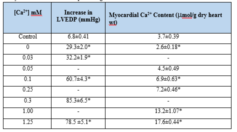
Reperfusion of the Ca2+-depleted hearts with Ca2+-containing medium was found to result in loss of contractility, development of contracture, damage to ultrastructure and leakage of intracellular enzymes from the myocardium [41, 45-48]. The paradoxical effects of Ca2+-deprived hearts were reported to occur in different species [49] and were similar to those seen during the development of oxygen- paradox in normal hearts [50]. The Ca2+-paradox phenomenon was shown to be associated with irreversible changes in the surface electrical activity [41] and a marked increase in the left ventricular end-diastolic pressure (LVEDP) [41,42,51-53]. The occurrence of intracellular Ca2+-overload and the increase in LVEDP (Table 1) as well as the development of cardiac contracture in the Ca2+-paradoxic heart were found to be dependent upon the concentration of Ca2+ in the reperfusion medium [42,53,54].
Although some investigators failed to demonstrate Ca2+-paradox associated changes in isolated cardiomyocytes [55], others have shown these alterations upon successive exposure of cardiomyocytes to Ca2+-free medium and Ca2+-containing medium [48, 56-58]. Nonetheless, ischemic preconditioning has been observed to attenuate the Ca2+-paradox associated increase in LVEDP, depression in the left ventricular developed pressure and leakage of myoglobin from the heart [59]. The presence of low Na+ during perfusion of the heart with Ca2+-free medium was also found to prevent the development of cardiac dysfunction and the occurrence of intracellular Ca2+-overload upon reperfusion [41-43].
The ultrastructural changes in the Ca2+-deprived and reperfused hearts included swelling of mitochondria and sarcotubular system, occurrence of contractile bands, and partial separation of the intercalated disc as well as basement membrane from sarcolemma [41,43,45,60]. The alterations in ultrastructure of the myocardium were dependent upon the concentration of Ca2+ in the reperfusion medium [41,60] and were attenuated by reducing the concentration of Na+ during the Ca2+-free perfusion phase [41]. These ultrastructural changes are similar to those seen in the ischemic heart disease [27-28] and may be a consequence of increased activities of cardiac lysosomal hydrolases [61], different intracellular proteases [35] and phospholipases [62]. Although the occurrence of autophagy has been reported in ischemia-reperfused hearts and myocardial infarction [27,28], no information regarding autophagic changes in the Ca2+-paradoxic heart is available at present. It is pointed out that the activation of NFκB and increased production of TNF-α have also been reported to cause cardiac injury due to intracellular Ca2+-overload [63]. Furthermore, the occurrence of cell death (apoptosis) in the Ca2+-paradox heart has been associated with the activation of mitogen-activated protein kinases (p38 and ERK) as well as different apoptotic signal transduction pathways [64]. Thus, the development of cardiac dysfunction and cellular damage due to intracellular Ca2+-overload appears to be occurring as a consequence of complex and diverse mechanisms.
Mitochondrial Ca2+-overload and Energy Depletion
It is now well known that intracellular Ca2+-overload in the heart results in the development of mitochondrial Ca2+-overload and defects in energy production [9,47,65]. Although low concentrations of Ca2+ are required for the stimulation of mitochondrial oxidative phosphorylation, high concentrations of Ca2+ have been shown to impair the mitochondrial function for ATP production [9,53,65, 66]. Perfusion of hearts with Ca2+-free medium followed by reperfusion with Ca2+-containing medium for the induction of intracellular Ca2+-overload was found to be associated with depressed mitochondrial state 3 respiration, respiratory control index, ADP/O ratio and oxidative phosphorylation without any changes in state 4 respiration [53,67]. These alterations were prevented when the reperfusion was carried out at low concentrations (0.1-0.5 mM) of Ca2+ but were not affected by different antioxidants [55]. The impaired mitochondrial function in the Ca2+-paradoxic heart has been associated with elevated levels of citric acid cycle intermediates and is considered to be due to defects in mitochondrial membrane potentials [68,69].
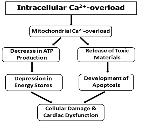
A dramatic decrease in high -energy phosphate stores in the heart has been shown to occur upon the induction intracellular Ca2+-overload [67,70,71]. It may be noted that Ca2+-binding and Ca2+-uptake activities of mitochondria, isolated from the Ca2+-paradoxic hearts, were found to be increased [72]. Such a change in the mitochondrial Ca2+-transport activity was suggested to contribute towards the occurrence of mitochondrial Ca2+-overload as it was attenuated when the perfusion with Ca2+-free medium was carried out in the presence of low Na+ [72]. It is also pointed out that mitochondrial Ca2+-overload may release several cytotoxic substances, which may also serve as signals for inducing apoptosis in the Ca2+-paradoxic hearts [64]. Thus, it appears that mitochondrial Ca2+-overload may be involved in cardiac dysfunction and cellular damage in the heart by depressing the high energy phosphate stores as well as inducing apoptosis in the myocardium. A schematic representation of these events is shown in Figure 1.
Subcellular Defects and Ca2+-handling Abnormalities
While the SL membrane is concerned with influx and efflux of Ca2+ for maintaining Ca2+-homeostasis in cardiomyocytes, the SR tubular system is involved in raising and lowering the concentration of Ca2+, whereas the interaction of Ca2+ with MF proteins determines the contractile status of the myocardium [3,4]. Reperfusion of Ca2+-deprived hearts with Ca2+-containing medium has been shown to exert profound effects on the activities of different subcellular organelles (Table 2) [73-75].
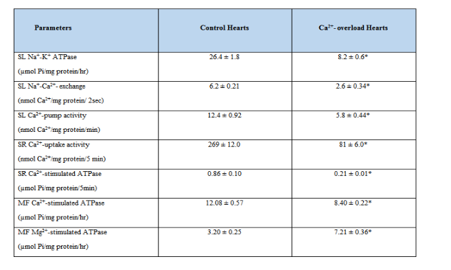
Depressions in the SL Na+-K+ ATPase, SL Na+-Ca2+ exchanger and SL Ca2+-pump ATPase activities in the Ca2+-paradoxic heart can be seen to contribute towards the occurrence of intracellular Ca2+-overload in cardiomyocytes [73,76,77]. These SL defects were attenuated when the perfusion with Ca2+-free medium was carried out in the presence of low Na+ (35mM) or at low temperature (210C) [42,78]. On the other hand, the density of SL Ca2+-channels was increased upon subjecting the heart to Ca2+-paradox and this change was also attenuated by carrying out the perfusion with Ca2+-free medium in the presence of low Na+ or at low temperature [79]. Furthermore, alterations in the SL membrane were also apparent because the activities of β-AR – G-protein – adenylyl cyclase complex were observed to be increased [80] and the activity of SL Ca2+/Mg2+-ecto ATPAse was decreased [81] in the Ca2+-paradoxic heart. Although the status of SL store-operated Ca2+-channels [6] in the Ca2+-paradoxic heart has not be determined, their participation in inducing intracellular Ca2+-overload can not be ruled out at present.
The induction of Ca2+-paradox in the heart upon perfusion with Ca2+-free medium followed by Ca2+-containing medium was seen to be associated with marked depression in the SR Ca2+-uptake and release activities [72,74]. These changes in Ca2+-handling by SR were dependent upon the concentration of Ca2+ in the reperfusion medium and were attenuated when the perfusion with Ca2+-free medium was carried out in the presence of low Na+ or at low temperature. Although MF Ca2+-stimulated ATPase activity was not altered during the initial (5 min) reperfusion phases of Ca2+-paradox development [67], reperfusion of Ca2+-deprived hearts with Ca2+-containing medium for 10 min was found to depress the MF Ca2+-stimulated ATPase activity and increase the MF Mg2+-ATPase activity [75]. These alterations were associated with degradation of MF α -myosin heavy chain and troponin T proteins in the Ca2+-paradoxic hearts. The activation of proteases such as calpain by elevated levels of intracellular Ca2+ in cardiomyocytes is considered to be involved in alterations of the SL, SR and MF activities upon reducing their protein content [35]. These events for inducing subcellular defects due to the occurrence of intracellular Ca2+-overload in the Ca2+-paradoxic hearts are shown in Figure 2.
It should be recognized that Ca2+-handling abnormalities in SL and SR due to intracellular Ca2+-overload may also be induced by changes in the phospholipid composition of these membranes [62]. It is also noteworthy that similar Ca2+-handling defects have also been observed in heart failure and ischemic heart disease [27,28,82-85].
Alterations in cardiac Gene Expression
In view of the role of cardiac gene expression in maintaining the function of different subcellular organelles in the heart [27,28, 85], it has been suggested that subcellular remodeling in the Ca2+-paradoxic heart may be due to changes in gene expression for different subcellular proteins [9,73,74]. Accordingly, subcellular remodeling due to intracellular Ca2+-overload may be occurring as a consequence of both the activation of calpain and the depression in mRNA levels for different cardiac genes (Figure 2).

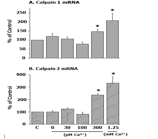
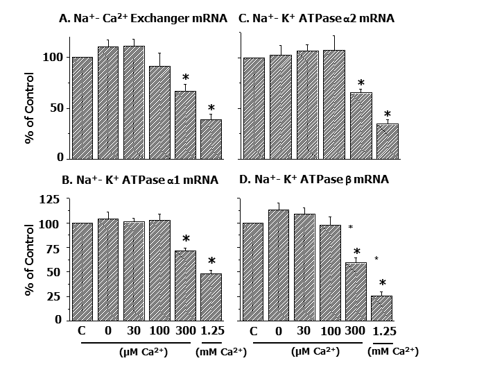
In this regard, it may be noted that the increase in mRNA levels for both calpain 1 and calpain 2 in the Ca2+-paradoxic heart was found to be dependent on the concentration of Ca2+ in the reperfusion medium (Figure 3) [54].
Furthermore, it was demonstrated that depressions in gene expression for SL Na+-Ca2+ exchanger as well as different isoforms of SL Na+- K+ ATPase protein due to Ca2+-paradox were dependent upon the concentration of Ca2+ in the reperfusion medium (Figure 4) [54].
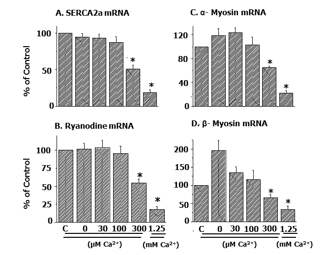
Likewise, alterations in mRNA levels for SR Ca2+-pump protein and Ca2+-release channels as well as MF α- and β- myosin proteins in the Ca2+-paradoxic heart were observed to be dependent upon the concentration of Ca2+ in the reperfusion medium (Figure 5) [54].
These observations provide evidence for a defect in the formation of subcellular proteins resulting in subcellular remodeling due to intracellular Ca2+-overload. Thus, cardiac genes can be seen as excellent molecular targets for the development of novel interventions for the improved therapy of heart disease.
From the forgoing discussion, it is evident that two major mechanisms, namely energy depletion due to mitochondrial Ca2+-overload and subcellular remodeling due to increased proteolysis and reduced gene expression, are likely to explain the development of cellular damage, metabolic alterations and cardiac dysfunction due to intracellular Ca2+-overload. It is emphasized that the occurrence of intracellular Ca2+-overload in heart disease may become apparent due to increase in Ca2+ entry as a consequence of depressions in SL Na+-K+ ATPase and Na+-Ca+ exchange activities as well as increase in Ca2+-channel density in the SL membrane. Depressions in SL Ca2+-pump ATPase as well as SR Ca2+-uptake and SR Ca2+-release activities in heart disease can also be seen to participate in the development of intracellular Ca2+-overload. Since the observed changes in subcellular Ca2+- handling due to intracellular Ca2+-overload are similar to those seen in failing hearts and thus may be responsible for the development of cardiac dysfunction in different types of heart types of heart disease. It may be noted that the SL and SR defects during the development of heart disease are also induced by prolonged exposure of the heart to elevated levels of vasoactive hormones such as catecholamines and angiotensin II in the circulation. The accumulation of Ca2+ by mitochondria under conditions of intracellular Ca2+-overload may be beneficial at initial stages but the resultant mitochondrial Ca2+-overload can be seen to impair ATP production and promote the development of cellular damage. Thus, different interventions which can attenuate the Ca2+ entry into cardiomyocytes, reduce the occurrence of mitochondrial Ca2+-overload, inhibit the activation of proteases and promote cardiac gene expression can be seen to exert beneficial effects in preventing the development as well as progression of heart disease.
The infrastructure support for this project was provided the St. Boniface Hospital Research Foundation, Winnipeg, Canada.
The authors declare that there was no conflict of interest.
Clearly Auctoresonline and particularly Psychology and Mental Health Care Journal is dedicated to improving health care services for individuals and populations. The editorial boards' ability to efficiently recognize and share the global importance of health literacy with a variety of stakeholders. Auctoresonline publishing platform can be used to facilitate of optimal client-based services and should be added to health care professionals' repertoire of evidence-based health care resources.

Journal of Clinical Cardiology and Cardiovascular Intervention The submission and review process was adequate. However I think that the publication total value should have been enlightened in early fases. Thank you for all.

Journal of Women Health Care and Issues By the present mail, I want to say thank to you and tour colleagues for facilitating my published article. Specially thank you for the peer review process, support from the editorial office. I appreciate positively the quality of your journal.

Journal of Clinical Research and Reports I would be very delighted to submit my testimonial regarding the reviewer board and the editorial office. The reviewer board were accurate and helpful regarding any modifications for my manuscript. And the editorial office were very helpful and supportive in contacting and monitoring with any update and offering help. It was my pleasure to contribute with your promising Journal and I am looking forward for more collaboration.

We would like to thank the Journal of Thoracic Disease and Cardiothoracic Surgery because of the services they provided us for our articles. The peer-review process was done in a very excellent time manner, and the opinions of the reviewers helped us to improve our manuscript further. The editorial office had an outstanding correspondence with us and guided us in many ways. During a hard time of the pandemic that is affecting every one of us tremendously, the editorial office helped us make everything easier for publishing scientific work. Hope for a more scientific relationship with your Journal.

The peer-review process which consisted high quality queries on the paper. I did answer six reviewers’ questions and comments before the paper was accepted. The support from the editorial office is excellent.

Journal of Neuroscience and Neurological Surgery. I had the experience of publishing a research article recently. The whole process was simple from submission to publication. The reviewers made specific and valuable recommendations and corrections that improved the quality of my publication. I strongly recommend this Journal.

Dr. Katarzyna Byczkowska My testimonial covering: "The peer review process is quick and effective. The support from the editorial office is very professional and friendly. Quality of the Clinical Cardiology and Cardiovascular Interventions is scientific and publishes ground-breaking research on cardiology that is useful for other professionals in the field.

Thank you most sincerely, with regard to the support you have given in relation to the reviewing process and the processing of my article entitled "Large Cell Neuroendocrine Carcinoma of The Prostate Gland: A Review and Update" for publication in your esteemed Journal, Journal of Cancer Research and Cellular Therapeutics". The editorial team has been very supportive.

Testimony of Journal of Clinical Otorhinolaryngology: work with your Reviews has been a educational and constructive experience. The editorial office were very helpful and supportive. It was a pleasure to contribute to your Journal.

Dr. Bernard Terkimbi Utoo, I am happy to publish my scientific work in Journal of Women Health Care and Issues (JWHCI). The manuscript submission was seamless and peer review process was top notch. I was amazed that 4 reviewers worked on the manuscript which made it a highly technical, standard and excellent quality paper. I appreciate the format and consideration for the APC as well as the speed of publication. It is my pleasure to continue with this scientific relationship with the esteem JWHCI.

This is an acknowledgment for peer reviewers, editorial board of Journal of Clinical Research and Reports. They show a lot of consideration for us as publishers for our research article “Evaluation of the different factors associated with side effects of COVID-19 vaccination on medical students, Mutah university, Al-Karak, Jordan”, in a very professional and easy way. This journal is one of outstanding medical journal.

Dear Hao Jiang, to Journal of Nutrition and Food Processing We greatly appreciate the efficient, professional and rapid processing of our paper by your team. If there is anything else we should do, please do not hesitate to let us know. On behalf of my co-authors, we would like to express our great appreciation to editor and reviewers.

As an author who has recently published in the journal "Brain and Neurological Disorders". I am delighted to provide a testimonial on the peer review process, editorial office support, and the overall quality of the journal. The peer review process at Brain and Neurological Disorders is rigorous and meticulous, ensuring that only high-quality, evidence-based research is published. The reviewers are experts in their fields, and their comments and suggestions were constructive and helped improve the quality of my manuscript. The review process was timely and efficient, with clear communication from the editorial office at each stage. The support from the editorial office was exceptional throughout the entire process. The editorial staff was responsive, professional, and always willing to help. They provided valuable guidance on formatting, structure, and ethical considerations, making the submission process seamless. Moreover, they kept me informed about the status of my manuscript and provided timely updates, which made the process less stressful. The journal Brain and Neurological Disorders is of the highest quality, with a strong focus on publishing cutting-edge research in the field of neurology. The articles published in this journal are well-researched, rigorously peer-reviewed, and written by experts in the field. The journal maintains high standards, ensuring that readers are provided with the most up-to-date and reliable information on brain and neurological disorders. In conclusion, I had a wonderful experience publishing in Brain and Neurological Disorders. The peer review process was thorough, the editorial office provided exceptional support, and the journal's quality is second to none. I would highly recommend this journal to any researcher working in the field of neurology and brain disorders.
Dear Agrippa Hilda, Journal of Neuroscience and Neurological Surgery, Editorial Coordinator, I trust this message finds you well. I want to extend my appreciation for considering my article for publication in your esteemed journal. I am pleased to provide a testimonial regarding the peer review process and the support received from your editorial office. The peer review process for my paper was carried out in a highly professional and thorough manner. The feedback and comments provided by the authors were constructive and very useful in improving the quality of the manuscript. This rigorous assessment process undoubtedly contributes to the high standards maintained by your journal.

International Journal of Clinical Case Reports and Reviews. I strongly recommend to consider submitting your work to this high-quality journal. The support and availability of the Editorial staff is outstanding and the review process was both efficient and rigorous.

Thank you very much for publishing my Research Article titled “Comparing Treatment Outcome Of Allergic Rhinitis Patients After Using Fluticasone Nasal Spray And Nasal Douching" in the Journal of Clinical Otorhinolaryngology. As Medical Professionals we are immensely benefited from study of various informative Articles and Papers published in this high quality Journal. I look forward to enriching my knowledge by regular study of the Journal and contribute my future work in the field of ENT through the Journal for use by the medical fraternity. The support from the Editorial office was excellent and very prompt. I also welcome the comments received from the readers of my Research Article.

Dear Erica Kelsey, Editorial Coordinator of Cancer Research and Cellular Therapeutics Our team is very satisfied with the processing of our paper by your journal. That was fast, efficient, rigorous, but without unnecessary complications. We appreciated the very short time between the submission of the paper and its publication on line on your site.

I am very glad to say that the peer review process is very successful and fast and support from the Editorial Office. Therefore, I would like to continue our scientific relationship for a long time. And I especially thank you for your kindly attention towards my article. Have a good day!

"We recently published an article entitled “Influence of beta-Cyclodextrins upon the Degradation of Carbofuran Derivatives under Alkaline Conditions" in the Journal of “Pesticides and Biofertilizers” to show that the cyclodextrins protect the carbamates increasing their half-life time in the presence of basic conditions This will be very helpful to understand carbofuran behaviour in the analytical, agro-environmental and food areas. We greatly appreciated the interaction with the editor and the editorial team; we were particularly well accompanied during the course of the revision process, since all various steps towards publication were short and without delay".

I would like to express my gratitude towards you process of article review and submission. I found this to be very fair and expedient. Your follow up has been excellent. I have many publications in national and international journal and your process has been one of the best so far. Keep up the great work.

We are grateful for this opportunity to provide a glowing recommendation to the Journal of Psychiatry and Psychotherapy. We found that the editorial team were very supportive, helpful, kept us abreast of timelines and over all very professional in nature. The peer review process was rigorous, efficient and constructive that really enhanced our article submission. The experience with this journal remains one of our best ever and we look forward to providing future submissions in the near future.

I am very pleased to serve as EBM of the journal, I hope many years of my experience in stem cells can help the journal from one way or another. As we know, stem cells hold great potential for regenerative medicine, which are mostly used to promote the repair response of diseased, dysfunctional or injured tissue using stem cells or their derivatives. I think Stem Cell Research and Therapeutics International is a great platform to publish and share the understanding towards the biology and translational or clinical application of stem cells.

I would like to give my testimony in the support I have got by the peer review process and to support the editorial office where they were of asset to support young author like me to be encouraged to publish their work in your respected journal and globalize and share knowledge across the globe. I really give my great gratitude to your journal and the peer review including the editorial office.

I am delighted to publish our manuscript entitled "A Perspective on Cocaine Induced Stroke - Its Mechanisms and Management" in the Journal of Neuroscience and Neurological Surgery. The peer review process, support from the editorial office, and quality of the journal are excellent. The manuscripts published are of high quality and of excellent scientific value. I recommend this journal very much to colleagues.

Dr.Tania Muñoz, My experience as researcher and author of a review article in The Journal Clinical Cardiology and Interventions has been very enriching and stimulating. The editorial team is excellent, performs its work with absolute responsibility and delivery. They are proactive, dynamic and receptive to all proposals. Supporting at all times the vast universe of authors who choose them as an option for publication. The team of review specialists, members of the editorial board, are brilliant professionals, with remarkable performance in medical research and scientific methodology. Together they form a frontline team that consolidates the JCCI as a magnificent option for the publication and review of high-level medical articles and broad collective interest. I am honored to be able to share my review article and open to receive all your comments.

“The peer review process of JPMHC is quick and effective. Authors are benefited by good and professional reviewers with huge experience in the field of psychology and mental health. The support from the editorial office is very professional. People to contact to are friendly and happy to help and assist any query authors might have. Quality of the Journal is scientific and publishes ground-breaking research on mental health that is useful for other professionals in the field”.

Dear editorial department: On behalf of our team, I hereby certify the reliability and superiority of the International Journal of Clinical Case Reports and Reviews in the peer review process, editorial support, and journal quality. Firstly, the peer review process of the International Journal of Clinical Case Reports and Reviews is rigorous, fair, transparent, fast, and of high quality. The editorial department invites experts from relevant fields as anonymous reviewers to review all submitted manuscripts. These experts have rich academic backgrounds and experience, and can accurately evaluate the academic quality, originality, and suitability of manuscripts. The editorial department is committed to ensuring the rigor of the peer review process, while also making every effort to ensure a fast review cycle to meet the needs of authors and the academic community. Secondly, the editorial team of the International Journal of Clinical Case Reports and Reviews is composed of a group of senior scholars and professionals with rich experience and professional knowledge in related fields. The editorial department is committed to assisting authors in improving their manuscripts, ensuring their academic accuracy, clarity, and completeness. Editors actively collaborate with authors, providing useful suggestions and feedback to promote the improvement and development of the manuscript. We believe that the support of the editorial department is one of the key factors in ensuring the quality of the journal. Finally, the International Journal of Clinical Case Reports and Reviews is renowned for its high- quality articles and strict academic standards. The editorial department is committed to publishing innovative and academically valuable research results to promote the development and progress of related fields. The International Journal of Clinical Case Reports and Reviews is reasonably priced and ensures excellent service and quality ratio, allowing authors to obtain high-level academic publishing opportunities in an affordable manner. I hereby solemnly declare that the International Journal of Clinical Case Reports and Reviews has a high level of credibility and superiority in terms of peer review process, editorial support, reasonable fees, and journal quality. Sincerely, Rui Tao.

Clinical Cardiology and Cardiovascular Interventions I testity the covering of the peer review process, support from the editorial office, and quality of the journal.

Clinical Cardiology and Cardiovascular Interventions, we deeply appreciate the interest shown in our work and its publication. It has been a true pleasure to collaborate with you. The peer review process, as well as the support provided by the editorial office, have been exceptional, and the quality of the journal is very high, which was a determining factor in our decision to publish with you.

The peer reviewers process is quick and effective, the supports from editorial office is excellent, the quality of journal is high. I would like to collabroate with Internatioanl journal of Clinical Case Reports and Reviews journal clinically in the future time.

Clinical Cardiology and Cardiovascular Interventions, I would like to express my sincerest gratitude for the trust placed in our team for the publication in your journal. It has been a true pleasure to collaborate with you on this project. I am pleased to inform you that both the peer review process and the attention from the editorial coordination have been excellent. Your team has worked with dedication and professionalism to ensure that your publication meets the highest standards of quality. We are confident that this collaboration will result in mutual success, and we are eager to see the fruits of this shared effort.

Dear Dr. Jessica Magne, Editorial Coordinator 0f Clinical Cardiology and Cardiovascular Interventions, I hope this message finds you well. I want to express my utmost gratitude for your excellent work and for the dedication and speed in the publication process of my article titled "Navigating Innovation: Qualitative Insights on Using Technology for Health Education in Acute Coronary Syndrome Patients." I am very satisfied with the peer review process, the support from the editorial office, and the quality of the journal. I hope we can maintain our scientific relationship in the long term.

Dear Monica Gissare, - Editorial Coordinator of Nutrition and Food Processing. ¨My testimony with you is truly professional, with a positive response regarding the follow-up of the article and its review, you took into account my qualities and the importance of the topic¨.

Dear Dr. Jessica Magne, Editorial Coordinator 0f Clinical Cardiology and Cardiovascular Interventions, The review process for the article “The Handling of Anti-aggregants and Anticoagulants in the Oncologic Heart Patient Submitted to Surgery” was extremely rigorous and detailed. From the initial submission to the final acceptance, the editorial team at the “Journal of Clinical Cardiology and Cardiovascular Interventions” demonstrated a high level of professionalism and dedication. The reviewers provided constructive and detailed feedback, which was essential for improving the quality of our work. Communication was always clear and efficient, ensuring that all our questions were promptly addressed. The quality of the “Journal of Clinical Cardiology and Cardiovascular Interventions” is undeniable. It is a peer-reviewed, open-access publication dedicated exclusively to disseminating high-quality research in the field of clinical cardiology and cardiovascular interventions. The journal's impact factor is currently under evaluation, and it is indexed in reputable databases, which further reinforces its credibility and relevance in the scientific field. I highly recommend this journal to researchers looking for a reputable platform to publish their studies.

Dear Editorial Coordinator of the Journal of Nutrition and Food Processing! "I would like to thank the Journal of Nutrition and Food Processing for including and publishing my article. The peer review process was very quick, movement and precise. The Editorial Board has done an extremely conscientious job with much help, valuable comments and advices. I find the journal very valuable from a professional point of view, thank you very much for allowing me to be part of it and I would like to participate in the future!”

Dealing with The Journal of Neurology and Neurological Surgery was very smooth and comprehensive. The office staff took time to address my needs and the response from editors and the office was prompt and fair. I certainly hope to publish with this journal again.Their professionalism is apparent and more than satisfactory. Susan Weiner

My Testimonial Covering as fellowing: Lin-Show Chin. The peer reviewers process is quick and effective, the supports from editorial office is excellent, the quality of journal is high. I would like to collabroate with Internatioanl journal of Clinical Case Reports and Reviews.

My experience publishing in Psychology and Mental Health Care was exceptional. The peer review process was rigorous and constructive, with reviewers providing valuable insights that helped enhance the quality of our work. The editorial team was highly supportive and responsive, making the submission process smooth and efficient. The journal's commitment to high standards and academic rigor makes it a respected platform for quality research. I am grateful for the opportunity to publish in such a reputable journal.

My experience publishing in International Journal of Clinical Case Reports and Reviews was exceptional. I Come forth to Provide a Testimonial Covering the Peer Review Process and the editorial office for the Professional and Impartial Evaluation of the Manuscript.

I would like to offer my testimony in the support. I have received through the peer review process and support the editorial office where they are to support young authors like me, encourage them to publish their work in your esteemed journals, and globalize and share knowledge globally. I really appreciate your journal, peer review, and editorial office.

Dear Agrippa Hilda- Editorial Coordinator of Journal of Neuroscience and Neurological Surgery, "The peer review process was very quick and of high quality, which can also be seen in the articles in the journal. The collaboration with the editorial office was very good."

I would like to express my sincere gratitude for the support and efficiency provided by the editorial office throughout the publication process of my article, “Delayed Vulvar Metastases from Rectal Carcinoma: A Case Report.” I greatly appreciate the assistance and guidance I received from your team, which made the entire process smooth and efficient. The peer review process was thorough and constructive, contributing to the overall quality of the final article. I am very grateful for the high level of professionalism and commitment shown by the editorial staff, and I look forward to maintaining a long-term collaboration with the International Journal of Clinical Case Reports and Reviews.

To Dear Erin Aust, I would like to express my heartfelt appreciation for the opportunity to have my work published in this esteemed journal. The entire publication process was smooth and well-organized, and I am extremely satisfied with the final result. The Editorial Team demonstrated the utmost professionalism, providing prompt and insightful feedback throughout the review process. Their clear communication and constructive suggestions were invaluable in enhancing my manuscript, and their meticulous attention to detail and dedication to quality are truly commendable. Additionally, the support from the Editorial Office was exceptional. From the initial submission to the final publication, I was guided through every step of the process with great care and professionalism. The team's responsiveness and assistance made the entire experience both easy and stress-free. I am also deeply impressed by the quality and reputation of the journal. It is an honor to have my research featured in such a respected publication, and I am confident that it will make a meaningful contribution to the field.

"I am grateful for the opportunity of contributing to [International Journal of Clinical Case Reports and Reviews] and for the rigorous review process that enhances the quality of research published in your esteemed journal. I sincerely appreciate the time and effort of your team who have dedicatedly helped me in improvising changes and modifying my manuscript. The insightful comments and constructive feedback provided have been invaluable in refining and strengthening my work".

I thank the ‘Journal of Clinical Research and Reports’ for accepting this article for publication. This is a rigorously peer reviewed journal which is on all major global scientific data bases. I note the review process was prompt, thorough and professionally critical. It gave us an insight into a number of important scientific/statistical issues. The review prompted us to review the relevant literature again and look at the limitations of the study. The peer reviewers were open, clear in the instructions and the editorial team was very prompt in their communication. This journal certainly publishes quality research articles. I would recommend the journal for any future publications.

Dear Jessica Magne, with gratitude for the joint work. Fast process of receiving and processing the submitted scientific materials in “Clinical Cardiology and Cardiovascular Interventions”. High level of competence of the editors with clear and correct recommendations and ideas for enriching the article.

We found the peer review process quick and positive in its input. The support from the editorial officer has been very agile, always with the intention of improving the article and taking into account our subsequent corrections.

My article, titled 'No Way Out of the Smartphone Epidemic Without Considering the Insights of Brain Research,' has been republished in the International Journal of Clinical Case Reports and Reviews. The review process was seamless and professional, with the editors being both friendly and supportive. I am deeply grateful for their efforts.

To Dear Erin Aust – Editorial Coordinator of Journal of General Medicine and Clinical Practice! I declare that I am absolutely satisfied with your work carried out with great competence in following the manuscript during the various stages from its receipt, during the revision process to the final acceptance for publication. Thank Prof. Elvira Farina

Dear Jessica, and the super professional team of the ‘Clinical Cardiology and Cardiovascular Interventions’ I am sincerely grateful to the coordinated work of the journal team for the no problem with the submission of my manuscript: “Cardiometabolic Disorders in A Pregnant Woman with Severe Preeclampsia on the Background of Morbid Obesity (Case Report).” The review process by 5 experts was fast, and the comments were professional, which made it more specific and academic, and the process of publication and presentation of the article was excellent. I recommend that my colleagues publish articles in this journal, and I am interested in further scientific cooperation. Sincerely and best wishes, Dr. Oleg Golyanovskiy.

Dear Ashley Rosa, Editorial Coordinator of the journal - Psychology and Mental Health Care. " The process of obtaining publication of my article in the Psychology and Mental Health Journal was positive in all areas. The peer review process resulted in a number of valuable comments, the editorial process was collaborative and timely, and the quality of this journal has been quickly noticed, resulting in alternative journals contacting me to publish with them." Warm regards, Susan Anne Smith, PhD. Australian Breastfeeding Association.

Dear Jessica Magne, Editorial Coordinator, Clinical Cardiology and Cardiovascular Interventions, Auctores Publishing LLC. I appreciate the journal (JCCI) editorial office support, the entire team leads were always ready to help, not only on technical front but also on thorough process. Also, I should thank dear reviewers’ attention to detail and creative approach to teach me and bring new insights by their comments. Surely, more discussions and introduction of other hemodynamic devices would provide better prevention and management of shock states. Your efforts and dedication in presenting educational materials in this journal are commendable. Best wishes from, Farahnaz Fallahian.

Dear Maria Emerson, Editorial Coordinator, International Journal of Clinical Case Reports and Reviews, Auctores Publishing LLC. I am delighted to have published our manuscript, "Acute Colonic Pseudo-Obstruction (ACPO): A rare but serious complication following caesarean section." I want to thank the editorial team, especially Maria Emerson, for their prompt review of the manuscript, quick responses to queries, and overall support. Yours sincerely Dr. Victor Olagundoye.

Dear Ashley Rosa, Editorial Coordinator, International Journal of Clinical Case Reports and Reviews. Many thanks for publishing this manuscript after I lost confidence the editors were most helpful, more than other journals Best wishes from, Susan Anne Smith, PhD. Australian Breastfeeding Association.

Dear Agrippa Hilda, Editorial Coordinator, Journal of Neuroscience and Neurological Surgery. The entire process including article submission, review, revision, and publication was extremely easy. The journal editor was prompt and helpful, and the reviewers contributed to the quality of the paper. Thank you so much! Eric Nussbaum, MD

Dr Hala Al Shaikh This is to acknowledge that the peer review process for the article ’ A Novel Gnrh1 Gene Mutation in Four Omani Male Siblings, Presentation and Management ’ sent to the International Journal of Clinical Case Reports and Reviews was quick and smooth. The editorial office was prompt with easy communication.

Dear Erin Aust, Editorial Coordinator, Journal of General Medicine and Clinical Practice. We are pleased to share our experience with the “Journal of General Medicine and Clinical Practice”, following the successful publication of our article. The peer review process was thorough and constructive, helping to improve the clarity and quality of the manuscript. We are especially thankful to Ms. Erin Aust, the Editorial Coordinator, for her prompt communication and continuous support throughout the process. Her professionalism ensured a smooth and efficient publication experience. The journal upholds high editorial standards, and we highly recommend it to fellow researchers seeking a credible platform for their work. Best wishes By, Dr. Rakhi Mishra.

Dear Jessica Magne, Editorial Coordinator, Clinical Cardiology and Cardiovascular Interventions, Auctores Publishing LLC. The peer review process of the journal of Clinical Cardiology and Cardiovascular Interventions was excellent and fast, as was the support of the editorial office and the quality of the journal. Kind regards Walter F. Riesen Prof. Dr. Dr. h.c. Walter F. Riesen.

Dear Ashley Rosa, Editorial Coordinator, International Journal of Clinical Case Reports and Reviews, Auctores Publishing LLC. Thank you for publishing our article, Exploring Clozapine's Efficacy in Managing Aggression: A Multiple Single-Case Study in Forensic Psychiatry in the international journal of clinical case reports and reviews. We found the peer review process very professional and efficient. The comments were constructive, and the whole process was efficient. On behalf of the co-authors, I would like to thank you for publishing this article. With regards, Dr. Jelle R. Lettinga.

Dear Clarissa Eric, Editorial Coordinator, Journal of Clinical Case Reports and Studies, I would like to express my deep admiration for the exceptional professionalism demonstrated by your journal. I am thoroughly impressed by the speed of the editorial process, the substantive and insightful reviews, and the meticulous preparation of the manuscript for publication. Additionally, I greatly appreciate the courteous and immediate responses from your editorial office to all my inquiries. Best Regards, Dariusz Ziora

Dear Chrystine Mejia, Editorial Coordinator, Journal of Neurodegeneration and Neurorehabilitation, Auctores Publishing LLC, We would like to thank the editorial team for the smooth and high-quality communication leading up to the publication of our article in the Journal of Neurodegeneration and Neurorehabilitation. The reviewers have extensive knowledge in the field, and their relevant questions helped to add value to our publication. Kind regards, Dr. Ravi Shrivastava.

Dear Clarissa Eric, Editorial Coordinator, Journal of Clinical Case Reports and Studies, Auctores Publishing LLC, USA Office: +1-(302)-520-2644. I would like to express my sincere appreciation for the efficient and professional handling of my case report by the ‘Journal of Clinical Case Reports and Studies’. The peer review process was not only fast but also highly constructive—the reviewers’ comments were clear, relevant, and greatly helped me improve the quality and clarity of my manuscript. I also received excellent support from the editorial office throughout the process. Communication was smooth and timely, and I felt well guided at every stage, from submission to publication. The overall quality and rigor of the journal are truly commendable. I am pleased to have published my work with Journal of Clinical Case Reports and Studies, and I look forward to future opportunities for collaboration. Sincerely, Aline Tollet, UCLouvain.

Dear Ms. Mayra Duenas, Editorial Coordinator, International Journal of Clinical Case Reports and Reviews. “The International Journal of Clinical Case Reports and Reviews represented the “ideal house” to share with the research community a first experience with the use of the Simeox device for speech rehabilitation. High scientific reputation and attractive website communication were first determinants for the selection of this Journal, and the following submission process exceeded expectations: fast but highly professional peer review, great support by the editorial office, elegant graphic layout. Exactly what a dynamic research team - also composed by allied professionals - needs!" From, Chiara Beccaluva, PT - Italy.

Dear Maria Emerson, Editorial Coordinator, we have deeply appreciated the professionalism demonstrated by the International Journal of Clinical Case Reports and Reviews. The reviewers have extensive knowledge of our field and have been very efficient and fast in supporting the process. I am really looking forward to further collaboration. Thanks. Best regards, Dr. Claudio Ligresti

Dear Chrystine Mejia, Editorial Coordinator, Journal of Neurodegeneration and Neurorehabilitation. “The peer review process was efficient and constructive, and the editorial office provided excellent communication and support throughout. The journal ensures scientific rigor and high editorial standards, while also offering a smooth and timely publication process. We sincerely appreciate the work of the editorial team in facilitating the dissemination of innovative approaches such as the Bonori Method.” Best regards, Dr. Giselle Pentón-Rol.
