AUCTORES
Globalize your Research

Review | DOI: https://doi.org/10.31579/2768-0487/181
1Medical Microbiology Department, College of Health Sciences, Hawler Medical University.
2Department of Biology, College of Education, Salahaddin University-Erbil, Kurdistan Region, Iraq.
3Midwifery department, Erbil technical medical institute, Erbil polytechnic university, Kurdistan region/Erbil/ Iraq.
4Medical Laboratory Technology Department, Kalar Technical College, Garmian Polytechnic University, Kalar, Iraq.
*Corresponding Author: Fattma Abody., Medical Microbiology Department, College of Health Sciences, Hawler Medical University.
Citation: Al-Daoody AA, Fattma A. Ali, Sawsan M. Sorche, Media A. Othman, Dlawar Q. Ali, (2025), Echinococcus Granulosus Infection and Detection in Human and Animals, Journal of Clinical and Laboratory Research, 8(3); DOI:10.31579/2768-0487/181
Copyright: © 2025, Fattma Abody. This is an open-access article distributed under the terms of the Creative Commons Attribution License, which permits unrestricted use, distribution, and reproduction in any medium, provided the original author and source are credited.
Received: 04 June 2025 | Accepted: 20 June 2025 | Published: 04 July 2025
Keywords: granulosus echinococcus; zoonosis; cystic echinococcosis
Cystic echinococcosis The disease is commonly seen in rural areas where the local population is in close contact with livestock and dogs. The larval stage of Echinococcus granulosus, known as metacestodes, is the cause of cystic echinococcosis (CE), a zoonosis. Despite being classified as a neglected illness by the World Health Organization (WHO), CE is the second most common foodborne parasite disease and, given its potential morbidity and zonal endemicity, continues to be a significant public health concern. One important WHO goal is the prevention and control of CE, particularly from a One Health standpoint. , since the illness impacts the food chain in addition to humans and animals. There is continuing research regarding the precise geographic prevalence of the disease, as there are relatively little recent epidemiological data available due to the fact that not all nations have a CE surveillance strategy, reporting system, or particular management protocols. We have examined and gathered data from national guidelines and numerous medical databases to provide fresh insights on the topic. After certain inclusion and exclusion criteria were applied, only 52 of the 751 research articles that were initially found were included in the analysis. Featured are noteworthy global initiatives that have made important contributions and had a good effect. The available statistics were compared to WHO guidelines on the matter, highlighting the steps that have been taken and those that remain to effectively restrict the disease's spread.
According to recent investigations, the genus Echinococcosis has a well-established taxonomy with 8–10 species, 6 of which are considered harmful to humans: Echinococcus canadensis, Echinococcus ortleppi, Echinococcus vogelii, Echinococcus multilocularis, Echinococcus granulosus sensu stricto, and Echinococcus oligarthr (Knapp et al., 2022) Global zoonotic disease cystic echinococcosis (CE) is brought on by the larval and metacestode stages of Echinococcus granulosus. The zoonotic cestodes of the species complex Echinococcus granulosus sensu lato (Taeniidae: Cestoda) are the cause of cystic echinococcosis (CE), also referred to as hydatid disease or hydatidosis. E. granulosus can have a domestic or sylvatic life cycle, with a carnivore and a herbivore usually involved. People may unintentionally become dead-end hosts and get CE. According to the Foodborne Disease Burden Epidemiology Reference Group of the World Health Organization (WHO), CE costs 184,000 disability-adjusted life years worldwide per year in humans. An estimated $2 billion is lost annually in producing animals due to decreases in carcass weight, milk production, fecundity, and wool/hide production (Tamarozzi et al., 2020). To finish its life cycle, Echinococcus granulosus needs two hosts: a canid definitive host and an intermediate host. E. granulosus eggs excreted in the feces of infected canine hosts are what give humans and other intermediate hosts the illness. The parasite's metacestode develop in intermediate hosts, This is called a hydatid cyst. Humans are not infected by the cysts; only canid definitive hosts are. In the United States, dogs (WHO, 2015) According to estimates, the prevalence of E. granulosus s.l. in Iran is 4.2% in humans, 15.6% in intermediate hosts, and 23.6% in definitive hosts. This illness is endemic in Iran, where many instances of infection are documented each year. Thus, the goal of the current investigation was to ascertain the genotypes and prevalence of E. granulosus s.l. isolated from domestic and stray dogs in the northeastern Iranian province of Khorasan Razavi (Hejazi et al., 2024). Echinococcus granulosus infections in cats are rare because cats do not belong to the parasite life cycle that is represented by carnivorous and herbivorous animals. However, it can accidentally when consuming food or water tainted with the worm's larvae, particularly when the final host is present (Al-Ardi, 2024).
Literature review
Echinococcus spp. are Taeniidae cestode parasites. There are now eight species recognized in the genus Echinococcus, and only one genotypic cluster (E. canadensis) within it. The genus is worldwide in distribution, with the exception of Antarctica, and these parasites use a predator-prey interaction to spread. Certain Echinococcus species are spread by predator-prey relationships involving domestic animals, whereas other species rely on wildlife lifecycles, although domestic animals may also play a role in transmission (Romig et al., 2017). While all species in the genus have been identified as zoonotic, two species— Senu lato (s.l.) E. granulosus and E. multilocularis represent a serious threat to public health because they are known to cause alveolar echinococcosis (AE) and cystic echinococcosis (CE), respectively. These parasites are two of the most common zoonoses of medical significance, causing sickness in both humans and animals and causing major health and economic issues (Tamarozzi et al., 2020). The parasitic zoonotic Human cystic echinococcosis (CE) is caused by Echinococcus granulosus. Infection occurs when hosts eat Echinococcus eggs, which then develop into the larval (metacestode) stage. Canids are the only animals that the adult-stage parasite can parasitize in terms of host. The hydatid metacestode stage of the parasite is found in domestic ruminants, including sheep, cattle, and camels. The fecal–oral pathway is the means by which transmission from definitive to intermediate hosts happens. Humans are unintentionally dead-end intermediate hosts for the parasite since they are unable to support its life cycle biologically (Tamarozzi et al., 2020).
1.1 Transmission and life cycle
Echinococcosis is spread by a variety of hosts in endemic areas, including intermediate (cattle, pig, sheep, etc.) and definitive (domestic dogs, lions, etc.). Numerous routes of transmission have been identified, including the fecal-oral route, which involves consuming contaminated water, eating raw produce without washing it, and coming into contact with contaminated soil. Other routes of transmission include coming into contact with dogs and livestock (especially ruminants), either directly or by coming into contact with contaminated fur. The fact that the mode of transmission of echinococcosis varies geographically and is influenced by host availability, social and cultural norms, public health awareness, and environmental factors should be especially noted while discussing the disease. Although it has long been believed that direct contact with canine hosts is the primary means of transmission to humans, the association is not strong (Chaâbane-Banaoues et al., 2015). There are endemic areas where there is little to no contact with dogs despite their presence, or where the prevalence of the disease and the number of infected dogs are unrelated. This has led to the conclusion that, even while direct contact isn't usually the cause, soil pollution is probably the main method that the high environmental quantity of Echinococcus eggs contributes to transmission. The eggs go through their larval stage after transmission and become metacestodes. The organs of the intermediary hosts are then where the metacestodes develop; they especially like the liver and lungs. The cycle is completed when adult worms form as a result of dogs consuming these diseased organs Determining the exact moment and mode of transmission in a patient is challenging due to the various routes of transmission and the inability to precisely ascertain the time of infection based on the metacestode itself (Tamarozzi et al., 2020).
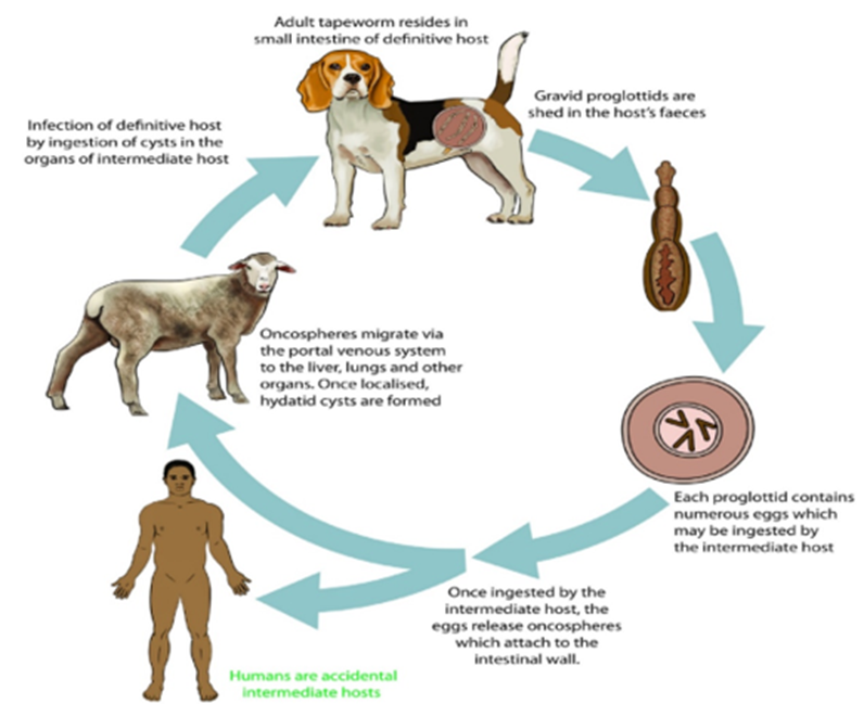
Figure 1: Life cycle of the Echinococcal granulosis.
The eggs release oncospheres, which adhere to the intestinal wall after being consumed by the intermediate host. Through the portal venous system, the oncospheres go from the colon to different organs where they form cysts and daughter cysts. 1-4 The liver accounts for 70% of cases,with the lungs coming in second with 20%. The kidneys, heart, and spleen, peritoneum, CNS , and bones are among the infrequent locations. The growth rate of hidatid cysts is sluggish, ranging from 1 to 10 mm annually; liver cysts grow more slowly than lung cysts (Tsoulfas et al., 2020). The CE cyst is a circular cystic lesion that is characterized by an exterior, acellular, laminated layer called the ectocyst and an interior, germinal layer called the endocyst. Little vesicles known as brood capsules are produced by the inner germinal layer; these capsules divide asexually to produce several protoscolices (Figure 2). The pericyst is a granulomatous, adventitial layer that surrounds the echinococcal laminated layer. This layer, which is frequently observed in imaging investigations, is created by the immunological system of the host to ward off the cystic infection (Calame et al., 2022).
Figure 2: Illustration of the layers of a liver hydatid cyst, comprising the outside (laminated) acellular layer, the inner (germinal) nucleated layer, and the granulomatous layer (adventitial layer), which is created by the host immune system to fend off the cystic infection. Here, the daughter cyst is visible floating inside the primary cyst, and the protocoled—the future head of the adult worm—is observed budding from the germinal layer. A sonographic finding that combines cystic fluid with protocolizes is called the hydatid sand (Souflas et al., 2020).
1.2 Echinococcal granuloses in livestock
The hydatid cysts in livestock's bodies develop after they consume these eggs through food, water, or vegetables. Hydatidosis is caused by the worm's larvae (cysts) establishing themselves in several organs, including the liver and lungs, and occasionally the brain, heart, and spinal cord of the intermediate host, such as humans, even though infection of carnivores with the worm's mature stage does not pose a specific threat. But a ruptured cyst can also lead to more serious problems, such as internal injuries and trauma. The quantity, size, and location of the developed cysts determine the clinical signs and symptoms of hydatidosis in people and animals. In humans, the disease is relevant because it affects vital organs like the liver and lungs, but in domestic cattle and livestock, it is significant because of the financial loss . Hydatidosis is regarded as one of the main health and economic issues because of the significant financial losses it causes in the cattle and public health sectors (Hosseini-Safa et al., 2016).
1.3 Clinical prestation
In the early stages of the disease, patients may have nonspecific signs and symptoms or be completely asymptomatic. Patients with intra-abdominal CE often show symptoms much later in the course of the illness. This could be because the cysts are growing slowly or because the granulomatous adventitial layer was initially used by the immunological system of the host to seal off the infection with cysts. Cystic development is more rapid in AIDS-positive CE patients, suggesting that an immunological condition may contribute to the progression of the disease (Wen et al., 2019). Hepatic mass, nausea, vomiting, and right hypochondrial discomfort are examples of chronic presenting problems. If the biliary system is compressed by a nearby liver cyst, obstructive jaundice may result. 2,4 Abdominal distension or hepatomegaly may be observed on a physical examination. When a cyst is large (diameter greater than 10 cm) or occupies 70% of the organ volume, signs of liver involvement usually appear ) (El Nakeeb et al., 2017). Patients with complex illnesses may also have spontaneous or sporadic cystic rupture following forceful abdominal trauma. Rupture is a potentially lethal complication, despite its uncommon manifestation. If burst cysts communicate with the biliary system, cholangiolitis may be present as a result of biliary tree obstruction. An immune response to a spontaneous intraperitoneal rupture may cause an allergic reaction, the most serious of which is anaphylactic shock. Additionally, live protoscolices may "seed" in the peritoneum as a result of cystic rupture, leading to secondary hydatidosis Rarely the breaking open of a liver cyst by the diaphragm may result in subsequent pulmonary involvement (Kern et al., 2017; Keong et al., 2018).
Diagnosis
To diagnose hepatic CE, a thorough clinical assessment of the patient is essential. This entails a comprehensive history that highlights any interactions with dogs, wildlife, or trips to endemic regions, as well as a targeted physical examination. When a patient presents with questionable clinical findings, additional laboratory and radiographic tests may be necessary to confirm the diagnosis (Junghanss et al., 2008).
Laboratory investigations
A useful supplement to radiographic studies in the diagnosis of liver hydatid disease is serology for CE. It does have certain drawbacks, though, such as serology's incapacity to differentiate between active and dormant cysts when the ultrasound results are unclear. Western blotting (WB), indirect hemagglutination assay (IHA), and enzyme-linked immunosorbent assay (ELISA) are three laboratory techniques that may be used to diagnose liver CE. For liver-only hydatid involvement, the gold standard serological test is thought to be the enzyme-linked immunosorbent assay (ELISA). E. granulosus-specific antigen is detected by immunoglobulin IgG (G) ELISA, which has a sensitivity of 93.5% and specificity of 89.7%., and 18 Nonetheless, cysts in their early stages wherein the endocyst contains E. granulosus antigens frequently have seronegative findings. The host's immune system's reaction to the parasite infection is then cut off from the antigens. When the cysts calcify in the later stages of the illness process, there is an identical lack of immune response that frequently yields a seronegative result (Keong et al., 2018; Calame et al., 2022). Eighty to one hundred percent sensitivity and the specificity of 88–96% are associated with WB serology for liver CE. The high cost of the test and the sharp decline in sensitivity rates in extra-hepatic illness are the drawbacks of WB.IHA testing has a 90% sensitivity rate, yet if the result is positive, it can stay that way for a number of years. Regular blood tests may reveal vague alterations. Liver function tests are abnormal in only around 40% of individuals. Alkaline phosphatase is frequently increased in cases of derangement, whether or not hyperbilirubinemia is present. In 25–40% of patients, a differential white cell count may reveal eosinophilia (Rashid et al., 2018; Tsoulfas et al., 2020).
Imaging
The preferred diagnostic method for liver CE is ultrasound imaging, which has a sensitivity of 90% to 95%. Liver CE can show up on ultrasonography as a solid mass or as a single, anechoic lens cyst with characteristics of a basic cyst, depending on the situation.20 Real-time imaging reveals a "falling snowflake pattern" as the patient shifts positions. This pattern is made by several echogenic foci that are created when cystic fluid and protoscolices are combined, commonly referred to as "hydatid sand (Kern et al., 2017). "Floating membranes" are the result of the endocyst's separation from the pericyst. Furthermore, a "wheel-spoke" form may result from a multivesicular mother cyst wit Hydatid cysts have been categorized by the World Health Organization Informal Working Groups about the WHO-IWGE Echinococosis according on sonographic characteristics. Cysts are categorized from CE1 to CE5 according to this categorization, which is based on many ultrasonography findings (Table 1). The CE1 and CE2 types correlate with the disease's "active" stage. According to Figure 3, CE3a and CE3b are in the "transitional" stage, whereas CE4 and CE5 are in the "inactive" stage. This method directs additional management in addition to aiding in the general classification of liver hydatid cysts. offspring cysts divided by radiating septae that depict the hydatid sand/matrix and the cyst walls (Fadel et al., 2019).

Figure 3: Imaging of liver cystic echinococcosis (Illuri et al., 2018).
Treatment
Routine care of hepatic patients involves considerations of the patient's symptoms, radiological stage, size and location of the cyst(s), presence of comorbidities, and treating physicians' ability (Velasco-Tirado et al., 2018). Medical therapy, a "watch and wait" strategy, percutaneous therapy, and surgical procedures are among the available management options (Tsoulfas et al., 2020).
Watch and wait’ approach
With interval ultrasonography monitoring, inactive, degenerating CE4 and all CE5 cysts can be seen without the need for medication or surgery (Mönnink et al., 2021).
Surgery
Options for liver surgery There are two types of CE approaches: radical and conservative (Figure). The radical treatment has drawn criticism for being a severe kind of therapy associated with high morbidity for a relatively benign disease process. It includes total cystectomy and hepatic resection. Additionally, patients must typically have surgery in a specialized hepatobiliary unit due to the high technical difficulty of the procedure.These factors make the conservative strategy with a partial cystectomy more popular. Under albendazole cover, this entails removing the cyst's contents, sterilizing the cavity that remains, and partially resecting the cyst (Vagholkar et al., 2016; Tsoulfas et al., 2020). The hepatic cyst(s) are accessed and exposed by a suitable incision in the partial cystectomy technique. Abdominal swabs soaked in a scolicidal substance (20% hypertonic saline) are used to protect the operative field. In the unlikely case of a spill during the evacuation of cyst-content, this helps to prevent contamination. Next, a selected scolicidal substance is injected into the cyst after it has been pierced and aspirated. Similar to PAIR, if the cyst aspirates bile, There is biliary communication and avoidance of the scolicidal agent. The contents are re-aspirated after 15 minutes, and the cyst is then opened and the endocyst's contents are suctioned out, Subsequently, the cyst is exposed by removing the cyst wall external to the liver parenchyma. A cysto-biliary fistula can be sutured closed if it is discovered during surgery. After the contents of the cystic cavity are removed, the cavity must be completely destroyed using either omentoplasty or capitonnage (Mihmanli et al., 2020). An intraoperative picture of a partial cystectomy with biliary connections for a liver hydatid cyst (Deo et al., 2020)

Figure 4: bile-stained floating membrane removed after a partial cystectomy is seen in an intraoperative picture (Tsoulfas et al., 2020).
Preventive strategies
Enacting legislation requiring the use of safe slaughter procedures and efficient canine deworming programs, as observed in New Zealand and Tasmania, can lead to the eradication of CE. By deworming their dogs, other nations including Uruguay, Chile, and Argentina saw a comparable decline in CE. The programs used supervised praziquantel deworming for dogs four to eight times a year (Craig et al., 2017). Vaccinating sheep with the EG95 vaccine is a more recent method to stop the spread of CE. As an extra intervention to lessen CE transmission, it is presently registered for usage in Argentina and China. For poorer nations where CE is typically endemic, this strategy is not feasible due to the high cost of the immunization. Although dog vaccination attempts have been documented, no quantifiable results have been demonstrated in terms of reducing the spread of CE (Wen et al., 2019). It is possible to take preventive actions to help lower the spread of CE. Public health initiatives including routine canine deworming programs, tightly enforced safe slaughter procedures reinforced by local laws, and public awareness campaigns regarding the illness process could help reduce the spread of the disease among endemic populations (Junghanss et al., 2008; Keong et al., 2018).
The true worldwide burden of CE is significantly underestimated since epidemiological data from endemic areas are scarce. Over two-thirds of cases of this disease process involve hepatic infection, making humans the unintentional intermediate hosts. Most symptoms and signs are non-specific, particularly in the early stages of the illness Consequently, especially in endemic areas, doctors ought to have a low bar for considering the diagnosis in patients with positive serology and suggestive radiological features. The patient's symptoms and the radiographic stage determine the standard course of treatment for liver CE , the cyst's location and size, any problems, and the knowledge of the medical professionals. The WHO's well-established public health protocols must be put into practice in order to reduce the substantial yearly expenses related to CE. Even though the condition is most common in developing nations, increased migration and travel force medical professionals to include CE in their differential diagnosis for any worrisome liver cyst.
Clearly Auctoresonline and particularly Psychology and Mental Health Care Journal is dedicated to improving health care services for individuals and populations. The editorial boards' ability to efficiently recognize and share the global importance of health literacy with a variety of stakeholders. Auctoresonline publishing platform can be used to facilitate of optimal client-based services and should be added to health care professionals' repertoire of evidence-based health care resources.

Journal of Clinical Cardiology and Cardiovascular Intervention The submission and review process was adequate. However I think that the publication total value should have been enlightened in early fases. Thank you for all.

Journal of Women Health Care and Issues By the present mail, I want to say thank to you and tour colleagues for facilitating my published article. Specially thank you for the peer review process, support from the editorial office. I appreciate positively the quality of your journal.

Journal of Clinical Research and Reports I would be very delighted to submit my testimonial regarding the reviewer board and the editorial office. The reviewer board were accurate and helpful regarding any modifications for my manuscript. And the editorial office were very helpful and supportive in contacting and monitoring with any update and offering help. It was my pleasure to contribute with your promising Journal and I am looking forward for more collaboration.

We would like to thank the Journal of Thoracic Disease and Cardiothoracic Surgery because of the services they provided us for our articles. The peer-review process was done in a very excellent time manner, and the opinions of the reviewers helped us to improve our manuscript further. The editorial office had an outstanding correspondence with us and guided us in many ways. During a hard time of the pandemic that is affecting every one of us tremendously, the editorial office helped us make everything easier for publishing scientific work. Hope for a more scientific relationship with your Journal.

The peer-review process which consisted high quality queries on the paper. I did answer six reviewers’ questions and comments before the paper was accepted. The support from the editorial office is excellent.

Journal of Neuroscience and Neurological Surgery. I had the experience of publishing a research article recently. The whole process was simple from submission to publication. The reviewers made specific and valuable recommendations and corrections that improved the quality of my publication. I strongly recommend this Journal.

Dr. Katarzyna Byczkowska My testimonial covering: "The peer review process is quick and effective. The support from the editorial office is very professional and friendly. Quality of the Clinical Cardiology and Cardiovascular Interventions is scientific and publishes ground-breaking research on cardiology that is useful for other professionals in the field.

Thank you most sincerely, with regard to the support you have given in relation to the reviewing process and the processing of my article entitled "Large Cell Neuroendocrine Carcinoma of The Prostate Gland: A Review and Update" for publication in your esteemed Journal, Journal of Cancer Research and Cellular Therapeutics". The editorial team has been very supportive.

Testimony of Journal of Clinical Otorhinolaryngology: work with your Reviews has been a educational and constructive experience. The editorial office were very helpful and supportive. It was a pleasure to contribute to your Journal.

Dr. Bernard Terkimbi Utoo, I am happy to publish my scientific work in Journal of Women Health Care and Issues (JWHCI). The manuscript submission was seamless and peer review process was top notch. I was amazed that 4 reviewers worked on the manuscript which made it a highly technical, standard and excellent quality paper. I appreciate the format and consideration for the APC as well as the speed of publication. It is my pleasure to continue with this scientific relationship with the esteem JWHCI.

This is an acknowledgment for peer reviewers, editorial board of Journal of Clinical Research and Reports. They show a lot of consideration for us as publishers for our research article “Evaluation of the different factors associated with side effects of COVID-19 vaccination on medical students, Mutah university, Al-Karak, Jordan”, in a very professional and easy way. This journal is one of outstanding medical journal.

Dear Hao Jiang, to Journal of Nutrition and Food Processing We greatly appreciate the efficient, professional and rapid processing of our paper by your team. If there is anything else we should do, please do not hesitate to let us know. On behalf of my co-authors, we would like to express our great appreciation to editor and reviewers.

As an author who has recently published in the journal "Brain and Neurological Disorders". I am delighted to provide a testimonial on the peer review process, editorial office support, and the overall quality of the journal. The peer review process at Brain and Neurological Disorders is rigorous and meticulous, ensuring that only high-quality, evidence-based research is published. The reviewers are experts in their fields, and their comments and suggestions were constructive and helped improve the quality of my manuscript. The review process was timely and efficient, with clear communication from the editorial office at each stage. The support from the editorial office was exceptional throughout the entire process. The editorial staff was responsive, professional, and always willing to help. They provided valuable guidance on formatting, structure, and ethical considerations, making the submission process seamless. Moreover, they kept me informed about the status of my manuscript and provided timely updates, which made the process less stressful. The journal Brain and Neurological Disorders is of the highest quality, with a strong focus on publishing cutting-edge research in the field of neurology. The articles published in this journal are well-researched, rigorously peer-reviewed, and written by experts in the field. The journal maintains high standards, ensuring that readers are provided with the most up-to-date and reliable information on brain and neurological disorders. In conclusion, I had a wonderful experience publishing in Brain and Neurological Disorders. The peer review process was thorough, the editorial office provided exceptional support, and the journal's quality is second to none. I would highly recommend this journal to any researcher working in the field of neurology and brain disorders.
Dear Agrippa Hilda, Journal of Neuroscience and Neurological Surgery, Editorial Coordinator, I trust this message finds you well. I want to extend my appreciation for considering my article for publication in your esteemed journal. I am pleased to provide a testimonial regarding the peer review process and the support received from your editorial office. The peer review process for my paper was carried out in a highly professional and thorough manner. The feedback and comments provided by the authors were constructive and very useful in improving the quality of the manuscript. This rigorous assessment process undoubtedly contributes to the high standards maintained by your journal.

International Journal of Clinical Case Reports and Reviews. I strongly recommend to consider submitting your work to this high-quality journal. The support and availability of the Editorial staff is outstanding and the review process was both efficient and rigorous.

Thank you very much for publishing my Research Article titled “Comparing Treatment Outcome Of Allergic Rhinitis Patients After Using Fluticasone Nasal Spray And Nasal Douching" in the Journal of Clinical Otorhinolaryngology. As Medical Professionals we are immensely benefited from study of various informative Articles and Papers published in this high quality Journal. I look forward to enriching my knowledge by regular study of the Journal and contribute my future work in the field of ENT through the Journal for use by the medical fraternity. The support from the Editorial office was excellent and very prompt. I also welcome the comments received from the readers of my Research Article.

Dear Erica Kelsey, Editorial Coordinator of Cancer Research and Cellular Therapeutics Our team is very satisfied with the processing of our paper by your journal. That was fast, efficient, rigorous, but without unnecessary complications. We appreciated the very short time between the submission of the paper and its publication on line on your site.

I am very glad to say that the peer review process is very successful and fast and support from the Editorial Office. Therefore, I would like to continue our scientific relationship for a long time. And I especially thank you for your kindly attention towards my article. Have a good day!

"We recently published an article entitled “Influence of beta-Cyclodextrins upon the Degradation of Carbofuran Derivatives under Alkaline Conditions" in the Journal of “Pesticides and Biofertilizers” to show that the cyclodextrins protect the carbamates increasing their half-life time in the presence of basic conditions This will be very helpful to understand carbofuran behaviour in the analytical, agro-environmental and food areas. We greatly appreciated the interaction with the editor and the editorial team; we were particularly well accompanied during the course of the revision process, since all various steps towards publication were short and without delay".

I would like to express my gratitude towards you process of article review and submission. I found this to be very fair and expedient. Your follow up has been excellent. I have many publications in national and international journal and your process has been one of the best so far. Keep up the great work.

We are grateful for this opportunity to provide a glowing recommendation to the Journal of Psychiatry and Psychotherapy. We found that the editorial team were very supportive, helpful, kept us abreast of timelines and over all very professional in nature. The peer review process was rigorous, efficient and constructive that really enhanced our article submission. The experience with this journal remains one of our best ever and we look forward to providing future submissions in the near future.

I am very pleased to serve as EBM of the journal, I hope many years of my experience in stem cells can help the journal from one way or another. As we know, stem cells hold great potential for regenerative medicine, which are mostly used to promote the repair response of diseased, dysfunctional or injured tissue using stem cells or their derivatives. I think Stem Cell Research and Therapeutics International is a great platform to publish and share the understanding towards the biology and translational or clinical application of stem cells.

I would like to give my testimony in the support I have got by the peer review process and to support the editorial office where they were of asset to support young author like me to be encouraged to publish their work in your respected journal and globalize and share knowledge across the globe. I really give my great gratitude to your journal and the peer review including the editorial office.

I am delighted to publish our manuscript entitled "A Perspective on Cocaine Induced Stroke - Its Mechanisms and Management" in the Journal of Neuroscience and Neurological Surgery. The peer review process, support from the editorial office, and quality of the journal are excellent. The manuscripts published are of high quality and of excellent scientific value. I recommend this journal very much to colleagues.

Dr.Tania Muñoz, My experience as researcher and author of a review article in The Journal Clinical Cardiology and Interventions has been very enriching and stimulating. The editorial team is excellent, performs its work with absolute responsibility and delivery. They are proactive, dynamic and receptive to all proposals. Supporting at all times the vast universe of authors who choose them as an option for publication. The team of review specialists, members of the editorial board, are brilliant professionals, with remarkable performance in medical research and scientific methodology. Together they form a frontline team that consolidates the JCCI as a magnificent option for the publication and review of high-level medical articles and broad collective interest. I am honored to be able to share my review article and open to receive all your comments.

“The peer review process of JPMHC is quick and effective. Authors are benefited by good and professional reviewers with huge experience in the field of psychology and mental health. The support from the editorial office is very professional. People to contact to are friendly and happy to help and assist any query authors might have. Quality of the Journal is scientific and publishes ground-breaking research on mental health that is useful for other professionals in the field”.

Dear editorial department: On behalf of our team, I hereby certify the reliability and superiority of the International Journal of Clinical Case Reports and Reviews in the peer review process, editorial support, and journal quality. Firstly, the peer review process of the International Journal of Clinical Case Reports and Reviews is rigorous, fair, transparent, fast, and of high quality. The editorial department invites experts from relevant fields as anonymous reviewers to review all submitted manuscripts. These experts have rich academic backgrounds and experience, and can accurately evaluate the academic quality, originality, and suitability of manuscripts. The editorial department is committed to ensuring the rigor of the peer review process, while also making every effort to ensure a fast review cycle to meet the needs of authors and the academic community. Secondly, the editorial team of the International Journal of Clinical Case Reports and Reviews is composed of a group of senior scholars and professionals with rich experience and professional knowledge in related fields. The editorial department is committed to assisting authors in improving their manuscripts, ensuring their academic accuracy, clarity, and completeness. Editors actively collaborate with authors, providing useful suggestions and feedback to promote the improvement and development of the manuscript. We believe that the support of the editorial department is one of the key factors in ensuring the quality of the journal. Finally, the International Journal of Clinical Case Reports and Reviews is renowned for its high- quality articles and strict academic standards. The editorial department is committed to publishing innovative and academically valuable research results to promote the development and progress of related fields. The International Journal of Clinical Case Reports and Reviews is reasonably priced and ensures excellent service and quality ratio, allowing authors to obtain high-level academic publishing opportunities in an affordable manner. I hereby solemnly declare that the International Journal of Clinical Case Reports and Reviews has a high level of credibility and superiority in terms of peer review process, editorial support, reasonable fees, and journal quality. Sincerely, Rui Tao.

Clinical Cardiology and Cardiovascular Interventions I testity the covering of the peer review process, support from the editorial office, and quality of the journal.

Clinical Cardiology and Cardiovascular Interventions, we deeply appreciate the interest shown in our work and its publication. It has been a true pleasure to collaborate with you. The peer review process, as well as the support provided by the editorial office, have been exceptional, and the quality of the journal is very high, which was a determining factor in our decision to publish with you.

The peer reviewers process is quick and effective, the supports from editorial office is excellent, the quality of journal is high. I would like to collabroate with Internatioanl journal of Clinical Case Reports and Reviews journal clinically in the future time.

Clinical Cardiology and Cardiovascular Interventions, I would like to express my sincerest gratitude for the trust placed in our team for the publication in your journal. It has been a true pleasure to collaborate with you on this project. I am pleased to inform you that both the peer review process and the attention from the editorial coordination have been excellent. Your team has worked with dedication and professionalism to ensure that your publication meets the highest standards of quality. We are confident that this collaboration will result in mutual success, and we are eager to see the fruits of this shared effort.

Dear Dr. Jessica Magne, Editorial Coordinator 0f Clinical Cardiology and Cardiovascular Interventions, I hope this message finds you well. I want to express my utmost gratitude for your excellent work and for the dedication and speed in the publication process of my article titled "Navigating Innovation: Qualitative Insights on Using Technology for Health Education in Acute Coronary Syndrome Patients." I am very satisfied with the peer review process, the support from the editorial office, and the quality of the journal. I hope we can maintain our scientific relationship in the long term.

Dear Monica Gissare, - Editorial Coordinator of Nutrition and Food Processing. ¨My testimony with you is truly professional, with a positive response regarding the follow-up of the article and its review, you took into account my qualities and the importance of the topic¨.

Dear Dr. Jessica Magne, Editorial Coordinator 0f Clinical Cardiology and Cardiovascular Interventions, The review process for the article “The Handling of Anti-aggregants and Anticoagulants in the Oncologic Heart Patient Submitted to Surgery” was extremely rigorous and detailed. From the initial submission to the final acceptance, the editorial team at the “Journal of Clinical Cardiology and Cardiovascular Interventions” demonstrated a high level of professionalism and dedication. The reviewers provided constructive and detailed feedback, which was essential for improving the quality of our work. Communication was always clear and efficient, ensuring that all our questions were promptly addressed. The quality of the “Journal of Clinical Cardiology and Cardiovascular Interventions” is undeniable. It is a peer-reviewed, open-access publication dedicated exclusively to disseminating high-quality research in the field of clinical cardiology and cardiovascular interventions. The journal's impact factor is currently under evaluation, and it is indexed in reputable databases, which further reinforces its credibility and relevance in the scientific field. I highly recommend this journal to researchers looking for a reputable platform to publish their studies.

Dear Editorial Coordinator of the Journal of Nutrition and Food Processing! "I would like to thank the Journal of Nutrition and Food Processing for including and publishing my article. The peer review process was very quick, movement and precise. The Editorial Board has done an extremely conscientious job with much help, valuable comments and advices. I find the journal very valuable from a professional point of view, thank you very much for allowing me to be part of it and I would like to participate in the future!”

Dealing with The Journal of Neurology and Neurological Surgery was very smooth and comprehensive. The office staff took time to address my needs and the response from editors and the office was prompt and fair. I certainly hope to publish with this journal again.Their professionalism is apparent and more than satisfactory. Susan Weiner

My Testimonial Covering as fellowing: Lin-Show Chin. The peer reviewers process is quick and effective, the supports from editorial office is excellent, the quality of journal is high. I would like to collabroate with Internatioanl journal of Clinical Case Reports and Reviews.

My experience publishing in Psychology and Mental Health Care was exceptional. The peer review process was rigorous and constructive, with reviewers providing valuable insights that helped enhance the quality of our work. The editorial team was highly supportive and responsive, making the submission process smooth and efficient. The journal's commitment to high standards and academic rigor makes it a respected platform for quality research. I am grateful for the opportunity to publish in such a reputable journal.

My experience publishing in International Journal of Clinical Case Reports and Reviews was exceptional. I Come forth to Provide a Testimonial Covering the Peer Review Process and the editorial office for the Professional and Impartial Evaluation of the Manuscript.

I would like to offer my testimony in the support. I have received through the peer review process and support the editorial office where they are to support young authors like me, encourage them to publish their work in your esteemed journals, and globalize and share knowledge globally. I really appreciate your journal, peer review, and editorial office.

Dear Agrippa Hilda- Editorial Coordinator of Journal of Neuroscience and Neurological Surgery, "The peer review process was very quick and of high quality, which can also be seen in the articles in the journal. The collaboration with the editorial office was very good."

I would like to express my sincere gratitude for the support and efficiency provided by the editorial office throughout the publication process of my article, “Delayed Vulvar Metastases from Rectal Carcinoma: A Case Report.” I greatly appreciate the assistance and guidance I received from your team, which made the entire process smooth and efficient. The peer review process was thorough and constructive, contributing to the overall quality of the final article. I am very grateful for the high level of professionalism and commitment shown by the editorial staff, and I look forward to maintaining a long-term collaboration with the International Journal of Clinical Case Reports and Reviews.

To Dear Erin Aust, I would like to express my heartfelt appreciation for the opportunity to have my work published in this esteemed journal. The entire publication process was smooth and well-organized, and I am extremely satisfied with the final result. The Editorial Team demonstrated the utmost professionalism, providing prompt and insightful feedback throughout the review process. Their clear communication and constructive suggestions were invaluable in enhancing my manuscript, and their meticulous attention to detail and dedication to quality are truly commendable. Additionally, the support from the Editorial Office was exceptional. From the initial submission to the final publication, I was guided through every step of the process with great care and professionalism. The team's responsiveness and assistance made the entire experience both easy and stress-free. I am also deeply impressed by the quality and reputation of the journal. It is an honor to have my research featured in such a respected publication, and I am confident that it will make a meaningful contribution to the field.

"I am grateful for the opportunity of contributing to [International Journal of Clinical Case Reports and Reviews] and for the rigorous review process that enhances the quality of research published in your esteemed journal. I sincerely appreciate the time and effort of your team who have dedicatedly helped me in improvising changes and modifying my manuscript. The insightful comments and constructive feedback provided have been invaluable in refining and strengthening my work".

I thank the ‘Journal of Clinical Research and Reports’ for accepting this article for publication. This is a rigorously peer reviewed journal which is on all major global scientific data bases. I note the review process was prompt, thorough and professionally critical. It gave us an insight into a number of important scientific/statistical issues. The review prompted us to review the relevant literature again and look at the limitations of the study. The peer reviewers were open, clear in the instructions and the editorial team was very prompt in their communication. This journal certainly publishes quality research articles. I would recommend the journal for any future publications.

Dear Jessica Magne, with gratitude for the joint work. Fast process of receiving and processing the submitted scientific materials in “Clinical Cardiology and Cardiovascular Interventions”. High level of competence of the editors with clear and correct recommendations and ideas for enriching the article.

We found the peer review process quick and positive in its input. The support from the editorial officer has been very agile, always with the intention of improving the article and taking into account our subsequent corrections.

My article, titled 'No Way Out of the Smartphone Epidemic Without Considering the Insights of Brain Research,' has been republished in the International Journal of Clinical Case Reports and Reviews. The review process was seamless and professional, with the editors being both friendly and supportive. I am deeply grateful for their efforts.

To Dear Erin Aust – Editorial Coordinator of Journal of General Medicine and Clinical Practice! I declare that I am absolutely satisfied with your work carried out with great competence in following the manuscript during the various stages from its receipt, during the revision process to the final acceptance for publication. Thank Prof. Elvira Farina

Dear Jessica, and the super professional team of the ‘Clinical Cardiology and Cardiovascular Interventions’ I am sincerely grateful to the coordinated work of the journal team for the no problem with the submission of my manuscript: “Cardiometabolic Disorders in A Pregnant Woman with Severe Preeclampsia on the Background of Morbid Obesity (Case Report).” The review process by 5 experts was fast, and the comments were professional, which made it more specific and academic, and the process of publication and presentation of the article was excellent. I recommend that my colleagues publish articles in this journal, and I am interested in further scientific cooperation. Sincerely and best wishes, Dr. Oleg Golyanovskiy.

Dear Ashley Rosa, Editorial Coordinator of the journal - Psychology and Mental Health Care. " The process of obtaining publication of my article in the Psychology and Mental Health Journal was positive in all areas. The peer review process resulted in a number of valuable comments, the editorial process was collaborative and timely, and the quality of this journal has been quickly noticed, resulting in alternative journals contacting me to publish with them." Warm regards, Susan Anne Smith, PhD. Australian Breastfeeding Association.

Dear Jessica Magne, Editorial Coordinator, Clinical Cardiology and Cardiovascular Interventions, Auctores Publishing LLC. I appreciate the journal (JCCI) editorial office support, the entire team leads were always ready to help, not only on technical front but also on thorough process. Also, I should thank dear reviewers’ attention to detail and creative approach to teach me and bring new insights by their comments. Surely, more discussions and introduction of other hemodynamic devices would provide better prevention and management of shock states. Your efforts and dedication in presenting educational materials in this journal are commendable. Best wishes from, Farahnaz Fallahian.

Dear Maria Emerson, Editorial Coordinator, International Journal of Clinical Case Reports and Reviews, Auctores Publishing LLC. I am delighted to have published our manuscript, "Acute Colonic Pseudo-Obstruction (ACPO): A rare but serious complication following caesarean section." I want to thank the editorial team, especially Maria Emerson, for their prompt review of the manuscript, quick responses to queries, and overall support. Yours sincerely Dr. Victor Olagundoye.

Dear Ashley Rosa, Editorial Coordinator, International Journal of Clinical Case Reports and Reviews. Many thanks for publishing this manuscript after I lost confidence the editors were most helpful, more than other journals Best wishes from, Susan Anne Smith, PhD. Australian Breastfeeding Association.

Dear Agrippa Hilda, Editorial Coordinator, Journal of Neuroscience and Neurological Surgery. The entire process including article submission, review, revision, and publication was extremely easy. The journal editor was prompt and helpful, and the reviewers contributed to the quality of the paper. Thank you so much! Eric Nussbaum, MD

Dr Hala Al Shaikh This is to acknowledge that the peer review process for the article ’ A Novel Gnrh1 Gene Mutation in Four Omani Male Siblings, Presentation and Management ’ sent to the International Journal of Clinical Case Reports and Reviews was quick and smooth. The editorial office was prompt with easy communication.

Dear Erin Aust, Editorial Coordinator, Journal of General Medicine and Clinical Practice. We are pleased to share our experience with the “Journal of General Medicine and Clinical Practice”, following the successful publication of our article. The peer review process was thorough and constructive, helping to improve the clarity and quality of the manuscript. We are especially thankful to Ms. Erin Aust, the Editorial Coordinator, for her prompt communication and continuous support throughout the process. Her professionalism ensured a smooth and efficient publication experience. The journal upholds high editorial standards, and we highly recommend it to fellow researchers seeking a credible platform for their work. Best wishes By, Dr. Rakhi Mishra.

Dear Jessica Magne, Editorial Coordinator, Clinical Cardiology and Cardiovascular Interventions, Auctores Publishing LLC. The peer review process of the journal of Clinical Cardiology and Cardiovascular Interventions was excellent and fast, as was the support of the editorial office and the quality of the journal. Kind regards Walter F. Riesen Prof. Dr. Dr. h.c. Walter F. Riesen.

Dear Ashley Rosa, Editorial Coordinator, International Journal of Clinical Case Reports and Reviews, Auctores Publishing LLC. Thank you for publishing our article, Exploring Clozapine's Efficacy in Managing Aggression: A Multiple Single-Case Study in Forensic Psychiatry in the international journal of clinical case reports and reviews. We found the peer review process very professional and efficient. The comments were constructive, and the whole process was efficient. On behalf of the co-authors, I would like to thank you for publishing this article. With regards, Dr. Jelle R. Lettinga.

Dear Clarissa Eric, Editorial Coordinator, Journal of Clinical Case Reports and Studies, I would like to express my deep admiration for the exceptional professionalism demonstrated by your journal. I am thoroughly impressed by the speed of the editorial process, the substantive and insightful reviews, and the meticulous preparation of the manuscript for publication. Additionally, I greatly appreciate the courteous and immediate responses from your editorial office to all my inquiries. Best Regards, Dariusz Ziora

Dear Chrystine Mejia, Editorial Coordinator, Journal of Neurodegeneration and Neurorehabilitation, Auctores Publishing LLC, We would like to thank the editorial team for the smooth and high-quality communication leading up to the publication of our article in the Journal of Neurodegeneration and Neurorehabilitation. The reviewers have extensive knowledge in the field, and their relevant questions helped to add value to our publication. Kind regards, Dr. Ravi Shrivastava.

Dear Clarissa Eric, Editorial Coordinator, Journal of Clinical Case Reports and Studies, Auctores Publishing LLC, USA Office: +1-(302)-520-2644. I would like to express my sincere appreciation for the efficient and professional handling of my case report by the ‘Journal of Clinical Case Reports and Studies’. The peer review process was not only fast but also highly constructive—the reviewers’ comments were clear, relevant, and greatly helped me improve the quality and clarity of my manuscript. I also received excellent support from the editorial office throughout the process. Communication was smooth and timely, and I felt well guided at every stage, from submission to publication. The overall quality and rigor of the journal are truly commendable. I am pleased to have published my work with Journal of Clinical Case Reports and Studies, and I look forward to future opportunities for collaboration. Sincerely, Aline Tollet, UCLouvain.

Dear Ms. Mayra Duenas, Editorial Coordinator, International Journal of Clinical Case Reports and Reviews. “The International Journal of Clinical Case Reports and Reviews represented the “ideal house” to share with the research community a first experience with the use of the Simeox device for speech rehabilitation. High scientific reputation and attractive website communication were first determinants for the selection of this Journal, and the following submission process exceeded expectations: fast but highly professional peer review, great support by the editorial office, elegant graphic layout. Exactly what a dynamic research team - also composed by allied professionals - needs!" From, Chiara Beccaluva, PT - Italy.

Dear Maria Emerson, Editorial Coordinator, we have deeply appreciated the professionalism demonstrated by the International Journal of Clinical Case Reports and Reviews. The reviewers have extensive knowledge of our field and have been very efficient and fast in supporting the process. I am really looking forward to further collaboration. Thanks. Best regards, Dr. Claudio Ligresti

Dear Chrystine Mejia, Editorial Coordinator, Journal of Neurodegeneration and Neurorehabilitation. “The peer review process was efficient and constructive, and the editorial office provided excellent communication and support throughout. The journal ensures scientific rigor and high editorial standards, while also offering a smooth and timely publication process. We sincerely appreciate the work of the editorial team in facilitating the dissemination of innovative approaches such as the Bonori Method.” Best regards, Dr. Matteo Bonori.

I recommend without hesitation submitting relevant papers on medical decision making to the International Journal of Clinical Case Reports and Reviews. I am very grateful to the editorial staff. Maria Emerson was a pleasure to communicate with. The time from submission to publication was an extremely short 3 weeks. The editorial staff submitted the paper to three reviewers. Two of the reviewers commented positively on the value of publishing the paper. The editorial staff quickly recognized the third reviewer’s comments as an unjust attempt to reject the paper. I revised the paper as recommended by the first two reviewers.

Dear Maria Emerson, Editorial Coordinator, Journal of Clinical Research and Reports. Thank you for publishing our case report: "Clinical Case of Effective Fetal Stem Cells Treatment in a Patient with Autism Spectrum Disorder" within the "Journal of Clinical Research and Reports" being submitted by the team of EmCell doctors from Kyiv, Ukraine. We much appreciate a professional and transparent peer-review process from Auctores. All research Doctors are so grateful to your Editorial Office and Auctores Publishing support! I amiably wish our article publication maintained a top quality of your International Scientific Journal. My best wishes for a prosperity of the Journal of Clinical Research and Reports. Hope our scientific relationship and cooperation will remain long lasting. Thank you very much indeed. Kind regards, Dr. Andriy Sinelnyk Cell Therapy Center EmCell

Dear Editorial Team, Clinical Cardiology and Cardiovascular Interventions. It was truly a rewarding experience to work with the journal “Clinical Cardiology and Cardiovascular Interventions”. The peer review process was insightful and encouraging, helping us refine our work to a higher standard. The editorial office offered exceptional support with prompt and thoughtful communication. I highly value the journal’s role in promoting scientific advancement and am honored to be part of it. Best regards, Meng-Jou Lee, MD, Department of Anesthesiology, National Taiwan University Hospital.

Dear Editorial Team, Journal-Clinical Cardiology and Cardiovascular Interventions, “Publishing my article with Clinical Cardiology and Cardiovascular Interventions has been a highly positive experience. The peer-review process was rigorous yet supportive, offering valuable feedback that strengthened my work. The editorial team demonstrated exceptional professionalism, prompt communication, and a genuine commitment to maintaining the highest scientific standards. I am very pleased with the publication quality and proud to be associated with such a reputable journal.” Warm regards, Dr. Mahmoud Kamal Moustafa Ahmed

Dear Maria Emerson, Editorial Coordinator of ‘International Journal of Clinical Case Reports and Reviews’, I appreciate the opportunity to publish my article with your journal. The editorial office provided clear communication during the submission and review process, and I found the overall experience professional and constructive. Best regards, Elena Salvatore.

Dear Mayra Duenas, Editorial Coordinator of ‘International Journal of Clinical Case Reports and Reviews Herewith I confirm an optimal peer review process and a great support of the editorial office of the present journal

Dear Editorial Team, Clinical Cardiology and Cardiovascular Interventions. I am really grateful for the peers review; their feedback gave me the opportunity to reflect on the message and impact of my work and to ameliorate the article. The editors did a great job in addition by encouraging me to continue with the process of publishing.

Dear Cecilia Lilly, Editorial Coordinator, Endocrinology and Disorders, Thank you so much for your quick response regarding reviewing and all process till publishing our manuscript entitled: Prevalence of Pre-Diabetes and its Associated Risk Factors Among Nile College Students, Sudan. Best regards, Dr Mamoun Magzoub.

International Journal of Clinical Case Reports and Reviews is a high quality journal that has a clear and concise submission process. The peer review process was comprehensive and constructive. Support from the editorial office was excellent, since the administrative staff were responsive. The journal provides a fast and timely publication timeline.

Dear Maria Emerson, Editorial Coordinator of International Journal of Clinical Case Reports and Reviews, What distinguishes International Journal of Clinical Case Report and Review is not only the scientific rigor of its publications, but the intellectual climate in which research is evaluated. The submission process is refreshingly free of unnecessary formal barriers and bureaucratic rituals that often complicate academic publishing without adding real value. The peer-review system is demanding yet constructive, guided by genuine scientific dialogue rather than hierarchical or authoritarian attitudes. Reviewers act as collaborators in improving the manuscript, not as gatekeepers imposing arbitrary standards. This journal offers a rare balance: high methodological standards combined with a respectful, transparent, and supportive editorial approach. In an era where publishing can feel more burdensome than research itself, this platform restores the original purpose of peer review — to refine ideas, not to obstruct them Prof. Perlat Kapisyzi, FCCP PULMONOLOGIST AND THORACIC IMAGING.

Dear Mayra Duenas, Editorial Coordinator of the journal IJCCR, I write here a little on my experience as an author submitting to the International Journal of Clinical Case Reports and Reviews (IJCCR). This was my first submission to IJCCR and my manuscript was inherently an outsider’s effort. It attempted to broadly identify and then make some sense of life’s under-appreciated mysteries. I initially had responded to a request for possible submissions. I then contacted IJCCR with a tentative topic for a manuscript. They quickly got back with an approval for the submission, but with a particular requirement that it be medically relevant. I then put together a manuscript and submitted it. After the usual back-and-forth over forms and formality, the manuscript was sent off for reviews. Within 2 weeks I got back 4 reviews which were both helpful and also surprising. Surprising in that the topic was somewhat foreign to medical literature. My subsequent updates in response to the reviewer comments went smoothly and in short order I had a series of proofs to evaluate. All in all, the whole publication process seemed outstanding. It was both helpful in terms of the paper’s content and also in terms of its efficient and friendly communications. Thank you all very much. Sincerely, Ted Christopher, Rochester, NY.

Dear Grace Pierce, Editorial Coordinator of the journal IJCCR, I had a very positive experience with Auctores - Journal throughout the publication process. The Editorial Team was highly responsive, professional, and supportive at every stage. I would like to extend my sincere thanks to the Editor: Grace Pierce, for her guidance and assistance. The peer-review process was smooth and constructive, helping improve the quality of my work. I would gladly recommend Auctores Journal to fellow researchers and authors. Dr. SABITA SINHA, Medical Oncologist, MD (Electro Homeopathy).

Dear Maria Emerson, Editorial Coordinator of - Journal of Clinical Research and Reports. ''I am pleased to provide this testimonial following the publication of our recent case report in this journal. The peer review process was rigorous, constructive, thorough, and conducted in a timely manner. The reviewers’ comments were thoughtful, detailed, and highly constructive, contributing substantially to the refinement, clarity, and scientific robustness of our manuscript. The process was conducted with professionalism and academic integrity throughout. The support provided by the editorial office was exemplary. Communication was consistently prompt, clear, and courteous at all stages of the submission and publication process. The editorial team demonstrated a high level of organization and responsiveness, ensuring that all queries were addressed efficiently and that the process remained transparent and well-coordinated. The overall quality of the journal is reflected in its strong editorial standards, commitment to scientific excellence, and dedication to publishing clinically meaningful research. It has been a privilege to publish our work in this journal, and we would welcome the opportunity to contribute further in the future.'' Best wishes from, Dr. Efstratios Trogkanis, Cardiologist.

Dear Reader: We have published several articles in the Auctores Publishing, LLC, journal, Clinical Medical Reviews and Reports in recent years (CMRR). This is an ‘open access’ journal and the following are our observations. From the initial invitation to submit an article, to the final edits of galley proofs, we have found CMRR personnel to be professional, responsive, rapid and thorough. This entire process begins with Catherine Mitchell, Editorial Coordinator. She is simply outstanding, and, I believe, unparalleled in her capacity. I cannot imagine a more responsive and dedicated Editorial Coordinator. As I read the dates and timing of her correspondence with us, it seems that she never sleeps. I hope Auctores Publishing, LLC, appreciates her efforts as much as these authors do. Thank you to Auctores Publishing, LLC, to the Editorial Staff/Board, and to Catherine Mitchell from a grateful author(s).
