AUCTORES
Globalize your Research

Research Article | DOI: https://doi.org/10.31579/2641-0419/085
1 Cardiothoracic Sciences Centre All India Institute of Medical Sciences, New Delhi, India
*Corresponding Author: Ujjwal Kumar Chowdhury, M.Ch., Diplomate NB Professor Department of Cardiothoracic and Vascular Surgery AIIMS, New Delhi-110029, INDIA.
Citation: Ujjwal K. Chowdhury., Diplomate NB., Ramesh Menon, Niwin George, Lakshmi K. Sankhyan., (2020) Use of Transtracheal Oxygen following Decannulation of Pediatric Tracheostomy. J, Clinical Cardiology and Cardiovascular Interventions, 3(10); Doi:10.31579/2641-0419/085
Copyright: © 2020 Ujjwal Kumar Chowdhury, This is an open access article distributed under the Creative Commons Attribution License, which permits unrestricted use, distribution, and reproduction in any medium, provided the original work is properly cited.
Received: 22 September 2020 | Accepted: 30 October 2020 | Published: 04 November 2020
Keywords: congenital heart disease; congestive heart failure; tracheostomy; decannulation of tracheostomy tube; transtracheal oxygenation; tracheobronchomalacia
Purpose: Uninterrupted sustained oxygenation is paramount in neonates and infants with cyanotic/acyanotic congenital heart diseases (CHD) undergoing closed or open heart surgeries and tracheostomy tube decannulation to avoid hypoxic events.
Description: We describe here-in a new device, permitting uninterrupted delivery of oxygen through the tracheostomy stoma, allowing continuation of enteral feeds and suctioning of the endotracheal secretions through the tracheostomy stoma.
Evaluation: Eighty-four neonates and infants with a median age of four months (IQR:23 days-9 months) undergoing different closed and open heart surgeries for cyanotic/acyanotic CHD with or without pulmonary arterial hypertension were treated with a device permitting uninterrupted oxygenation following tracheostomy tube decannulation. There were 11 (13.1%) deaths due to multifactorial etiologies, and one was lost to follow-up. Seventy-two children were successfully decannulated using this protocol. At a median follow-up of 166(IQR:82.5-216) months, the actuarial survival was 86.61% (SE±0.04%; 95% CI: 77.1-92.3).
Conclusions: Transtracheal oxygenation through the tracheostomy stoma via a thin catheter allows uninterrupted oxygenation following tracheostomy tube decannulation, continuation of enteral feeds, and allows intermittent endotracheal suctioning, thus avoiding post decannulation hypoxic events.
Running title: Transtracheal oxygenation
Despite advances in pediatric anaesthesia, intensive care and myocardial protection during the past decade, mortality attributed to pediatric tracheostomy ranges from 0.5 to 5% with European and American reviews.[1-3]
Decannulation as soon as the child’s condition permit is always desirable to avert the extubation-reintubation cycle, mediastinitis, tracheostomy site breakdown, peristomal granulation, suprastomal granuloma, tracheitis, subglottic stenosis, tracheobronchomalacia, tracheobrachiocephalic artery fistula, and tracheoesophageal fistula. [1-3] Acute decannulation failures can be catastrophic and this risk should be minimized. Literature documents decannulation failure rates between 6.5% to 21.4%. [1-7]
Over the past 23 years, we encountered a series of 84 neonates and infants requiring tracheostomy for prolonged mechanical ventilation and other causes. The leading causative factors for extubation failure and requirement of tracheostomy were cardiac cachexia (46.4%), severe cardiac-related pulmonary arterial hypertension (46.4%), unstable hemodynamics (41.6%), persistent pulmonary infection, lung collapse and sepsis (22.6%), renal failure requiring peritoneal dialysis and hemodialysis (20.2%) and craniofacial syndromes with upper airway problems (19%). Maintenance of sustained oxygenation post-decannulation is paramount to avoid hypoxic adverse events in these critically ill infants (Tables 1-3).[4]
In order to avert post-decannulation hypoxia, the corresponding author has developed a technique of supplementation of oxygen through the tracheostomy stoma with the aim of maintaining uninterrupted oxygenation for a varying period ranging between 1-5 days. In this report, we examined the effects of dual source of sustained oxygenation (transtracheostomy and oxygen hood) following decannulation of the tracheostomy tube to avert decannulation failure and hypoxic adverse events in these neonates and infants with cardiac cachexia and cyanotic/acyanotic congenital heart diseases (CHD) and pulmonary hypertension.
Technical Considerations
This study conforms to the principles outlined in the declaration of Helsinki and was approved by the Institutional Ethics Committee. Patients were enrolled in the study after obtaining informed consent from parents/guardians. The demographic characteristics of all 84 neonates and infants are presented in (tables 1 and 2).
Decisions for extubation, reintubation, tracheostomy, and decannulation were made collectively by a panel of reviewers comprising of the cardiac intensivist, cardiac surgeon, respiratory therapist and otolaryngologist.
Between January 1997 and December 2019, the corresponding author have performed tracheostomy on 84 neonates and infants undergoing different types of closed and open heart surgical procedures (primary operation).
Tracheostomy was performed in the operating room using a midline vertical incision through the second to fourth tracheal cartilages in all patients. Utmost precautions were taken to avoid injury to the cricoid cartilage, esophagus and recurrent laryngeal nerve. The tube was not sutured to the skin. We minimized the pretracheal and paratracheal dissection between deep and superficial fascia and injury to the pleura to avoid the potentially lethal complications like tension pneumothorax and pneumomediastinum.
Decannulation was considered when patients had: i) no requirement of ventilator support for a period of 7 days; ii) minimal requirement for endotracheal suctioning; iii) no pulmonary atelectasis/lobar collapse; iv) stable hemodynamics with reduced/nil requirement of inotropes; v) no diaphragmatic paresis/paralysis; vi) normal kidney function; vii) patent upper airway, ascertained with laryngoscopy; viii) removal of any obstructing suprastomal granulation tissue; and ix) no comorbidities necessitating tracheostomy.
Our decannulation protocol contained the following: tracheostomy size reduction, and clinical observation, complete airway evaluation (flexible laryngoscopy, and direct tracheostomy and bronchoscopy), and intermittent non-invasive ventilation. The “capping trial” i.e. progressive downsizing of the tracheostomy tube followed by blocking of the tube stoma for extended periods of time, although practiced in adults, is difficult and risky in neonates and infants, hence was not practiced in our center.
The child’s tracheostomy tube was down-sized to the smallest uncuffed tube (smallest 3mm) whenever possible. The lumen of a 2.5mm tracheostomy tube is too small and there is difficulty in suctioning in the event of mucous plugging.
After proper suctioning of the posterior nasopharynx, suprastomal area, tracheobronchial tree, and preoxygenation, the tracheostomy tube was removed. A thin silastic catheter was inserted through the tracheostomy stoma for administering oxygen in an uninterrupted manner (Figure 1).
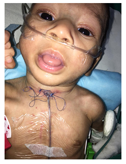
Figure 1: Photograph of the child who underwent reimplantation of anomalous origin of the left coronary artery from pulmonary artery with transtracheal oxygen through the tracheostomy stoma following decanulation of the tracheostomy tube. Note the dual source of oxygenation through the nasal prongs and tracheostomy stoma.
The infants head was placed in oxygen hood, thus providing dual source of oxygen for a period ranging between 1-5 days. During this period, the enteral feed was continued, and intermittent endotracheal suction was given through the tracheostomy stoma until peristomal edema subsided and natural non-obstructive airway was restored.
Original cohort
The median age of patients at operation was 4months (IQR: 23days-9 months). Twenty-five (29.8%) patients were younger than 1 month, and 59 (70.2%) were between 1 and 12 months.
Thirty (35.7%) infants in this study group were premature and thirty-nine (46.4%) weighed less than 50th percentile of predicted weight by national standards (Tables 1 and 2).
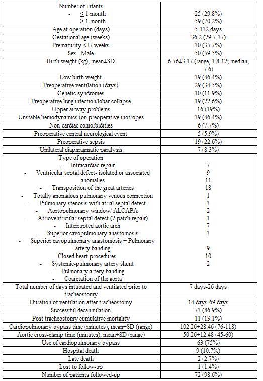
Table 1: Demographics of the patients in the study population (n=84)

Table 2: Age at primary operation, diagnosis, type of operation of all patients in the study group undergoing tracheostomy (n=84)
Twenty-nine (34.5%) patients required preoperative ventilation. A combination of three or more risk factors requiring prolonged mechanical ventilation were present in 58.3% (n=49) patients (Table 3).
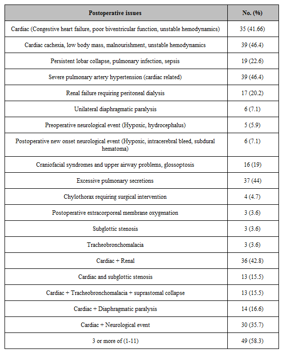
Table 3: Reasons for prolonged ventilation of all patients in the study group undergoing tracheostomy (n=84)
Ventilation could not be weaned sufficiently to allow a trial of extubation in 39 (46.4%) patients. In 38 (45.2%) cases extubation had failed on single occasion, and in 7 (8.3%) cases on two occasions.
Among original cohort of 84 patients, there were 9 (10.8%) perioperative deaths after tracheostomy while still ventilated through the tracheostomy within 30 post-operative days due to a combination of 3 or more risk factors. There were 2(2.7%) late deaths 39 and 81 months after surgery due to renal failure (n=1), and cerebral thrombosis (n=1) respectively. One patient was lost to follow-up.
Cohort of survivors
One infant with tracheobronchomalacia had local infection at the tracheostomy site and the tracheostomy was exchanged for a period of nasotracheal ventilation. After controlling the infection, the tracheostomy wound was revised without additional complications. Two children with subglottic stenosis who were preoperatively ventilated for 15 and 20 days respectively underwent excision of subglottic membrane and cricoid splinting and were ventilated for a period of 3 weeks in two sessions. There were no other tracheostomy-related complications.
Preoperative ventilation before the “primary operation”, pulmonary infection, subglottic stenosis, tracheobronchomalacia, diaphragmatic paralysis, cardiac cachexia, renal failure, and sepsis were associated with a longer period of post tracheostomy ventilation.
Decannulation was successful in all 72 infants. All neonates and infants received dual source of oxygenation (transtracheostomy) and nasal prongs or oxygen hood) for a period between 1-5 days. There were no instances of hypoxic adverse events. A patent stoma allowed intermittent endotracheal suction and nutrition was attended to during this critical period through nasogastric feeling. Temporary stomal manipulation / instrumentation for 1-5 days did not affect stomal healing and there were no post decannulation complications.
The median duration of mechanical ventilation after tracheostomy was 22 days (range 11-35 days) and the median duration of tracheostomy to decannulation in survivors was 31 days (30-52 days). Sixty-eight (94.4%) survivors were in Ross’s clinical score of 2 and without antifailure cardiac medications. There was no reoperation following decannulation. At the end of the follow-up, all survivors were successfully decannulated.
Comment
Tracheostomy is performed in 1.3% to 2.7% of children following cardiac surgery due to the requirement of prolonged ventilation and various other reasons. [1-3] The predisposing factors include younger age, prematurity, low birth weight, cardiac cachexia, preoperative mechanical ventilation, longer CPB time, premature extubation, the need for reoperation, phrenic nerve injury, diaphragmatic paralysis and sepsis. [1-7]
Tracheostomy has been shown to reduce total mechanical ventilation time, decrease the occurrence of lower respiratory tract infection, improve oral and dental hygiene, reduce upper airway injury including vocal cord ulceration, reduce in-hospital mortality and hospital cost. [8-10]Additionally, it decreases the number of self extubations, requirement of sedation, decrease dead space ventilation, airway resistance and work of breathing, allows weight gain through enteral/parenteral feeds, thereby facilitating separation from ventilator support, shorten ICU and hospital stays. [8-10]
There is no consensus in the published literature on the indications, and timing of pediatric tracheostomy and on the optimal decannulation protocol. Data from the pediatric population are limited to small, single-center reviews, and it is indeed difficult to extrapolate data from other centers. [6,7]. Additionally, in the developing world, there is a high incidence of post-operative sepsis.
Children requiring tracheostomy after surgery for CHD are at significant risk or poor outcomes with less than half still alive at a median follow-up of 3.9 years. [1-3] Mastropietro reported an overall mortality rate of 25.2% among 606 tracheostomies in neonates and infants in the STS congenital Heart Surgery database. [1] Johnson and colleagues in a multi institutional study reported an overall mortality rate of 21.6% on 1292 pediatric tracheostomies. [2] Our overall mortality rate of 10% is in accordance with the published investigations which documents a tracheostomy- related mortality between 3.2% and 25% after 1985. [1-10]
Both anatomic and physiologic characteristics of the infant trachea require special surgical techniques and adequate postoperative care. Infants have shorter and fatter necks than adults. The infant larynx is situated more superior and anterior in the neck at the level of the third or fourth cervical vertebra, and it starts to descend at around 2 years of age. Its size is approximately 1/3rd that of the adult larynx. The hyoid frequently overlies the thyroid cartilage notch, making palpation of the anatomic landmarks difficult. The infant thyrohyoid membrane is also much shorter. The cricoid cartilage is the narrowest part of the airway in a child, and in adults, it is vocal cords.
If a tracheostomy is being planned for upper airway obstruction due to subglottic stenosis or complete tracheal rings, tracheostomy may be difficult, with risk of damage to the posterior tracheal wall. Although debatable, a midline vertical incision in infants through the 2nd or 4th tracheal cartilages is the most preferred technique.
The important issues which require consideration while performing pediatric tracheostomy are: i) the underlying indication of tracheostomy incision, ii) prevention of accidental decanulation; iii) avoidance of suturing the tracheostomy tube to the skin; and iv) prevention of long-term tracheal stenosis.
Following placement of the tracheostomy tube, one of the concerns is the duration of time that the patients remain dependent on the device. In our study, we have been able to decannulate all infants prior to discharge. Due to lack of health care resources, hospital discharge with a tracheostomy or ventilator is not a viable option in India, therefore in hospital decannulation is preferable in our setup.
Stomaplasty techniques introduced by Eliachar, Koltai, and Bjork involves the resection of the tracheal cartilages and are recommended when longer duration of tracheostomy is expected.1-4 We did not use any of these methods, since we expected our patients to be decannulated before discharge.
The reported incidence of pediatric tracheostomy-related complications is around 15% -19%.[1-10] In this study, there was one case of tracheitis and two patients had sub glottic stenosis. In order to avert decannulation failure in these compromised neonates and infants, we developed this technique of dual source oxygenation following decannulation. Thus, there were five forces driving our decision-making for technical modifications to facilitate decannulation of the tracheostomy tube;
We have been able to address all our desires by this technique presented herein and we conclude that this technical modification is simple, reproducible, and inexpensive, allows sustained oxygenation, thus averts decannulation failure and hypoxic post decannulation adverse events.
Since we have achieved step one-demonstration of safety, a comparative trial of conventional decannulation and “dual-source” oxygenation following decannulation is the topic of our future investigation.
We conclude that transtracheal oxygenation via a thin silastic catheter in neonates and infants undergoing decannulation of tracheostomy tube provides uninterrupted route of oxygenation, allows continuation of enteral feeds, facilitates intermittent endotracheal suction until peristomal oedema subsides and natural non-obstructive airway is restored.
This “dual-source” strategy of oxygen administration is safe, expedient and obviates the need to remove the nasogastric tube for oxygen administration via nasal cannula. Knowledge of this approach should contribute to the armamentarium of cardiac surgeons faced with decannulation of the tracheostomy tube.
Clearly Auctoresonline and particularly Psychology and Mental Health Care Journal is dedicated to improving health care services for individuals and populations. The editorial boards' ability to efficiently recognize and share the global importance of health literacy with a variety of stakeholders. Auctoresonline publishing platform can be used to facilitate of optimal client-based services and should be added to health care professionals' repertoire of evidence-based health care resources.

Journal of Clinical Cardiology and Cardiovascular Intervention The submission and review process was adequate. However I think that the publication total value should have been enlightened in early fases. Thank you for all.

Journal of Women Health Care and Issues By the present mail, I want to say thank to you and tour colleagues for facilitating my published article. Specially thank you for the peer review process, support from the editorial office. I appreciate positively the quality of your journal.

Journal of Clinical Research and Reports I would be very delighted to submit my testimonial regarding the reviewer board and the editorial office. The reviewer board were accurate and helpful regarding any modifications for my manuscript. And the editorial office were very helpful and supportive in contacting and monitoring with any update and offering help. It was my pleasure to contribute with your promising Journal and I am looking forward for more collaboration.

We would like to thank the Journal of Thoracic Disease and Cardiothoracic Surgery because of the services they provided us for our articles. The peer-review process was done in a very excellent time manner, and the opinions of the reviewers helped us to improve our manuscript further. The editorial office had an outstanding correspondence with us and guided us in many ways. During a hard time of the pandemic that is affecting every one of us tremendously, the editorial office helped us make everything easier for publishing scientific work. Hope for a more scientific relationship with your Journal.

The peer-review process which consisted high quality queries on the paper. I did answer six reviewers’ questions and comments before the paper was accepted. The support from the editorial office is excellent.

Journal of Neuroscience and Neurological Surgery. I had the experience of publishing a research article recently. The whole process was simple from submission to publication. The reviewers made specific and valuable recommendations and corrections that improved the quality of my publication. I strongly recommend this Journal.

Dr. Katarzyna Byczkowska My testimonial covering: "The peer review process is quick and effective. The support from the editorial office is very professional and friendly. Quality of the Clinical Cardiology and Cardiovascular Interventions is scientific and publishes ground-breaking research on cardiology that is useful for other professionals in the field.

Thank you most sincerely, with regard to the support you have given in relation to the reviewing process and the processing of my article entitled "Large Cell Neuroendocrine Carcinoma of The Prostate Gland: A Review and Update" for publication in your esteemed Journal, Journal of Cancer Research and Cellular Therapeutics". The editorial team has been very supportive.

Testimony of Journal of Clinical Otorhinolaryngology: work with your Reviews has been a educational and constructive experience. The editorial office were very helpful and supportive. It was a pleasure to contribute to your Journal.

Dr. Bernard Terkimbi Utoo, I am happy to publish my scientific work in Journal of Women Health Care and Issues (JWHCI). The manuscript submission was seamless and peer review process was top notch. I was amazed that 4 reviewers worked on the manuscript which made it a highly technical, standard and excellent quality paper. I appreciate the format and consideration for the APC as well as the speed of publication. It is my pleasure to continue with this scientific relationship with the esteem JWHCI.

This is an acknowledgment for peer reviewers, editorial board of Journal of Clinical Research and Reports. They show a lot of consideration for us as publishers for our research article “Evaluation of the different factors associated with side effects of COVID-19 vaccination on medical students, Mutah university, Al-Karak, Jordan”, in a very professional and easy way. This journal is one of outstanding medical journal.

Dear Hao Jiang, to Journal of Nutrition and Food Processing We greatly appreciate the efficient, professional and rapid processing of our paper by your team. If there is anything else we should do, please do not hesitate to let us know. On behalf of my co-authors, we would like to express our great appreciation to editor and reviewers.

As an author who has recently published in the journal "Brain and Neurological Disorders". I am delighted to provide a testimonial on the peer review process, editorial office support, and the overall quality of the journal. The peer review process at Brain and Neurological Disorders is rigorous and meticulous, ensuring that only high-quality, evidence-based research is published. The reviewers are experts in their fields, and their comments and suggestions were constructive and helped improve the quality of my manuscript. The review process was timely and efficient, with clear communication from the editorial office at each stage. The support from the editorial office was exceptional throughout the entire process. The editorial staff was responsive, professional, and always willing to help. They provided valuable guidance on formatting, structure, and ethical considerations, making the submission process seamless. Moreover, they kept me informed about the status of my manuscript and provided timely updates, which made the process less stressful. The journal Brain and Neurological Disorders is of the highest quality, with a strong focus on publishing cutting-edge research in the field of neurology. The articles published in this journal are well-researched, rigorously peer-reviewed, and written by experts in the field. The journal maintains high standards, ensuring that readers are provided with the most up-to-date and reliable information on brain and neurological disorders. In conclusion, I had a wonderful experience publishing in Brain and Neurological Disorders. The peer review process was thorough, the editorial office provided exceptional support, and the journal's quality is second to none. I would highly recommend this journal to any researcher working in the field of neurology and brain disorders.
Dear Agrippa Hilda, Journal of Neuroscience and Neurological Surgery, Editorial Coordinator, I trust this message finds you well. I want to extend my appreciation for considering my article for publication in your esteemed journal. I am pleased to provide a testimonial regarding the peer review process and the support received from your editorial office. The peer review process for my paper was carried out in a highly professional and thorough manner. The feedback and comments provided by the authors were constructive and very useful in improving the quality of the manuscript. This rigorous assessment process undoubtedly contributes to the high standards maintained by your journal.

International Journal of Clinical Case Reports and Reviews. I strongly recommend to consider submitting your work to this high-quality journal. The support and availability of the Editorial staff is outstanding and the review process was both efficient and rigorous.

Thank you very much for publishing my Research Article titled “Comparing Treatment Outcome Of Allergic Rhinitis Patients After Using Fluticasone Nasal Spray And Nasal Douching" in the Journal of Clinical Otorhinolaryngology. As Medical Professionals we are immensely benefited from study of various informative Articles and Papers published in this high quality Journal. I look forward to enriching my knowledge by regular study of the Journal and contribute my future work in the field of ENT through the Journal for use by the medical fraternity. The support from the Editorial office was excellent and very prompt. I also welcome the comments received from the readers of my Research Article.

Dear Erica Kelsey, Editorial Coordinator of Cancer Research and Cellular Therapeutics Our team is very satisfied with the processing of our paper by your journal. That was fast, efficient, rigorous, but without unnecessary complications. We appreciated the very short time between the submission of the paper and its publication on line on your site.

I am very glad to say that the peer review process is very successful and fast and support from the Editorial Office. Therefore, I would like to continue our scientific relationship for a long time. And I especially thank you for your kindly attention towards my article. Have a good day!

"We recently published an article entitled “Influence of beta-Cyclodextrins upon the Degradation of Carbofuran Derivatives under Alkaline Conditions" in the Journal of “Pesticides and Biofertilizers” to show that the cyclodextrins protect the carbamates increasing their half-life time in the presence of basic conditions This will be very helpful to understand carbofuran behaviour in the analytical, agro-environmental and food areas. We greatly appreciated the interaction with the editor and the editorial team; we were particularly well accompanied during the course of the revision process, since all various steps towards publication were short and without delay".

I would like to express my gratitude towards you process of article review and submission. I found this to be very fair and expedient. Your follow up has been excellent. I have many publications in national and international journal and your process has been one of the best so far. Keep up the great work.

We are grateful for this opportunity to provide a glowing recommendation to the Journal of Psychiatry and Psychotherapy. We found that the editorial team were very supportive, helpful, kept us abreast of timelines and over all very professional in nature. The peer review process was rigorous, efficient and constructive that really enhanced our article submission. The experience with this journal remains one of our best ever and we look forward to providing future submissions in the near future.

I am very pleased to serve as EBM of the journal, I hope many years of my experience in stem cells can help the journal from one way or another. As we know, stem cells hold great potential for regenerative medicine, which are mostly used to promote the repair response of diseased, dysfunctional or injured tissue using stem cells or their derivatives. I think Stem Cell Research and Therapeutics International is a great platform to publish and share the understanding towards the biology and translational or clinical application of stem cells.

I would like to give my testimony in the support I have got by the peer review process and to support the editorial office where they were of asset to support young author like me to be encouraged to publish their work in your respected journal and globalize and share knowledge across the globe. I really give my great gratitude to your journal and the peer review including the editorial office.

I am delighted to publish our manuscript entitled "A Perspective on Cocaine Induced Stroke - Its Mechanisms and Management" in the Journal of Neuroscience and Neurological Surgery. The peer review process, support from the editorial office, and quality of the journal are excellent. The manuscripts published are of high quality and of excellent scientific value. I recommend this journal very much to colleagues.

Dr.Tania Muñoz, My experience as researcher and author of a review article in The Journal Clinical Cardiology and Interventions has been very enriching and stimulating. The editorial team is excellent, performs its work with absolute responsibility and delivery. They are proactive, dynamic and receptive to all proposals. Supporting at all times the vast universe of authors who choose them as an option for publication. The team of review specialists, members of the editorial board, are brilliant professionals, with remarkable performance in medical research and scientific methodology. Together they form a frontline team that consolidates the JCCI as a magnificent option for the publication and review of high-level medical articles and broad collective interest. I am honored to be able to share my review article and open to receive all your comments.

“The peer review process of JPMHC is quick and effective. Authors are benefited by good and professional reviewers with huge experience in the field of psychology and mental health. The support from the editorial office is very professional. People to contact to are friendly and happy to help and assist any query authors might have. Quality of the Journal is scientific and publishes ground-breaking research on mental health that is useful for other professionals in the field”.

Dear editorial department: On behalf of our team, I hereby certify the reliability and superiority of the International Journal of Clinical Case Reports and Reviews in the peer review process, editorial support, and journal quality. Firstly, the peer review process of the International Journal of Clinical Case Reports and Reviews is rigorous, fair, transparent, fast, and of high quality. The editorial department invites experts from relevant fields as anonymous reviewers to review all submitted manuscripts. These experts have rich academic backgrounds and experience, and can accurately evaluate the academic quality, originality, and suitability of manuscripts. The editorial department is committed to ensuring the rigor of the peer review process, while also making every effort to ensure a fast review cycle to meet the needs of authors and the academic community. Secondly, the editorial team of the International Journal of Clinical Case Reports and Reviews is composed of a group of senior scholars and professionals with rich experience and professional knowledge in related fields. The editorial department is committed to assisting authors in improving their manuscripts, ensuring their academic accuracy, clarity, and completeness. Editors actively collaborate with authors, providing useful suggestions and feedback to promote the improvement and development of the manuscript. We believe that the support of the editorial department is one of the key factors in ensuring the quality of the journal. Finally, the International Journal of Clinical Case Reports and Reviews is renowned for its high- quality articles and strict academic standards. The editorial department is committed to publishing innovative and academically valuable research results to promote the development and progress of related fields. The International Journal of Clinical Case Reports and Reviews is reasonably priced and ensures excellent service and quality ratio, allowing authors to obtain high-level academic publishing opportunities in an affordable manner. I hereby solemnly declare that the International Journal of Clinical Case Reports and Reviews has a high level of credibility and superiority in terms of peer review process, editorial support, reasonable fees, and journal quality. Sincerely, Rui Tao.

Clinical Cardiology and Cardiovascular Interventions I testity the covering of the peer review process, support from the editorial office, and quality of the journal.

Clinical Cardiology and Cardiovascular Interventions, we deeply appreciate the interest shown in our work and its publication. It has been a true pleasure to collaborate with you. The peer review process, as well as the support provided by the editorial office, have been exceptional, and the quality of the journal is very high, which was a determining factor in our decision to publish with you.

The peer reviewers process is quick and effective, the supports from editorial office is excellent, the quality of journal is high. I would like to collabroate with Internatioanl journal of Clinical Case Reports and Reviews journal clinically in the future time.

Clinical Cardiology and Cardiovascular Interventions, I would like to express my sincerest gratitude for the trust placed in our team for the publication in your journal. It has been a true pleasure to collaborate with you on this project. I am pleased to inform you that both the peer review process and the attention from the editorial coordination have been excellent. Your team has worked with dedication and professionalism to ensure that your publication meets the highest standards of quality. We are confident that this collaboration will result in mutual success, and we are eager to see the fruits of this shared effort.

Dear Dr. Jessica Magne, Editorial Coordinator 0f Clinical Cardiology and Cardiovascular Interventions, I hope this message finds you well. I want to express my utmost gratitude for your excellent work and for the dedication and speed in the publication process of my article titled "Navigating Innovation: Qualitative Insights on Using Technology for Health Education in Acute Coronary Syndrome Patients." I am very satisfied with the peer review process, the support from the editorial office, and the quality of the journal. I hope we can maintain our scientific relationship in the long term.

Dear Monica Gissare, - Editorial Coordinator of Nutrition and Food Processing. ¨My testimony with you is truly professional, with a positive response regarding the follow-up of the article and its review, you took into account my qualities and the importance of the topic¨.

Dear Dr. Jessica Magne, Editorial Coordinator 0f Clinical Cardiology and Cardiovascular Interventions, The review process for the article “The Handling of Anti-aggregants and Anticoagulants in the Oncologic Heart Patient Submitted to Surgery” was extremely rigorous and detailed. From the initial submission to the final acceptance, the editorial team at the “Journal of Clinical Cardiology and Cardiovascular Interventions” demonstrated a high level of professionalism and dedication. The reviewers provided constructive and detailed feedback, which was essential for improving the quality of our work. Communication was always clear and efficient, ensuring that all our questions were promptly addressed. The quality of the “Journal of Clinical Cardiology and Cardiovascular Interventions” is undeniable. It is a peer-reviewed, open-access publication dedicated exclusively to disseminating high-quality research in the field of clinical cardiology and cardiovascular interventions. The journal's impact factor is currently under evaluation, and it is indexed in reputable databases, which further reinforces its credibility and relevance in the scientific field. I highly recommend this journal to researchers looking for a reputable platform to publish their studies.

Dear Editorial Coordinator of the Journal of Nutrition and Food Processing! "I would like to thank the Journal of Nutrition and Food Processing for including and publishing my article. The peer review process was very quick, movement and precise. The Editorial Board has done an extremely conscientious job with much help, valuable comments and advices. I find the journal very valuable from a professional point of view, thank you very much for allowing me to be part of it and I would like to participate in the future!”

Dealing with The Journal of Neurology and Neurological Surgery was very smooth and comprehensive. The office staff took time to address my needs and the response from editors and the office was prompt and fair. I certainly hope to publish with this journal again.Their professionalism is apparent and more than satisfactory. Susan Weiner

My Testimonial Covering as fellowing: Lin-Show Chin. The peer reviewers process is quick and effective, the supports from editorial office is excellent, the quality of journal is high. I would like to collabroate with Internatioanl journal of Clinical Case Reports and Reviews.

My experience publishing in Psychology and Mental Health Care was exceptional. The peer review process was rigorous and constructive, with reviewers providing valuable insights that helped enhance the quality of our work. The editorial team was highly supportive and responsive, making the submission process smooth and efficient. The journal's commitment to high standards and academic rigor makes it a respected platform for quality research. I am grateful for the opportunity to publish in such a reputable journal.

My experience publishing in International Journal of Clinical Case Reports and Reviews was exceptional. I Come forth to Provide a Testimonial Covering the Peer Review Process and the editorial office for the Professional and Impartial Evaluation of the Manuscript.

I would like to offer my testimony in the support. I have received through the peer review process and support the editorial office where they are to support young authors like me, encourage them to publish their work in your esteemed journals, and globalize and share knowledge globally. I really appreciate your journal, peer review, and editorial office.

Dear Agrippa Hilda- Editorial Coordinator of Journal of Neuroscience and Neurological Surgery, "The peer review process was very quick and of high quality, which can also be seen in the articles in the journal. The collaboration with the editorial office was very good."

I would like to express my sincere gratitude for the support and efficiency provided by the editorial office throughout the publication process of my article, “Delayed Vulvar Metastases from Rectal Carcinoma: A Case Report.” I greatly appreciate the assistance and guidance I received from your team, which made the entire process smooth and efficient. The peer review process was thorough and constructive, contributing to the overall quality of the final article. I am very grateful for the high level of professionalism and commitment shown by the editorial staff, and I look forward to maintaining a long-term collaboration with the International Journal of Clinical Case Reports and Reviews.

To Dear Erin Aust, I would like to express my heartfelt appreciation for the opportunity to have my work published in this esteemed journal. The entire publication process was smooth and well-organized, and I am extremely satisfied with the final result. The Editorial Team demonstrated the utmost professionalism, providing prompt and insightful feedback throughout the review process. Their clear communication and constructive suggestions were invaluable in enhancing my manuscript, and their meticulous attention to detail and dedication to quality are truly commendable. Additionally, the support from the Editorial Office was exceptional. From the initial submission to the final publication, I was guided through every step of the process with great care and professionalism. The team's responsiveness and assistance made the entire experience both easy and stress-free. I am also deeply impressed by the quality and reputation of the journal. It is an honor to have my research featured in such a respected publication, and I am confident that it will make a meaningful contribution to the field.

"I am grateful for the opportunity of contributing to [International Journal of Clinical Case Reports and Reviews] and for the rigorous review process that enhances the quality of research published in your esteemed journal. I sincerely appreciate the time and effort of your team who have dedicatedly helped me in improvising changes and modifying my manuscript. The insightful comments and constructive feedback provided have been invaluable in refining and strengthening my work".

I thank the ‘Journal of Clinical Research and Reports’ for accepting this article for publication. This is a rigorously peer reviewed journal which is on all major global scientific data bases. I note the review process was prompt, thorough and professionally critical. It gave us an insight into a number of important scientific/statistical issues. The review prompted us to review the relevant literature again and look at the limitations of the study. The peer reviewers were open, clear in the instructions and the editorial team was very prompt in their communication. This journal certainly publishes quality research articles. I would recommend the journal for any future publications.

Dear Jessica Magne, with gratitude for the joint work. Fast process of receiving and processing the submitted scientific materials in “Clinical Cardiology and Cardiovascular Interventions”. High level of competence of the editors with clear and correct recommendations and ideas for enriching the article.

We found the peer review process quick and positive in its input. The support from the editorial officer has been very agile, always with the intention of improving the article and taking into account our subsequent corrections.

My article, titled 'No Way Out of the Smartphone Epidemic Without Considering the Insights of Brain Research,' has been republished in the International Journal of Clinical Case Reports and Reviews. The review process was seamless and professional, with the editors being both friendly and supportive. I am deeply grateful for their efforts.

To Dear Erin Aust – Editorial Coordinator of Journal of General Medicine and Clinical Practice! I declare that I am absolutely satisfied with your work carried out with great competence in following the manuscript during the various stages from its receipt, during the revision process to the final acceptance for publication. Thank Prof. Elvira Farina

Dear Jessica, and the super professional team of the ‘Clinical Cardiology and Cardiovascular Interventions’ I am sincerely grateful to the coordinated work of the journal team for the no problem with the submission of my manuscript: “Cardiometabolic Disorders in A Pregnant Woman with Severe Preeclampsia on the Background of Morbid Obesity (Case Report).” The review process by 5 experts was fast, and the comments were professional, which made it more specific and academic, and the process of publication and presentation of the article was excellent. I recommend that my colleagues publish articles in this journal, and I am interested in further scientific cooperation. Sincerely and best wishes, Dr. Oleg Golyanovskiy.

Dear Ashley Rosa, Editorial Coordinator of the journal - Psychology and Mental Health Care. " The process of obtaining publication of my article in the Psychology and Mental Health Journal was positive in all areas. The peer review process resulted in a number of valuable comments, the editorial process was collaborative and timely, and the quality of this journal has been quickly noticed, resulting in alternative journals contacting me to publish with them." Warm regards, Susan Anne Smith, PhD. Australian Breastfeeding Association.

Dear Jessica Magne, Editorial Coordinator, Clinical Cardiology and Cardiovascular Interventions, Auctores Publishing LLC. I appreciate the journal (JCCI) editorial office support, the entire team leads were always ready to help, not only on technical front but also on thorough process. Also, I should thank dear reviewers’ attention to detail and creative approach to teach me and bring new insights by their comments. Surely, more discussions and introduction of other hemodynamic devices would provide better prevention and management of shock states. Your efforts and dedication in presenting educational materials in this journal are commendable. Best wishes from, Farahnaz Fallahian.

Dear Maria Emerson, Editorial Coordinator, International Journal of Clinical Case Reports and Reviews, Auctores Publishing LLC. I am delighted to have published our manuscript, "Acute Colonic Pseudo-Obstruction (ACPO): A rare but serious complication following caesarean section." I want to thank the editorial team, especially Maria Emerson, for their prompt review of the manuscript, quick responses to queries, and overall support. Yours sincerely Dr. Victor Olagundoye.

Dear Ashley Rosa, Editorial Coordinator, International Journal of Clinical Case Reports and Reviews. Many thanks for publishing this manuscript after I lost confidence the editors were most helpful, more than other journals Best wishes from, Susan Anne Smith, PhD. Australian Breastfeeding Association.

Dear Agrippa Hilda, Editorial Coordinator, Journal of Neuroscience and Neurological Surgery. The entire process including article submission, review, revision, and publication was extremely easy. The journal editor was prompt and helpful, and the reviewers contributed to the quality of the paper. Thank you so much! Eric Nussbaum, MD

Dr Hala Al Shaikh This is to acknowledge that the peer review process for the article ’ A Novel Gnrh1 Gene Mutation in Four Omani Male Siblings, Presentation and Management ’ sent to the International Journal of Clinical Case Reports and Reviews was quick and smooth. The editorial office was prompt with easy communication.

Dear Erin Aust, Editorial Coordinator, Journal of General Medicine and Clinical Practice. We are pleased to share our experience with the “Journal of General Medicine and Clinical Practice”, following the successful publication of our article. The peer review process was thorough and constructive, helping to improve the clarity and quality of the manuscript. We are especially thankful to Ms. Erin Aust, the Editorial Coordinator, for her prompt communication and continuous support throughout the process. Her professionalism ensured a smooth and efficient publication experience. The journal upholds high editorial standards, and we highly recommend it to fellow researchers seeking a credible platform for their work. Best wishes By, Dr. Rakhi Mishra.

Dear Jessica Magne, Editorial Coordinator, Clinical Cardiology and Cardiovascular Interventions, Auctores Publishing LLC. The peer review process of the journal of Clinical Cardiology and Cardiovascular Interventions was excellent and fast, as was the support of the editorial office and the quality of the journal. Kind regards Walter F. Riesen Prof. Dr. Dr. h.c. Walter F. Riesen.

Dear Ashley Rosa, Editorial Coordinator, International Journal of Clinical Case Reports and Reviews, Auctores Publishing LLC. Thank you for publishing our article, Exploring Clozapine's Efficacy in Managing Aggression: A Multiple Single-Case Study in Forensic Psychiatry in the international journal of clinical case reports and reviews. We found the peer review process very professional and efficient. The comments were constructive, and the whole process was efficient. On behalf of the co-authors, I would like to thank you for publishing this article. With regards, Dr. Jelle R. Lettinga.

Dear Clarissa Eric, Editorial Coordinator, Journal of Clinical Case Reports and Studies, I would like to express my deep admiration for the exceptional professionalism demonstrated by your journal. I am thoroughly impressed by the speed of the editorial process, the substantive and insightful reviews, and the meticulous preparation of the manuscript for publication. Additionally, I greatly appreciate the courteous and immediate responses from your editorial office to all my inquiries. Best Regards, Dariusz Ziora

Dear Chrystine Mejia, Editorial Coordinator, Journal of Neurodegeneration and Neurorehabilitation, Auctores Publishing LLC, We would like to thank the editorial team for the smooth and high-quality communication leading up to the publication of our article in the Journal of Neurodegeneration and Neurorehabilitation. The reviewers have extensive knowledge in the field, and their relevant questions helped to add value to our publication. Kind regards, Dr. Ravi Shrivastava.

Dear Clarissa Eric, Editorial Coordinator, Journal of Clinical Case Reports and Studies, Auctores Publishing LLC, USA Office: +1-(302)-520-2644. I would like to express my sincere appreciation for the efficient and professional handling of my case report by the ‘Journal of Clinical Case Reports and Studies’. The peer review process was not only fast but also highly constructive—the reviewers’ comments were clear, relevant, and greatly helped me improve the quality and clarity of my manuscript. I also received excellent support from the editorial office throughout the process. Communication was smooth and timely, and I felt well guided at every stage, from submission to publication. The overall quality and rigor of the journal are truly commendable. I am pleased to have published my work with Journal of Clinical Case Reports and Studies, and I look forward to future opportunities for collaboration. Sincerely, Aline Tollet, UCLouvain.

Dear Ms. Mayra Duenas, Editorial Coordinator, International Journal of Clinical Case Reports and Reviews. “The International Journal of Clinical Case Reports and Reviews represented the “ideal house” to share with the research community a first experience with the use of the Simeox device for speech rehabilitation. High scientific reputation and attractive website communication were first determinants for the selection of this Journal, and the following submission process exceeded expectations: fast but highly professional peer review, great support by the editorial office, elegant graphic layout. Exactly what a dynamic research team - also composed by allied professionals - needs!" From, Chiara Beccaluva, PT - Italy.

Dear Maria Emerson, Editorial Coordinator, we have deeply appreciated the professionalism demonstrated by the International Journal of Clinical Case Reports and Reviews. The reviewers have extensive knowledge of our field and have been very efficient and fast in supporting the process. I am really looking forward to further collaboration. Thanks. Best regards, Dr. Claudio Ligresti

Dear Chrystine Mejia, Editorial Coordinator, Journal of Neurodegeneration and Neurorehabilitation. “The peer review process was efficient and constructive, and the editorial office provided excellent communication and support throughout. The journal ensures scientific rigor and high editorial standards, while also offering a smooth and timely publication process. We sincerely appreciate the work of the editorial team in facilitating the dissemination of innovative approaches such as the Bonori Method.” Best regards, Dr. Giselle Pentón-Rol.
