AUCTORES
Globalize your Research

Research Article | DOI: https://doi.org/10.31579/2641-0419/057
1 Department of cardiovascular diseases, Faculty of Medical Sciences-LU, Beirut, Lebanon.
3 Medical students, Faculty of Medical Sciences-LU, Beirut, Lebanon.
4 Department of Internal Medicine diseases, Faculty of Medical Sciences-LU, Beirut, Lebanon.
5 Department of Infectious Diseases, Head of Internal Medicine Department of MEIH affiliated with the Faculty of Medical Sciences-LU, Bsalim, Lebanon
*Corresponding Author: El Murr Tony, MD, Department of Infectious Diseases, Head of Internal Medicine Department of MEIH affiliated with the Faculty of Medical Sciences-LU, Bsalim, Lebanon.
Citation: Boutros Y, Abi R. Nagi ., George B., Ricardo W., Joelle F., El M. Tony (2020) The Lebanese Geitawi Hospital-University Medical Center Heart Failure Registry. J. Clinical Cardiology and Cardiovascular Interventions, 3(5); Doi:10.31579/2641-0419/057
Copyright: © 2020 El Murr Tony, This is an open-access article distributed under the terms of the Creative Commons Attribution License, which permits unrestricted use, distribution, and reproduction in any medium, provided the original author and source are credited.
Received: 12 March 2020 | Accepted: 20 March 2020 | Published: 27 March 2020
Keywords: heart failure; registry; awareness; indicators; TTE; variables; quality improvement.
Heart Failure (HF) has become a major cause of death and hospitalization among people older than 60 year. Lack of available data and registries from different countries that may aid in understanding the burden of the disease does exist.
The aim of the lebanese Geitawi Hospital-University Medical Center (LGH-UMC) heart failure registry is to point toward the incidence of heart failure, in patients with suspected dyspnea, during a 7 months period in a single university medical center based on Trans-thoracic echocardiography (TTE) findings with emphasis on its etiology.
Study population
The LGH-HFR includes inpatients and outpatients (≥18 years) presenting for further evaluation of possible HF by performing a TTE after suspicious clinical findings. Patients with clinical impression of possible heart failure were reported to LGH-HFR by their doctors.
The final decision to register a patient in the LGH-HFR is made by a single cardiologist to ensure the validity of the clinical suspicion. Approximately 1422 patients with clinically suspicion HF were registered in the LGH-HFR between 1.1.2018 and 31.7.2018.
Main variables and descriptive data
Our study will be a retrospective cohort analysis of data collected from the LGH-HFR between 1.1.2018 and 31.7.2018. Our number of studied patients is about 1422. The main variables recorded in the LGH-HFR are related to the TTE findings: LVEDV, LVEF, presence of LVH, the presence of diastolic dysfunction, documentation of any valvulopathies, measurements of PAPs. Furthermore, pro-BNP level and basic patient characteristics (age and sex) will be recorded.
The findings then will be pooled according to the documentation of heart failure, either systolic or diastolic. Furthermore, specific TTE findings will be mentioned in each case after correlation with the HF type pointing toward the possible etiology of the cardiac function degradation.
Results of our study will be reported back to clinicians to promote awareness for HF and communicated with the HFRs of different hospitals and LSC in order to optimize the standards of care regarding HF and discussing the cost effectiveness issue if possible.
Conclusion
The LGH-HFR is a valuable tool for continuous improvement of quality of care in patients and awareness regarding HF in Lebanon. Furthermore, it will be an important resource for the Lebanese registry-based HF research once available.
Heart failure (HF) is an heterogeneous clinical syndrome in terms of symptoms (dyspnea, orthopnea, lower limb swelling), signs (elevated jugular venous pressure, pulmonary congestion), associated structural and/or functional cardiac dysfunction that may result in reduced cardiac output and/or elevated intracardiac pressures [1]. Both prevalence and mortality rate of HF have remained unacceptably high making its early detection so crucial in order to decrease the disease burden. Based on an 2013 update from the American Heart Association (AHA) we concluded that there were 5.1 million people with HF in the United States in 2006 [2] and 23 million people with HF worldwide [3]. These numbers reflect not only a major health issue but make HF a prime subject for cost/effectiveness debate. For example, its total annual expenditures in the USA are expected to rise to $70 billion by 2030 [4].
Despite improvements in medical therapy and techniques, the extent of this disease has not been identified with precision because the lack of population based studies or global heart failure registries that may point toward specific etiologies or medical therapy regimen leading to a new perspective to deal with this world health issue. In spite of the fact that there is no clear cut estimation, the prevalence of HF according to several studies is approximately 1–2% and rises to >10% among people over the age of 70 years [5]. These data were supported by the Framingham Study showing us that the prevalence of HF in men aged between 50 and 59 years is 8 per 1000, increasing to 66 per 1000 at ages 80 to 89 years; add that similar results exist in women [6]. The incidence of HF, as its prevalence, increases with age too as described by the same study (figure 1). For instance, the predicted number of new HF cases in the USA by 2040 will be around 774,000 raising the alarm for working on new policies in order to prevent and decrease the burden of this disease [7].

Data from Lloyd-Jones DM, Leip EP, Larson MG, et al. Circulation 2006; 113:791.
Graphic 69663 Version 2.0
Signs and symptoms of heart failure consist of a wide range of manifestations that reflect either a decrease in heart contractility or conditions where demands overcome the ability of the heart to pump enough blood. One of the most common symptoms is shortness of breath which intensity may point toward a progressive decline in the left ventricle function.
The breathlessness occurring in heart failure patients has some features that make it well defined from other conditions which may lead to the same clinical profile. First, it occurs upon exertion and then it appears in recumbent position necessitating head elevation (by increasing the number of pillows while sleeping for example), progressing to paroxysmal nocturnal dyspnea not easily relieved by changing positions, ending up at rest interfering with daily life occupations .Other manifestations include palpitations, chest pain, pulmonary edema, confusion, oliguria found in patients as the disease progresses with markedly reduced left ventricular function.
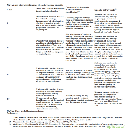
Concerning heart failure classification and staging, different systems have been issued making its diagnosis and prognosis easier to predict. The New York Heart Association (NYHA) for heart failure has four classes according to the degree of effort needed to provoke symptoms (table 1), complemented by the American College of Cardiology/American Heart Association
(ACC/AHA) heart failure stages that classify patients in four stages based on the presence or absence of a structural heart disease (figure 2).

Moreover, diagnosis of heart failure should be supported by objective data that confirm our clinical suspicion. It consists of complete blood count (CBC), serum iron level, electrolyte levels, and hepatorenal function studies. CBC (which usually is of little diagnostic help) may reveal a severe anemia causing or aggravating heart functions [8,9]. Serum iron deficiency is associated with a poor outcome and may lead to further deterioration in patient’s conditions by impairing the contractility of cardiomyocytes as stated by different experts opinions and published in a study establishing the fact that transferrin saturation (TSAT) up to 19.8% or a serum iron level up to 13 μmol/L were the cut points for selecting patients with the highest mortality rate associated with low serum ferritin level [10,11,12].
Some electrolytes disturbances may point also toward a regression in heart capacities. For example, dilutional hyponatremia (due to severe water retention and strict sodium restriction) or hyperkalemia (due to deterioration in kidney functions). Add that different factors can contribute to a reduction in GFR in patients with HF, including neurohormonal axis, renal hypoperfusion, increased renal venous pressure, and right ventricular dysfunction. Both lower GFR and higher blood urea nitrogen (BUN) have been associated with increased mortality in HF and make the diuresis of these patients a big issue [13-16].
Congestive hepatopathy is proven to be also an indicator of an increased risk of death in patient known to have HF. Usually it manifests by an elevation in serum concentration of total bilirubin (<3mg/dL) or mild elevation of LFTs and alkaline phosphatase [17].
In some cases, diagnosing HF appears to be challenging, that’s why more sophisticated testing would be highly recommended. So rapid measurement of B-type natriuretic peptide (BNP) or N-terminal proBNP (NT-proBNP) levels can guide clinicians in differentiating between cardiac and noncardiac causes of dyspnea especially upon atypical presentations. Usually, the major source of BNP is cardiac ventricles secreted after an overload in ventricular pressure or volume. Some studies have showed that BNP level aids in differentiating between systolic and diastolic heart dysfunction but contradictory data mention that it does not reliably differentiate between heart failure with preserved ejection fraction and heart failure with reduced ejection fraction [18-24]. In our study, we will correlate the level of serum pro-BNP to either systolic or diastolic heart failure based on TTE.
Chest radiography helps in supporting our diagnosis of HF by assessing the size and shape of the cardiac silhouette and edema at the lung bases knowing that 50% of patients with heart failure and documented elevation of pulmonary capillary wedge pressure (PCWP) do not manifest typical radiographic findings of pulmonary congestion.
One of the most used diagnostic tool in cardiac diseases is the EKG. In HF, there is no specific EKG findings. Q waves, non specific ST-T wave changes may be present in patients who suffer from a cardiomyopathy or already diagnosed with a prior myocardial infarction .
In addition to all of the above , two-dimensional (2-D) echocardiography is recommended in the initial evaluation of patients with known or suspected heart failure [18,19]. Doppler imaging helps also in further assessment of diastolic heart dysfunction and it is a main clue in the whole diagnostic process as almost 50% of patients presenting with symptoms highly suggestive of HF have normal systolic function. The primary finding, based on TTE, that helps us to differentiate between systolic and diastolic HF is the EF. This detection is so crucial and done by taking accurate measurements while performing the echography in order to target different etiologies so that we can discuss appropriate therapy with our patient.
Other features discovered while performing Doppler and 2D-echocardiography are ventricular filling and pulmonary artery pressures, heart cavity diameters and volumes, LV systolic and diastolic function, valvular anatomy and kinetics.
Add that, transesophageal echocardiography (TEE) may be very useful in conditions where TTE is not diagnostic or technically difficult to perform (patients morbidly obese or on mechanical ventilation) [18]. Its helps us then to assess accurately the heart kinetics, EF, valvular motion, any abnormal communication between cavities leading us to an unambiguous diagnosis.
Multiple heart failure registries have been conducted in different countries but an effort should be made in order to have a global HF registry so that we can understand this disease burden and help in improving patients conditions after studying the effect of different factors (age, sex, race, prevalence, adherence to therapy, length of hospital stay…) during the whole process.
For instance, a heart failure registry in Asia published in the journal of cardiac failure (volume 22, Issue 9, Supplement, Page S153) showed that HF in Korea is a serious condition with in hospital mortality rate of 5%, length of hospital stay in the hospital around 9 days and 1-year mortality rate of 20%.
Another Heart failure registry done in Denmark has shown a substantial improvement in the DHFR process indicators from 2003 to 2010 among patients diagnosed with incident HF. In the same period, the 1-year mortality decreased from 20.5% to 12.8% [25]. The same registry has shown that HF in elderly population (>75years) is not diagnosed and treated adequately.
In our study, we correlate different TTE findings (LVEDV, LVEF, presence of LVH, the presence of diastolic dysfunction, documentation of any valvulopathies, measurements of PAPs) and pro-BNP level in order to estimate the percentage of HF and its possible etiology during a 7 months period so that we can initiate a nucleus for a heart failure registry in our university medical center and hopefully nationwide
We retrospectively analyzed the results of 1520 TTE performed for 1422 patients between January and July 2018 in a university medical center in order to assess the prevalence of HF and its possible etiology in patients with clinical suspicion of HF.
The data was collected from the medical records department of our tertiary care center in Lebanon (Lebanese Geitawi Hospital-University medical center), regardless sex, age or any other condition.
Our data was taken from hospital EMF at the echocardiography lab. Then we plotted all these data on an excel sheet and statistically analyzed them using SPSS according to different TTE parameters, age, sex and pro-BNP level.
All TTEs were done using the same machine “Vivid E9 General Electric” and performed by different cardiologists.. All retrieved and consolidated data were then statistically analyzed.
HF documentation was done using TTE findings correlated to the pro-BNP level of the same patient.
Most of the TTEs were performed using same technique and when necessary, more sophisticated visual assessment done by experts.
Most of the measurements were repeated many times once in doubt and pro-BNP level ordered in many patients for further investigation and documentation of the heart dysfunction.
The number of TTE analyzed for possible HF between January and July 2018 was 1520 cases. The majority was complete in term of necessary data to document HF. Data were plotted into the excel sheet then analyzed more sophistically using IBM SPSS statistics software version 22 according to different parameters (LVEDV , LVEF , presence of LVH , the presence of diastolic dysfunction , documentation of any valvulopathies , measurements of PAPs , pro-BNP level). Means, frequency, cross-tabulation tables were then generated based on chi-square Theory to detect the correlations.
The study population was of 1422 patients and 68 with full data.
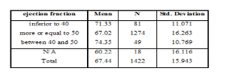
Patients age ranged between a minimum of 18 years and a maximum of 99 years, with a mean of 67.44 years (n=1422) and a standard deviation of 15.943.
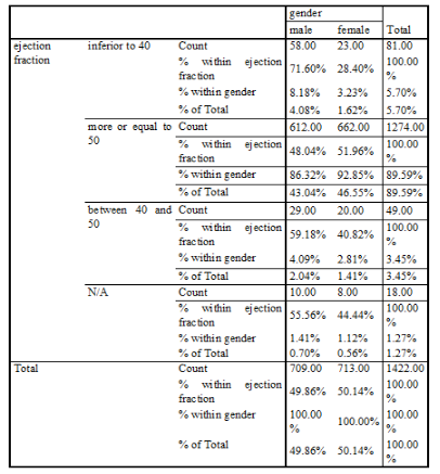
There were 709 males (49.86%) and 713 females (50.14%).
Out of the 1422 patients, 5.7% of our patients have an EF less than 40%, 89.59% have an EF more than 50%, 3.45% have an EF ranging between 40 and 50%, 1.27% of patients had no cut value for the EF.
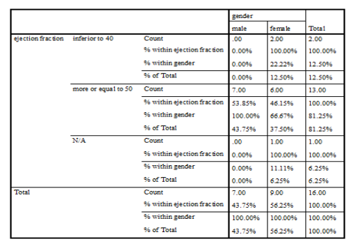
For the group of patients with EF less than 40% (HFrEF), their mean age was 71.33 years with a SD of 11.071 and 71.6% were male while 28.4% were female. Their distribution on our studied parameters is as the following:
For the group of patients with EF more or equal to 50% (HFpEF), their mean age was 67.02 years with a SD of 16.263 and 48% were male while 52% were female. Their distribution on our studied parameters is as the following:
For the group of patients with EF ranging between 40 and 50%, their mean age was 74.35 years with a SD of 10.769 and 59.2% were male while 40.8% were female. Their distribution on our studied parameters is as the following:
Our records showed that 16 of our patients had prosthetic valves, 10 of them aortic while the remaining were mitral. Among these patients, the minimum age was 31 and the maximum age was 87. Thus, 43.8% were male while 56.3% were female. Their distribution on our studied parameters is as the following:
- 12.5% (2 patients) had an EF less than 40% (HFrEF), among them 100% were female, 100% had a mild diastolic dysfunction, 100% had valvulopathies, 100% had a LVH, 100% had an elevated PASP, 100% had no documentation for the pro-BNP level while half of them had a LVEDd more than 55mm and the other half had a LVEDd less than 55mm.
- 81.25% (13 patients) had an EF more or equal to 50% (HFpEF), among them 53.85% were male and 46.15% were female, 23.08% had no diastolic dysfunction, 38.46% had no documentation and the remaining had a diastolic heart dysfuntion; 46.15% had a valvular disorder while 53.85% had none; 69.23% had elevated PASP, 7.69% had normal pressures while the remaining had no documentation; 61.54% had no LVH and 38.46% had a LVH; 30.77% had a LVEDd more than 55mm while the remaining had a diameter less than 55mm; 7.69% had a positive pro-BNP level while the remaining had no documentation


Although HF is caused by a myocardial dysfunction, but it may occur in the presence of near-normal cardiac function. To maintain this function, our heart may use different compensatory mechanisms (increasing in HR, blood volume, filling pressures,…) leading sometimes to a progressive decline in its ability to either contract or relax.
Epidemiologic studies indicate that HF occur with either a reduced (≤40%) or preserved (≥50%) EF and up to 50% of patients with heart failure have a preserved ejection fraction, knowing that this proportion has increased over time [26]. Clinical trials have showed that outcomes are better in HFpEF group and death of non cardiovascular causes are more
common in this group of patients who have heart failure with a preserved ejection fraction[27-29].
Concerning the main causes of HF, hypertension and CAD were on top of the list by 1970 but different statistical analysis have showed that diabetes and CAD (mainly MI) have become responsible for this issue due to improvement in management and early detection of hypertension and valvular disorders[6,31-34].
Conditions commonly associated with HFpEF and HFrEF include older age, hypertension, coronary disease, and diabetes mellitus. Some of these factors (age, hypertension, obesity, female gender, atrial fibrillation, increased urinary albumin excretion, and increased cystatin-C ) favour HFpEF for HFrEF which main etiologic factors are male sex, smoking, hs-TnT, and prior MI. The predictors of these two types of HF were evaluated in the PREVEND community-based, cohort study of middle-aged subjects[30].
In one review, the prevalence of HFpEF increased proportionally with age. It was 15 per cent for patients aged less than 50, 33 per cent for those aged between 30 and 50 and 50 per cent for those aged above 70. Add that systolic dysfunction contributed a little bit in the heart malfunction in few of these patients especially the elderly[35,36].
So studies have confirmed that aging may lead independently to a HFpEF. For instance, both the Baltimore Longitudinal Study on Aging and the Framingham Heart Study showed that age plays a major role in diastolic heart failure. Comorbid conditions may occur also with age such hypertension complicating our aim to clearly understand the etiology of HFpEF [37-39]. Our registry disclosed that the mean age of our patients (either with preserved or reduced EF) is 67.44 years with a standard deviation of 15.943 divided as follow: 71.33 years (with a SD of 11.0711) for patients with EF less than 40% and 67.02 years (with a SD of 16.263) for patients with EF more or equal to 50%.
Same studies confirmed the fact that blood pressure elevation above or equal to 160/100, after age of 40 especially, double the lifetime risk of development of HF in these subjects.
In a Mendelian randomization study, a relation of causality between high BMI and HF was made focusing on younger population (hazard ratio, 1.19 per BMI-unit increase, 95% CI 1.03-1.39) [40], so this entity of obesity related HFpEF has been growing over years especially in people with metabolic disorder making promises in the development of new regimen that may reduce both morbidity and mortality of this entity of HF(mineralocorticoid receptor antagonists, neprilysin inhibitor, SGLT2 inhibitor).
Different other studies indicated that women are more likely to be affected with HFpEF than men[41] as confirmed in our registry. This finding was based on different facts (greater aortic stiffness and arterial pulsatility in older postmenopausal women than men, higher indexed LV wall thicknesses[42], higher elastin deposition and lesser collagen production in arteries due to estrogen impregnation in premenopausal women[43], greater general adiposity and circulating inflammatory cells)[44]. These numerous risk factors explain the greater risk of development of HFpEF in women than in men so specific sex-based therapy for HF should be issued in order to reduce both morbidity and mortality of this HF entity[45]. In our registry, for the group of patients with HFrEF, 71.6% were male while 28.4% were female and for the patients who suffered from HFpEF, 48.04% were male while 51.96% were female.
On another hand , HFrEF may occur after a massive MI or multivessel disease(first described by Killip in the 1960s)[47] due to the impairment of myocytes to contract properly. This condition may be reversed if restoration of blood flow is made as soon as possible [46]. This latter form of HF has shown to be reduced after the introduction of primary PCI as suggested by the HORIZONS-AMI cohort of 3602 patients recruited between 2005-2007 treated with PPCI (8% of patients developed HF as Killip II-IV upon presentation, reduced to 4.6% at day 30 then rising to 5.1% at 2 years) [48].
Different diagnostic laboratory tools and imagery may aid in the diagnosis of HF as mentioned above. One of the most useful laboratory hormone level in HF are BNP and NT-proBNP useful in distinguishing HF from other causes of dyspnea.
Measurement of plasma BNP or NT-proBNP is recommended by the 2013 American College of Cardiology/American Heart Association guidelines as well as the 2010 Heart Failure Society of America, 2012 European Society of Cardiology, and 2012 Canadian Cardiovascular Society guidelines [49-52] when the diagnosis of HF is uncertain.
Although NT-proBNP level is fourfold higher than BNP in patients with LV dysfunction, their level are similar in normal subjects[53]. Most symptomatic HF patients have a BNP level above 400 pg/mL while values less than 100 pg/mL have higher negative predictive value for HF[54]. Our pro-BNP level shows the following values for non-heart failure population:
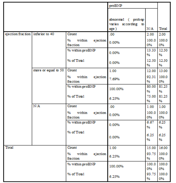
According to our statistics, the pro-BNP level was performed on 129 patients with 23 patients presented a normal level (21 patients with EF>or equal to 50%, 1 patient with EF between 40 and 50 and 1 patient with an undefined EF) and 106 patients presented an abnormal level according to our reference range (21 patients with EF<40, 76 patients with EF>or equal to 50, 8 patients with an EF between 40 and 50 and 1 patient with an undefined EF). So, none of our patients showed a normal level of pro-BNP with an EF less than 40% and its level was fourfold in HFpEF than in HFrEF(76 vs 21 patients).
In contrast, the NT-pro-BNP level required to diagnose HF varies with patient’s age and concentrations less than 300 pg/mL have high negative predictive value of 98% to exclude HF as cause of dyspnea. For instance, a normal level of NT-proBNP, based on Cleveland Clinic’s Reference Range is less than 125 pg/mL for patients aged 0-74 years and less than 450 pg/mL for patients aged 75-99 years. In case of heart failure, NT-proBNP levels that mean an unstable heart function are higher than 450 pg/mL for patients under age 50 and higher than 900 pg.mL for patients age 50 and older[55-57].
Since heart failure diagnosis is based mostly on the clinical presentation of the patient, TTE is performed to suggest possible etiologic factors of this condition (valvulopathies, systolic vs diastolic heart dysfunction…). Some of these findings studied in our registry include LVEDV, LVEF, presence of LVH, presence of diastolic dysfunction, documentation of any valvular disorder, measurements of PASP.
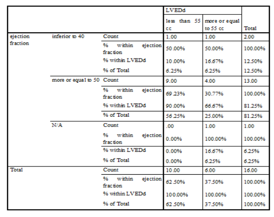
Since diastolic dysfunction results from impaired ventricular relaxation and filling patterns leading to a higher end-diastolic pressure for a given end-diastolic volume, LVEDd and LVEDV should be evaluated using ultrasonography for further investigation of the HF. Usually, its normal value is between 36 and 55mm at end diastole and between 23 and 40 at end systole in healthy subjects. Our numbers showed that 445 patients have a LVEDd more or equal to 55mm divided as following: 55 of them have an EF<40%, 344 with an EF>or equal to 50%, 36 with an EF between 40 and 50%, 10 patients with an undetermined EF.
Moreover, one of the most important and valuable variable measured while performing an echocardiography is the EF which plays a pivotal role in the diagnosis, risk stratification and therapeutic guidance of any suspected cardiac disease especially in term of HF. Although some clinical trials used to define HFpEF with an EF ranging between 40 and 55% but current guidelines recommend a cut value of 50% and an ejection fraction between 40 and 49% would be considered as gray area, while a “recovered hear failure with reduced ejection fraction” is the term given for HF in patients who had an EF less than 40% but recovered under medical therapy. As indicated by epidemiologic studies and HF registries, half of patients have a preserved ejection fraction and this proportion has increased over time [18,19]. Back to our numbers and for the 7 months period during which our study was conducted, we realize that 5.7% of our patients have an EF less than 40%, 89.49% have an EF more than 50%, 3.45% have an EF ranging between 40 and 50%, 1.27% of patients had no cut value for the EF. So, HFpEF is an emerging condition in our Lebanese society, as in other societies, that has been evolving over time.
Another commonly assessed echocardiographic feature is the presence of a left ventricular hypertrophy which may be caused by severe uncontrolled hypertension, aortic stenosis, hypertrophic cardiomyopathy or less commonly ventricular septal defects. Although EKG is a useful and cost-effective tool in detecting LVH, its moderate sensitivity or specificity due to different diagnostic sets, points toward the TTE or cardiac MRI for further confirmation[58-59].
LVH is defined as an increase in the mass of the LV obtained by 2D-echocardiography and is defined by the following criteria issued by the the American Society of Echocardiography, with the European Association of Echocardiography:
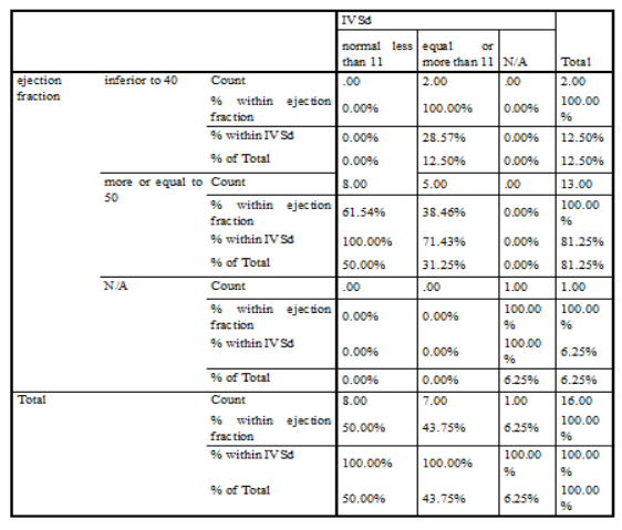
In our study, LVH is defined as IVS at the very end of diastole more or equal to 11 mm.
Patients with LVH may suffer from different cardiovascular complications such as heart failure and arrhythmia as shown by the Losartan Intervention for Endpoint Reduction in Hypertension (LIFE) study, where ST-T changes ("strain") on the baseline ECG in concert with voltage criteria for LVH increased the five-year risk of HF by more than threefold and the risk of HF-related mortality by fourfold, add that all-cause mortality were increased due to LVH in the middle aged population as shown by a large prospective study “ARIC”. Further studies confirmed the major role of echocardiography in detecting the risk for either atrial or ventricular arrhythmias caused by the LVH[61-64]. Our statistics showed that for the group of patients with EF less than 40%, 33.33 % had a LVH, 54.32% had no hypertrophy and 12.35% had no measurement; while for the group of patients with EF equal or more to 50%, 48.12 % had a LVH, 46.94% had no hypertrophy and 4.95% had no measurement.
Left ventricular diastolic dysfunction is one of the major features analyzed in our study. It should be suspected in any patient with symptoms suggestive of heart failure or even asymptomatic patients previously diagnosed with hypertension in whom screening with echocardiography may help us predicting complications or even death due to HFpEF. Thus, assessment of diastolic dysfunction should be considered an important step while evaluating a patient with suspicion of heart failure.
Patients with LVEF <50 percent, either symptomatic or not, may have diastolic dysfunction due to impaired LV relaxation while patients with LVEF >50 can have normal or abnormal diastolic function. For echocardiographic features that help us in diagnosing diastolic dysfunction, we consider the following criteria described in the 2016 American Society of Echocardiography and European Association of Cardiovascular Imaging guidelines:
The following rules are applied to determine if diastolic dysfunction is present:
-If <50 percent of measurable parameters for a patient meet the criteria above, then diastolic function is deemed normal.
-If >50 percent of measurable parameters meet the criteria above, then diastolic dysfunction is deemed present.
-If 50 percent of measurable parameters meet the criteria above, then the diagnosis of diastolic dysfunction is indeterminate [65].
In our study, we divided the diastolic dysfunction into three different categories:
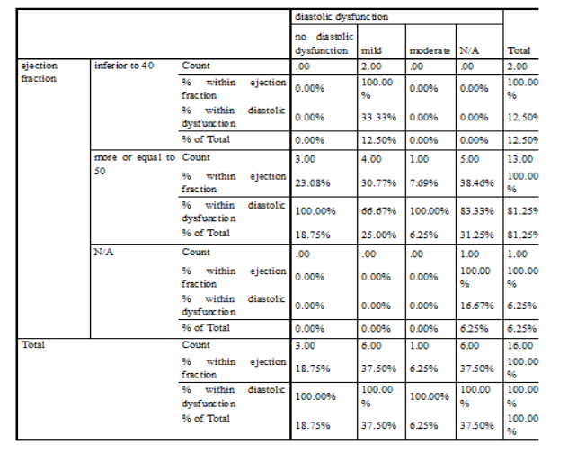
We found that for patients with an EF less than 40%, 17.28% had no diastolic dysfunction, while 65.43% had it, 2.47% with uncategorized diastolic dysfunction and data not fully clear for 14.81% of this population. While for patients with an EF more than 50%, one quarter of them showed no diastolic dysfunction and more than the half had one (the majority as grade I).
Moreover, registries have made valve disorders an important cause of heart failure either with preserved or reduced ejection fraction. For instance, the Euro Heart Survey showed that 69.8 % of patients with significant VHD (most commonly aortic stenosis and mitral regurgitation) presented with HF symptoms and 19.3 % of people with severe AS undergoing surgical aortic valve replacement (SAVR) had LVEF <50 % [66]. Further data from the German aortic valve registry indicated that 26.6% of patients presented for SAVR had an EF less than 50% [68] while the American Transcatheter Valve Therapy (TVT) registry showed a prevalence of 25.6% of patients with LVEF less than 45% among those who underwent a TAVR [69]. Last but not least, a survey involving huge number of patients with suspected HF 14% of patients suspected with HF and referred to echocardiography for confirmation showed moderate to severe coexistent valvular heart disease [67].
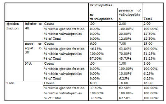
In one study involving 70,043 patients with suspected HF referred for echocardiography, MR of any severity was found in 12.5 % and moderate or severe MR in 3.1 % of patients [67]. In comparison to our numbers, we found that among patients with an EF less than 40%, 71.6% of them had a documented valvular disorder and the remaining ones had none. Add that half of our patients with an EF more or equal to 50% showed a valvulopathy regardless its severity, while the other half were valve disorders free.
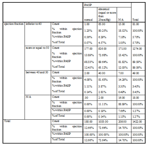
The last measured parameter in our study is the pulmonary artery systolic pressure (PASP). Its estimation is done by applying the following equation (based on the simplified Bernoulli equation): PASP ≈ RVSP = 4(peak TRV2) + RAP (estimated at 5-10mmHg).
When the TR signal is not reliably interpretable, echocardiography should be investigated for other parameters that may be responsible for pulmonary hypertension (right heart cavity sizes, non-TR-dependent estimators of PAP..).
Pulmonary hypertension (PH) is defined as an elevated mean arterial pressure ≥25 mmHg at rest and it is divided into 5 different categories by the World Health Organization (WHO) according to its etiology:
Identification of patients with PH-LHD is highly recommended because it is associated with a high rate of morbidity and mortality; patients may have either a HFrEF or HFpEF.
Most of series have indicated that 70 percent of PH is caused by LHD based on TTE [72]. PH-LHD is defined hemodynamically as an mPAP ≥25 mmHg and a pulmonary capillary wedge pressure (PCWP) ≥15 mmHg [73].
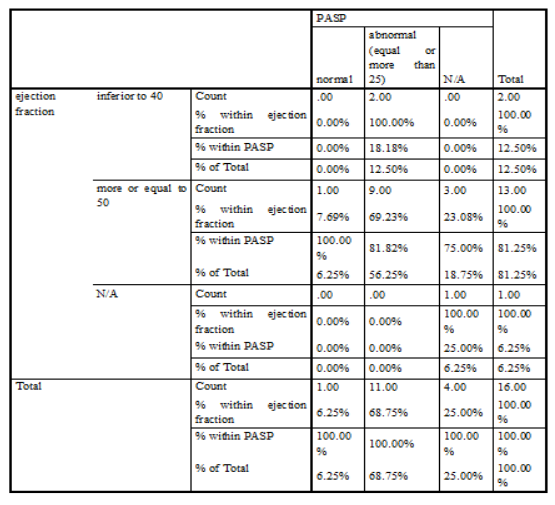
In some conditions, PH-LHD is confused with PAH that’s why investigating other factors (age, hypertensive, obesity,..) and TTE features of diastolic dysfunction favor HFpEF over PAH. Our numbers revealed that 80.25% of patients with EF less than 40% have an abnormal PASP (more or equal to 25mmHg) while only 1.23% have normal PASP. For patients with EF more or equal to 50%, 72.68% of them have an abnormal PASP and 13.89% have a normal pulmonary pressures.
LIMITATION AND PERSPECTIVE
The main limitation is that our study was conducted in only one medical center in Lebanon (LGH-UMC), which renders the generalization of the results to the Lebanese population rather difficult. But this makes the results more reliable in terms of echocardiography measurements as it is operator dependent.
Furthermore, patients information were taken from the EMF from the hospital and from the patient himself (before referring him to TTE) which reduces the likelihood of the results being biased.
Moreover, the cardiac sonography was performed by using the same machine by all the cardiologists and expert opinion was taken once in doubt which lead to a homogenous medical and imaging outcom
In this study, the prevalence of HF and its possible etiologies have been reviewed. As showed by numbers, aging play a major role in development of HF especially after the age of 65 years. The availability of cardiac sonography which is considered the most important tool in diagnosing HF, makes it easier to detect any possible disturbance that may lead to deterioration of the heart function, helping us in fighting either development or progression of a pre-existant HF and detecting its possible causes.
Add that, HFpEF should be deeply investigated while evaluating a patient with suspicion of HF because as our number showed its prevalence has been increasing over time in our community and in the international one.
All of this leads to a deduction in the overall cost by minimizing the rate of hospitalization and drug abuse while confirmation of HF is done using TTE which is available in almost all Lebanese hospitals.
We would like to thank everyone who made this work possible and paved the road for us.
Therefore we would like to thank very much the medical director who has reviewed this project.
Last but not least, we would like to thank the staff of the LGH for their constant help.
Clearly Auctoresonline and particularly Psychology and Mental Health Care Journal is dedicated to improving health care services for individuals and populations. The editorial boards' ability to efficiently recognize and share the global importance of health literacy with a variety of stakeholders. Auctoresonline publishing platform can be used to facilitate of optimal client-based services and should be added to health care professionals' repertoire of evidence-based health care resources.

Journal of Clinical Cardiology and Cardiovascular Intervention The submission and review process was adequate. However I think that the publication total value should have been enlightened in early fases. Thank you for all.

Journal of Women Health Care and Issues By the present mail, I want to say thank to you and tour colleagues for facilitating my published article. Specially thank you for the peer review process, support from the editorial office. I appreciate positively the quality of your journal.

Journal of Clinical Research and Reports I would be very delighted to submit my testimonial regarding the reviewer board and the editorial office. The reviewer board were accurate and helpful regarding any modifications for my manuscript. And the editorial office were very helpful and supportive in contacting and monitoring with any update and offering help. It was my pleasure to contribute with your promising Journal and I am looking forward for more collaboration.

We would like to thank the Journal of Thoracic Disease and Cardiothoracic Surgery because of the services they provided us for our articles. The peer-review process was done in a very excellent time manner, and the opinions of the reviewers helped us to improve our manuscript further. The editorial office had an outstanding correspondence with us and guided us in many ways. During a hard time of the pandemic that is affecting every one of us tremendously, the editorial office helped us make everything easier for publishing scientific work. Hope for a more scientific relationship with your Journal.

The peer-review process which consisted high quality queries on the paper. I did answer six reviewers’ questions and comments before the paper was accepted. The support from the editorial office is excellent.

Journal of Neuroscience and Neurological Surgery. I had the experience of publishing a research article recently. The whole process was simple from submission to publication. The reviewers made specific and valuable recommendations and corrections that improved the quality of my publication. I strongly recommend this Journal.

Dr. Katarzyna Byczkowska My testimonial covering: "The peer review process is quick and effective. The support from the editorial office is very professional and friendly. Quality of the Clinical Cardiology and Cardiovascular Interventions is scientific and publishes ground-breaking research on cardiology that is useful for other professionals in the field.

Thank you most sincerely, with regard to the support you have given in relation to the reviewing process and the processing of my article entitled "Large Cell Neuroendocrine Carcinoma of The Prostate Gland: A Review and Update" for publication in your esteemed Journal, Journal of Cancer Research and Cellular Therapeutics". The editorial team has been very supportive.

Testimony of Journal of Clinical Otorhinolaryngology: work with your Reviews has been a educational and constructive experience. The editorial office were very helpful and supportive. It was a pleasure to contribute to your Journal.

Dr. Bernard Terkimbi Utoo, I am happy to publish my scientific work in Journal of Women Health Care and Issues (JWHCI). The manuscript submission was seamless and peer review process was top notch. I was amazed that 4 reviewers worked on the manuscript which made it a highly technical, standard and excellent quality paper. I appreciate the format and consideration for the APC as well as the speed of publication. It is my pleasure to continue with this scientific relationship with the esteem JWHCI.

This is an acknowledgment for peer reviewers, editorial board of Journal of Clinical Research and Reports. They show a lot of consideration for us as publishers for our research article “Evaluation of the different factors associated with side effects of COVID-19 vaccination on medical students, Mutah university, Al-Karak, Jordan”, in a very professional and easy way. This journal is one of outstanding medical journal.

Dear Hao Jiang, to Journal of Nutrition and Food Processing We greatly appreciate the efficient, professional and rapid processing of our paper by your team. If there is anything else we should do, please do not hesitate to let us know. On behalf of my co-authors, we would like to express our great appreciation to editor and reviewers.

As an author who has recently published in the journal "Brain and Neurological Disorders". I am delighted to provide a testimonial on the peer review process, editorial office support, and the overall quality of the journal. The peer review process at Brain and Neurological Disorders is rigorous and meticulous, ensuring that only high-quality, evidence-based research is published. The reviewers are experts in their fields, and their comments and suggestions were constructive and helped improve the quality of my manuscript. The review process was timely and efficient, with clear communication from the editorial office at each stage. The support from the editorial office was exceptional throughout the entire process. The editorial staff was responsive, professional, and always willing to help. They provided valuable guidance on formatting, structure, and ethical considerations, making the submission process seamless. Moreover, they kept me informed about the status of my manuscript and provided timely updates, which made the process less stressful. The journal Brain and Neurological Disorders is of the highest quality, with a strong focus on publishing cutting-edge research in the field of neurology. The articles published in this journal are well-researched, rigorously peer-reviewed, and written by experts in the field. The journal maintains high standards, ensuring that readers are provided with the most up-to-date and reliable information on brain and neurological disorders. In conclusion, I had a wonderful experience publishing in Brain and Neurological Disorders. The peer review process was thorough, the editorial office provided exceptional support, and the journal's quality is second to none. I would highly recommend this journal to any researcher working in the field of neurology and brain disorders.
Dear Agrippa Hilda, Journal of Neuroscience and Neurological Surgery, Editorial Coordinator, I trust this message finds you well. I want to extend my appreciation for considering my article for publication in your esteemed journal. I am pleased to provide a testimonial regarding the peer review process and the support received from your editorial office. The peer review process for my paper was carried out in a highly professional and thorough manner. The feedback and comments provided by the authors were constructive and very useful in improving the quality of the manuscript. This rigorous assessment process undoubtedly contributes to the high standards maintained by your journal.

International Journal of Clinical Case Reports and Reviews. I strongly recommend to consider submitting your work to this high-quality journal. The support and availability of the Editorial staff is outstanding and the review process was both efficient and rigorous.

Thank you very much for publishing my Research Article titled “Comparing Treatment Outcome Of Allergic Rhinitis Patients After Using Fluticasone Nasal Spray And Nasal Douching" in the Journal of Clinical Otorhinolaryngology. As Medical Professionals we are immensely benefited from study of various informative Articles and Papers published in this high quality Journal. I look forward to enriching my knowledge by regular study of the Journal and contribute my future work in the field of ENT through the Journal for use by the medical fraternity. The support from the Editorial office was excellent and very prompt. I also welcome the comments received from the readers of my Research Article.

Dear Erica Kelsey, Editorial Coordinator of Cancer Research and Cellular Therapeutics Our team is very satisfied with the processing of our paper by your journal. That was fast, efficient, rigorous, but without unnecessary complications. We appreciated the very short time between the submission of the paper and its publication on line on your site.

I am very glad to say that the peer review process is very successful and fast and support from the Editorial Office. Therefore, I would like to continue our scientific relationship for a long time. And I especially thank you for your kindly attention towards my article. Have a good day!

"We recently published an article entitled “Influence of beta-Cyclodextrins upon the Degradation of Carbofuran Derivatives under Alkaline Conditions" in the Journal of “Pesticides and Biofertilizers” to show that the cyclodextrins protect the carbamates increasing their half-life time in the presence of basic conditions This will be very helpful to understand carbofuran behaviour in the analytical, agro-environmental and food areas. We greatly appreciated the interaction with the editor and the editorial team; we were particularly well accompanied during the course of the revision process, since all various steps towards publication were short and without delay".

I would like to express my gratitude towards you process of article review and submission. I found this to be very fair and expedient. Your follow up has been excellent. I have many publications in national and international journal and your process has been one of the best so far. Keep up the great work.

We are grateful for this opportunity to provide a glowing recommendation to the Journal of Psychiatry and Psychotherapy. We found that the editorial team were very supportive, helpful, kept us abreast of timelines and over all very professional in nature. The peer review process was rigorous, efficient and constructive that really enhanced our article submission. The experience with this journal remains one of our best ever and we look forward to providing future submissions in the near future.

I am very pleased to serve as EBM of the journal, I hope many years of my experience in stem cells can help the journal from one way or another. As we know, stem cells hold great potential for regenerative medicine, which are mostly used to promote the repair response of diseased, dysfunctional or injured tissue using stem cells or their derivatives. I think Stem Cell Research and Therapeutics International is a great platform to publish and share the understanding towards the biology and translational or clinical application of stem cells.

I would like to give my testimony in the support I have got by the peer review process and to support the editorial office where they were of asset to support young author like me to be encouraged to publish their work in your respected journal and globalize and share knowledge across the globe. I really give my great gratitude to your journal and the peer review including the editorial office.

I am delighted to publish our manuscript entitled "A Perspective on Cocaine Induced Stroke - Its Mechanisms and Management" in the Journal of Neuroscience and Neurological Surgery. The peer review process, support from the editorial office, and quality of the journal are excellent. The manuscripts published are of high quality and of excellent scientific value. I recommend this journal very much to colleagues.

Dr.Tania Muñoz, My experience as researcher and author of a review article in The Journal Clinical Cardiology and Interventions has been very enriching and stimulating. The editorial team is excellent, performs its work with absolute responsibility and delivery. They are proactive, dynamic and receptive to all proposals. Supporting at all times the vast universe of authors who choose them as an option for publication. The team of review specialists, members of the editorial board, are brilliant professionals, with remarkable performance in medical research and scientific methodology. Together they form a frontline team that consolidates the JCCI as a magnificent option for the publication and review of high-level medical articles and broad collective interest. I am honored to be able to share my review article and open to receive all your comments.

“The peer review process of JPMHC is quick and effective. Authors are benefited by good and professional reviewers with huge experience in the field of psychology and mental health. The support from the editorial office is very professional. People to contact to are friendly and happy to help and assist any query authors might have. Quality of the Journal is scientific and publishes ground-breaking research on mental health that is useful for other professionals in the field”.

Dear editorial department: On behalf of our team, I hereby certify the reliability and superiority of the International Journal of Clinical Case Reports and Reviews in the peer review process, editorial support, and journal quality. Firstly, the peer review process of the International Journal of Clinical Case Reports and Reviews is rigorous, fair, transparent, fast, and of high quality. The editorial department invites experts from relevant fields as anonymous reviewers to review all submitted manuscripts. These experts have rich academic backgrounds and experience, and can accurately evaluate the academic quality, originality, and suitability of manuscripts. The editorial department is committed to ensuring the rigor of the peer review process, while also making every effort to ensure a fast review cycle to meet the needs of authors and the academic community. Secondly, the editorial team of the International Journal of Clinical Case Reports and Reviews is composed of a group of senior scholars and professionals with rich experience and professional knowledge in related fields. The editorial department is committed to assisting authors in improving their manuscripts, ensuring their academic accuracy, clarity, and completeness. Editors actively collaborate with authors, providing useful suggestions and feedback to promote the improvement and development of the manuscript. We believe that the support of the editorial department is one of the key factors in ensuring the quality of the journal. Finally, the International Journal of Clinical Case Reports and Reviews is renowned for its high- quality articles and strict academic standards. The editorial department is committed to publishing innovative and academically valuable research results to promote the development and progress of related fields. The International Journal of Clinical Case Reports and Reviews is reasonably priced and ensures excellent service and quality ratio, allowing authors to obtain high-level academic publishing opportunities in an affordable manner. I hereby solemnly declare that the International Journal of Clinical Case Reports and Reviews has a high level of credibility and superiority in terms of peer review process, editorial support, reasonable fees, and journal quality. Sincerely, Rui Tao.

Clinical Cardiology and Cardiovascular Interventions I testity the covering of the peer review process, support from the editorial office, and quality of the journal.

Clinical Cardiology and Cardiovascular Interventions, we deeply appreciate the interest shown in our work and its publication. It has been a true pleasure to collaborate with you. The peer review process, as well as the support provided by the editorial office, have been exceptional, and the quality of the journal is very high, which was a determining factor in our decision to publish with you.

The peer reviewers process is quick and effective, the supports from editorial office is excellent, the quality of journal is high. I would like to collabroate with Internatioanl journal of Clinical Case Reports and Reviews journal clinically in the future time.

Clinical Cardiology and Cardiovascular Interventions, I would like to express my sincerest gratitude for the trust placed in our team for the publication in your journal. It has been a true pleasure to collaborate with you on this project. I am pleased to inform you that both the peer review process and the attention from the editorial coordination have been excellent. Your team has worked with dedication and professionalism to ensure that your publication meets the highest standards of quality. We are confident that this collaboration will result in mutual success, and we are eager to see the fruits of this shared effort.

Dear Dr. Jessica Magne, Editorial Coordinator 0f Clinical Cardiology and Cardiovascular Interventions, I hope this message finds you well. I want to express my utmost gratitude for your excellent work and for the dedication and speed in the publication process of my article titled "Navigating Innovation: Qualitative Insights on Using Technology for Health Education in Acute Coronary Syndrome Patients." I am very satisfied with the peer review process, the support from the editorial office, and the quality of the journal. I hope we can maintain our scientific relationship in the long term.

Dear Monica Gissare, - Editorial Coordinator of Nutrition and Food Processing. ¨My testimony with you is truly professional, with a positive response regarding the follow-up of the article and its review, you took into account my qualities and the importance of the topic¨.

Dear Dr. Jessica Magne, Editorial Coordinator 0f Clinical Cardiology and Cardiovascular Interventions, The review process for the article “The Handling of Anti-aggregants and Anticoagulants in the Oncologic Heart Patient Submitted to Surgery” was extremely rigorous and detailed. From the initial submission to the final acceptance, the editorial team at the “Journal of Clinical Cardiology and Cardiovascular Interventions” demonstrated a high level of professionalism and dedication. The reviewers provided constructive and detailed feedback, which was essential for improving the quality of our work. Communication was always clear and efficient, ensuring that all our questions were promptly addressed. The quality of the “Journal of Clinical Cardiology and Cardiovascular Interventions” is undeniable. It is a peer-reviewed, open-access publication dedicated exclusively to disseminating high-quality research in the field of clinical cardiology and cardiovascular interventions. The journal's impact factor is currently under evaluation, and it is indexed in reputable databases, which further reinforces its credibility and relevance in the scientific field. I highly recommend this journal to researchers looking for a reputable platform to publish their studies.

Dear Editorial Coordinator of the Journal of Nutrition and Food Processing! "I would like to thank the Journal of Nutrition and Food Processing for including and publishing my article. The peer review process was very quick, movement and precise. The Editorial Board has done an extremely conscientious job with much help, valuable comments and advices. I find the journal very valuable from a professional point of view, thank you very much for allowing me to be part of it and I would like to participate in the future!”

Dealing with The Journal of Neurology and Neurological Surgery was very smooth and comprehensive. The office staff took time to address my needs and the response from editors and the office was prompt and fair. I certainly hope to publish with this journal again.Their professionalism is apparent and more than satisfactory. Susan Weiner

My Testimonial Covering as fellowing: Lin-Show Chin. The peer reviewers process is quick and effective, the supports from editorial office is excellent, the quality of journal is high. I would like to collabroate with Internatioanl journal of Clinical Case Reports and Reviews.

My experience publishing in Psychology and Mental Health Care was exceptional. The peer review process was rigorous and constructive, with reviewers providing valuable insights that helped enhance the quality of our work. The editorial team was highly supportive and responsive, making the submission process smooth and efficient. The journal's commitment to high standards and academic rigor makes it a respected platform for quality research. I am grateful for the opportunity to publish in such a reputable journal.

My experience publishing in International Journal of Clinical Case Reports and Reviews was exceptional. I Come forth to Provide a Testimonial Covering the Peer Review Process and the editorial office for the Professional and Impartial Evaluation of the Manuscript.

I would like to offer my testimony in the support. I have received through the peer review process and support the editorial office where they are to support young authors like me, encourage them to publish their work in your esteemed journals, and globalize and share knowledge globally. I really appreciate your journal, peer review, and editorial office.

Dear Agrippa Hilda- Editorial Coordinator of Journal of Neuroscience and Neurological Surgery, "The peer review process was very quick and of high quality, which can also be seen in the articles in the journal. The collaboration with the editorial office was very good."

I would like to express my sincere gratitude for the support and efficiency provided by the editorial office throughout the publication process of my article, “Delayed Vulvar Metastases from Rectal Carcinoma: A Case Report.” I greatly appreciate the assistance and guidance I received from your team, which made the entire process smooth and efficient. The peer review process was thorough and constructive, contributing to the overall quality of the final article. I am very grateful for the high level of professionalism and commitment shown by the editorial staff, and I look forward to maintaining a long-term collaboration with the International Journal of Clinical Case Reports and Reviews.

To Dear Erin Aust, I would like to express my heartfelt appreciation for the opportunity to have my work published in this esteemed journal. The entire publication process was smooth and well-organized, and I am extremely satisfied with the final result. The Editorial Team demonstrated the utmost professionalism, providing prompt and insightful feedback throughout the review process. Their clear communication and constructive suggestions were invaluable in enhancing my manuscript, and their meticulous attention to detail and dedication to quality are truly commendable. Additionally, the support from the Editorial Office was exceptional. From the initial submission to the final publication, I was guided through every step of the process with great care and professionalism. The team's responsiveness and assistance made the entire experience both easy and stress-free. I am also deeply impressed by the quality and reputation of the journal. It is an honor to have my research featured in such a respected publication, and I am confident that it will make a meaningful contribution to the field.

"I am grateful for the opportunity of contributing to [International Journal of Clinical Case Reports and Reviews] and for the rigorous review process that enhances the quality of research published in your esteemed journal. I sincerely appreciate the time and effort of your team who have dedicatedly helped me in improvising changes and modifying my manuscript. The insightful comments and constructive feedback provided have been invaluable in refining and strengthening my work".

I thank the ‘Journal of Clinical Research and Reports’ for accepting this article for publication. This is a rigorously peer reviewed journal which is on all major global scientific data bases. I note the review process was prompt, thorough and professionally critical. It gave us an insight into a number of important scientific/statistical issues. The review prompted us to review the relevant literature again and look at the limitations of the study. The peer reviewers were open, clear in the instructions and the editorial team was very prompt in their communication. This journal certainly publishes quality research articles. I would recommend the journal for any future publications.

Dear Jessica Magne, with gratitude for the joint work. Fast process of receiving and processing the submitted scientific materials in “Clinical Cardiology and Cardiovascular Interventions”. High level of competence of the editors with clear and correct recommendations and ideas for enriching the article.

We found the peer review process quick and positive in its input. The support from the editorial officer has been very agile, always with the intention of improving the article and taking into account our subsequent corrections.

My article, titled 'No Way Out of the Smartphone Epidemic Without Considering the Insights of Brain Research,' has been republished in the International Journal of Clinical Case Reports and Reviews. The review process was seamless and professional, with the editors being both friendly and supportive. I am deeply grateful for their efforts.

To Dear Erin Aust – Editorial Coordinator of Journal of General Medicine and Clinical Practice! I declare that I am absolutely satisfied with your work carried out with great competence in following the manuscript during the various stages from its receipt, during the revision process to the final acceptance for publication. Thank Prof. Elvira Farina

Dear Jessica, and the super professional team of the ‘Clinical Cardiology and Cardiovascular Interventions’ I am sincerely grateful to the coordinated work of the journal team for the no problem with the submission of my manuscript: “Cardiometabolic Disorders in A Pregnant Woman with Severe Preeclampsia on the Background of Morbid Obesity (Case Report).” The review process by 5 experts was fast, and the comments were professional, which made it more specific and academic, and the process of publication and presentation of the article was excellent. I recommend that my colleagues publish articles in this journal, and I am interested in further scientific cooperation. Sincerely and best wishes, Dr. Oleg Golyanovskiy.

Dear Ashley Rosa, Editorial Coordinator of the journal - Psychology and Mental Health Care. " The process of obtaining publication of my article in the Psychology and Mental Health Journal was positive in all areas. The peer review process resulted in a number of valuable comments, the editorial process was collaborative and timely, and the quality of this journal has been quickly noticed, resulting in alternative journals contacting me to publish with them." Warm regards, Susan Anne Smith, PhD. Australian Breastfeeding Association.

Dear Jessica Magne, Editorial Coordinator, Clinical Cardiology and Cardiovascular Interventions, Auctores Publishing LLC. I appreciate the journal (JCCI) editorial office support, the entire team leads were always ready to help, not only on technical front but also on thorough process. Also, I should thank dear reviewers’ attention to detail and creative approach to teach me and bring new insights by their comments. Surely, more discussions and introduction of other hemodynamic devices would provide better prevention and management of shock states. Your efforts and dedication in presenting educational materials in this journal are commendable. Best wishes from, Farahnaz Fallahian.

Dear Maria Emerson, Editorial Coordinator, International Journal of Clinical Case Reports and Reviews, Auctores Publishing LLC. I am delighted to have published our manuscript, "Acute Colonic Pseudo-Obstruction (ACPO): A rare but serious complication following caesarean section." I want to thank the editorial team, especially Maria Emerson, for their prompt review of the manuscript, quick responses to queries, and overall support. Yours sincerely Dr. Victor Olagundoye.

Dear Ashley Rosa, Editorial Coordinator, International Journal of Clinical Case Reports and Reviews. Many thanks for publishing this manuscript after I lost confidence the editors were most helpful, more than other journals Best wishes from, Susan Anne Smith, PhD. Australian Breastfeeding Association.

Dear Agrippa Hilda, Editorial Coordinator, Journal of Neuroscience and Neurological Surgery. The entire process including article submission, review, revision, and publication was extremely easy. The journal editor was prompt and helpful, and the reviewers contributed to the quality of the paper. Thank you so much! Eric Nussbaum, MD

Dr Hala Al Shaikh This is to acknowledge that the peer review process for the article ’ A Novel Gnrh1 Gene Mutation in Four Omani Male Siblings, Presentation and Management ’ sent to the International Journal of Clinical Case Reports and Reviews was quick and smooth. The editorial office was prompt with easy communication.

Dear Erin Aust, Editorial Coordinator, Journal of General Medicine and Clinical Practice. We are pleased to share our experience with the “Journal of General Medicine and Clinical Practice”, following the successful publication of our article. The peer review process was thorough and constructive, helping to improve the clarity and quality of the manuscript. We are especially thankful to Ms. Erin Aust, the Editorial Coordinator, for her prompt communication and continuous support throughout the process. Her professionalism ensured a smooth and efficient publication experience. The journal upholds high editorial standards, and we highly recommend it to fellow researchers seeking a credible platform for their work. Best wishes By, Dr. Rakhi Mishra.

Dear Jessica Magne, Editorial Coordinator, Clinical Cardiology and Cardiovascular Interventions, Auctores Publishing LLC. The peer review process of the journal of Clinical Cardiology and Cardiovascular Interventions was excellent and fast, as was the support of the editorial office and the quality of the journal. Kind regards Walter F. Riesen Prof. Dr. Dr. h.c. Walter F. Riesen.

Dear Ashley Rosa, Editorial Coordinator, International Journal of Clinical Case Reports and Reviews, Auctores Publishing LLC. Thank you for publishing our article, Exploring Clozapine's Efficacy in Managing Aggression: A Multiple Single-Case Study in Forensic Psychiatry in the international journal of clinical case reports and reviews. We found the peer review process very professional and efficient. The comments were constructive, and the whole process was efficient. On behalf of the co-authors, I would like to thank you for publishing this article. With regards, Dr. Jelle R. Lettinga.

Dear Clarissa Eric, Editorial Coordinator, Journal of Clinical Case Reports and Studies, I would like to express my deep admiration for the exceptional professionalism demonstrated by your journal. I am thoroughly impressed by the speed of the editorial process, the substantive and insightful reviews, and the meticulous preparation of the manuscript for publication. Additionally, I greatly appreciate the courteous and immediate responses from your editorial office to all my inquiries. Best Regards, Dariusz Ziora

Dear Chrystine Mejia, Editorial Coordinator, Journal of Neurodegeneration and Neurorehabilitation, Auctores Publishing LLC, We would like to thank the editorial team for the smooth and high-quality communication leading up to the publication of our article in the Journal of Neurodegeneration and Neurorehabilitation. The reviewers have extensive knowledge in the field, and their relevant questions helped to add value to our publication. Kind regards, Dr. Ravi Shrivastava.

Dear Clarissa Eric, Editorial Coordinator, Journal of Clinical Case Reports and Studies, Auctores Publishing LLC, USA Office: +1-(302)-520-2644. I would like to express my sincere appreciation for the efficient and professional handling of my case report by the ‘Journal of Clinical Case Reports and Studies’. The peer review process was not only fast but also highly constructive—the reviewers’ comments were clear, relevant, and greatly helped me improve the quality and clarity of my manuscript. I also received excellent support from the editorial office throughout the process. Communication was smooth and timely, and I felt well guided at every stage, from submission to publication. The overall quality and rigor of the journal are truly commendable. I am pleased to have published my work with Journal of Clinical Case Reports and Studies, and I look forward to future opportunities for collaboration. Sincerely, Aline Tollet, UCLouvain.

Dear Ms. Mayra Duenas, Editorial Coordinator, International Journal of Clinical Case Reports and Reviews. “The International Journal of Clinical Case Reports and Reviews represented the “ideal house” to share with the research community a first experience with the use of the Simeox device for speech rehabilitation. High scientific reputation and attractive website communication were first determinants for the selection of this Journal, and the following submission process exceeded expectations: fast but highly professional peer review, great support by the editorial office, elegant graphic layout. Exactly what a dynamic research team - also composed by allied professionals - needs!" From, Chiara Beccaluva, PT - Italy.

Dear Maria Emerson, Editorial Coordinator, we have deeply appreciated the professionalism demonstrated by the International Journal of Clinical Case Reports and Reviews. The reviewers have extensive knowledge of our field and have been very efficient and fast in supporting the process. I am really looking forward to further collaboration. Thanks. Best regards, Dr. Claudio Ligresti

Dear Chrystine Mejia, Editorial Coordinator, Journal of Neurodegeneration and Neurorehabilitation. “The peer review process was efficient and constructive, and the editorial office provided excellent communication and support throughout. The journal ensures scientific rigor and high editorial standards, while also offering a smooth and timely publication process. We sincerely appreciate the work of the editorial team in facilitating the dissemination of innovative approaches such as the Bonori Method.” Best regards, Dr. Giselle Pentón-Rol.
