AUCTORES
Globalize your Research

Research Article | DOI: https://doi.org/10.31579/2641-0419/010
1 Center for Diagnostic Nanosystems, Marshall University, Huntington, WV, USA.
2 Department of Pharmaceutical Science and Research, School of Pharmacy, Marshall University, Huntington, WV, USA.
3 Department of Internal Medicine, Joan C. Edwards School of Medicine, Marshall University, Huntington, WV, USA.
4 Biotechnology Graduate Program West Virginia State University, Institute, WV.
5 Department of Public Heath, Marshall University, Huntington, WV, USA.
6 Department of Health and Human Service, School of Kinesiology, Marshall University, Huntington, WV.
7 Department of Pharmacology, Physiology and Toxicology, Joan C. Edwards School of Medicine, Marshall University, Huntington, WV, USA.
8 Department of Cardiology, Joan C. Edwards School of Medicine, Marshall University, Huntington, WV, USA.
*Corresponding Author: Kevin M. Rice, Center for Diagnostic Nanosystems, Marshall University, USA
Citation: Kevin M. Rice, Short Communication: The Effects Chronic Acetaminophen Treatment on Age-Associated Alterations of Cardiac Function in the Female F344xBN Heart, J.Clinical Cardiology and Cardiovascular Interventions. 1(2);Doi: 10.31579/2641-0419/010
Copyright: © 2018. Kevin M. Rice This is an open-access article distributed under the terms of the Creative Commons Attribution License, which permits unrestricted use, distribution, and reproduction in any medium, provided the original author and source are credited.
Received: 09 September 2018 | Accepted: 10 October 2018 | Published: 15 October 2018
Keywords: Cardiac Function; Chronic Acetaminophen;Echocardiographic procedures
Background
Although several studies have investigated the age-associated changes in male and female F344 as well as male Fischer 344/NNiaHSD x Brown Norway/BiNia F1 (F344xBN), no study to our knowledge has examined the age-associated changes in structure and function in the female F344xBN heart using echocardiographic measures. This information is crucial in determining whether or not the female F344xBN is an appropriate aging model. Additional studies have also suggested that age-associated increases in levels of oxidative stress may cause cardiac dysfunction and that chronic acetaminophen (APAP) ingestion may be protective against increased oxidative stress. On the basis of these studies we examined the function and structure of the aged female F344xBN heart in the absence and presence of chronic APAP treatment.
Methods
To investigate if the aging-related changes in cardiovascular structure and function can be attenuated with APAP treatment, aged (22-month old) female F344xBN rats were administered APAP (30 mg/kg body weight/day) for 8 months in drinking water and echocardiograms assessments were performed.
Results
Aging was associated with evidence of diastolic (impaired left ventricle relaxation time) and systolic dysfunction (fractional shortening and end systolic volume). The incidence of arrhythmias was not different with age. However, valvular dysfunction was increased. Chronic APAP treatment did not attenuate the age-associated changes in cardiac structure and function in the female F344xBN. However the occurrence of valve dysfunction with aging was significantly lowered in the APAP treated 30-month female hearts.
The American Heart Association estimates that 80 million Americans have one or more types of cardiovascular disease (CVD). It is estimated that 38.1 million of those afflicted are aged 60 or older. As a person ages he or she has a higher risk of developing myocardial infarction, stroke, atherosclerosis, peripheral occlusive disease, diabetes, hypertension [1-3].Thepotential bmechanism of increase risk of disease with age is that there is an increase of reactive oxygen species (ROS) which leads to oxidative stress [4]. Recently acetaminophen (APAP), an antipyretic and analgesic, has shown to be protective against oxidative stress in the heart and the vasculature of the brain due to its antioxidant properties [5-7].
Due to these antioxidant effects APAP is considered to be cardioprotective during ischemia/reperfusion, hypoxia/reoxygenation, exogenous peroxynitrite administration, and experimentally induced myocardial infarction due to its ability to block or reduce the development of mitochondrial permeability transition pores, mitochondrial swelling, cytochrome c release, late stage apoptosis, protein oxidation, production of hydroxyl radicals, arrhythmias, and peroxynitrite induced dysfunction in hearts [5, 8-16].
The Fischer 344/NNiaHSD x Brown Norway/BiNia F1 (F344xBN) has been recommended by the National Institute of Aging as a rat model of aging due to its increased life span and fewer age-associated pathologies compared to the standard model of aging – the Fischer 344 rat. Although several studies have investigated the age-associated changes in male and female F344 rats as well as male F344xBN rats [17-21], no study to our knowledge has examined the age-associated changes in structure and function in the female F344xBN heart using echocardiographic measures.
This information is crucial in determining whether or not the female F344xBN is an appropriate aging model to study the development of cardiovascular disease. Due to evidence that age-associated increase in levels of oxidative stress may cause cardiac dysfunction, we also determined the function and structure of the aged female F344xBN heart with chronic APAP treatment. The purpose of this study was to determine the age-associated changes in the female F344xBN cardiac structure and function and whether chronic APAP treatment would attenuate these effects.
Materials and Methods
All procedures were performed in accordance with the Guide for the Care and Use of Laboratory Animals as approved by the Council of the American Physiological Society and the Animal Use Review Board of Marshall University. All procedures were conducted in strict accordance with the Public Health Service animal welfare policy. Young (6-month), adult (22-month), aged (26-month), and very aged (30-month) female F344xBN rats were obtained from the National Institute for Aging were housed two per cage in an Association for Assessment and Accreditation of Laboratory Animal Care International (AAALAC) approved vivarium. Housing conditions consisted of a 12 h-12 h light-dark cycle with a temperature maintained at 22 ± 2˚C. Animals were provided with food and water ad libitum. Rats were allowed to recover from shipment for at least two weeks before experimentation. During this time the animals were carefully observed and weighed weekly. Any rat showing signs of failure to thrive, such as precipitous weight loss, disinterest in the environment, or unexpected gait alterations were excluded from the study. Six Female F344xBN rats were randomly assigned to each group (6C – 6-month animal; 22C – 22-month animal; 26C – 26-month animal; 26T – 26-month APAP-treated animal; 30C – 30-month animal; 30T – 30-month APAP-treated animal).
A therapeutic dose of APAP (30 mg/kg/day) was administered through the drinking water for treated animals at 22-months of age. Changes in body weight and water intake were monitored throughout the study to maintain a therapeutic dose. The 6-, 22-, 26-, and 30-month age-matched control groups received tap water and were maintained under the same conditions as the APAP treated groups.
Echocardiographic procedures
Control (6-, 22-, 26-, and 30-month) and APAP treated (26- and 30-month) rats were anesthetized with an intraperitoneal (ip) injection of ketamine (40 mg/kg) and xyaline (10 kg/mg). The ventral thoraxes where shaved, and the rats were placed either on their backs or left side and covered with ultrasonic transmission gel for adequate sonic transference. A Phillips 5500 ECHO system with a 12 MHz transducer was used to take two-dimensional ECHO measurements, two-dimensional guided M-mode, Doppler M-mode, and other recordings from parasternal long- and short-axis views. Parasternal long- and short axis views were used to determine two-dimensional cardiac structural measurements. The echocardiographic views were then used to position the M-mode echocardiographic line. In order to determine the presence of valve dysfunctions, valvular blood flow velocities were evaluated using pulse wave Doppler with the probe toward the apex (x-axis) and the depth along the y-axis (long axis procedure). Ejection fraction and fractional shortening during systole was calculated using the evaluation of wall structure from short-axis procedures with the probe oriented toward the left ventricle and across the heart. M-mode displays were analyzed by a digital echocardiographic analysis system.
The following measurements were selected for each assessment of cardiac structure and function. The structural parameters included diastolic (IVSd) and systolic (IVSs) left ventricular septal thickness, diastolic (LVIDd) and systolic (LVIDs) left ventricular internal dimension, diastiolic (LVPWd) and systolic (LVPWs) left ventricular posterior wall thickness, and right ventricular diastolic internal dimension (RVd). Functional measurement included ejection fraction (EF) and left ventricular fractional shortening during systole (FS).
Additional echocardiographic measurements included mitral valve deceleration (MV decel time), left ventricular mass (LVM), end-systolic volume (ESV), end-diastolic volume (EDV), peak velocity of the A wave (Amax), and peak velocity of the E wave (Emax) were used to evaluate systolic function. Mitral valve deceleration, Emax, Amax, and MV E/A ratio were used to evaluate diastolic function.
Electrocardiogram (EKG) measurements and Heart Collection
After completion of treatment and echocardiographic procedures, animals were anesthetized with an ip injection of ketamine (40 mg/kg) and xyaline (10 mg/kg) and supplemented as necessary for reflexive responses. Before heart collection, electrocardiograms (EKGs) with leads I, II, and III were measured in age-matched control and treated animals using the Biopac Student Lab software (BIOPAC Systems, Inc., Microsoft). After completion of EKG measurements, the heart was removed after a midline laparotomy and was then placed in Krebs-Ringer bicarbonate buffer (KRB) containing the following: 118 mM NaCl, 4.7 mM KCl, 2.5 mM CaCl2, 1.2 mM KH2PO4, 1.2 mM MgSO4, 24.2 mM NaHCO3, and 10 mM a-D-glucose (pH 7.4) equilibrated with 5% CO2/95% O2 and maintained at 37° C. Isolated hearts were quickly massaged to remove any blood from the ventricles, cleaned of connective tissue, weighed, and immediately snap frozen in liquid nitrogen for further analysis.
Results were reported as mean ± SEM. Statistical analyses were performed using Sigma Stat 3.5 statistical software (Systat Software, Inc). Age comparisons between ECHO structural, functional parameters, and morphologic indices were evaluated by one-way ANOVA or Kruskal-Wallis one-way Analysis of Variance on Ranks with the Student-Newman-Keuls or Dunn's methods as the post hoc test, respectively. Differences between age-matched control and treated animals were determined by Student’s T-test. The level of significance accepted a priori was P < 0.05.
Echocardiographic Structural Parameters
Echocardiographic evaluation of cardiac structural parameters in female F344xBN rats are compared in Table 1. Left ventricular septal thickness (IVS) during systole (lVSs) significantly increased at 26-months (0.253 ± 0.003 cm) compared to 6-months (0.193 ± 0.008 cm; P < 0.05); however, no age-associated changes during diastole (lVSd) were found. Left ventricular internal dimension during systole (LVIDs) significantly increased at 22- (0.340 ± 0.006 cm) and 26-months (0.331 ± 0.009 cm) compared to 6-month (0.378 ± 0.017 cm; P < 0.05). Treatment with APAP significantly decreased LVIDs compared to control animals at 30-months (0.344 ± 0.015 vs 0.356 ± 0.011 cm, respectively; P = 0.004). During diastole LVID did not change with age but significantly increased with APAP treatment at 26-months (0.630 ± 0.009 cm) when compared to control (0.593 ± 0.009 cm; P < 0.001). Left ventricular posterior wall thickness (LVPW) during systole (LVPWs) and diastole (LVPWd) was significantly increased at 22- (0.273 ± 0.004 and 0.183 ± 0.004 cm, respectively) and 30-months (0.288 ± 0.009 and 0.189 ± 0.008 cm, respectively) compared to 6-months (0.193 ± 0.002 and 0.153 ± 0.010 cm, respectively; P < 0.05). APAP treatment increased both LVPWs and LVPWd thickness at 26-months compared to control (0.314 ± 0.007 and 0.185 ± 0.003 cm, respectively; P = 0.002). No changes were found in right ventricular dimension during diastole or left ventricular mass with age or APAP treatment.
0.340 ± 0.006a
0.273 ± 0.004a
0.183 ± 0.004a
0.253 ± 0.003a
0.331 ± 0.009a
0.630 ± 0.009d
0.314 ± 0.007d
0.185 ± 0.003d
0.288 ± 0.009a
0.189 ± 0.008a
0.334 ± 0.015d
Table1:Echocardiographic evaluation of cardiac structural parameters in female F344xBN rats (mean ± SEM; n = ## rats/group). 6C – 6-month animal; 22C – 22-month animal; 26C – 26-month animal; 26T – 26-month APAP-treated animal; 30C – 30-month animal; 30T – 30-month APAP-treated animal; aP < 0.05 significant difference from 6-month control animal; dP < 0.05 age-matched control vs. APAP treated animal.
Echocardiographic Functional Parameters
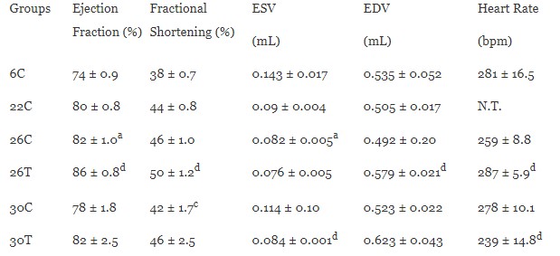
Echocardiographic evaluation of cardiac functional parameters in female F344xBN rats can be found in Table 2 and 3. As described in Table 2, ejection fraction was significantly increased at 26-months (82 ± 1.0%) compared to 6-months (74 ± 0.9%; P < 0.05). APAP treatment at 26-months (86 ± 0.8%) also significantly increased the ejection fraction compared to control (P = 0.009). Fractional shortening significantly decreased at 30-months (41.5 ± 1.7%) compared to 26-months (45.6 ± 1.0%; P < 0.05). APAP treatment at 26-months (49.8 ± 1.2%) significantly increased the fractional shortening when compared to control (P = 0.007). End systolic volume (ESV) was significantly decreased at 26-months (0.082 ± 0.005 mL) when compared to 6-months (0.143 ± 0.017 mL; P < 0.05). APAP treatment significantly decreased ESV at 30-months (0.084 ± 0.001 mL) when compared to control (0.114 ± 0.10 mL; P=0.034). APAP treatment at 26-months (0.579 ± 0.021 mL) significantly increased end diastolic volume when compared to control (0.492 ± 0.20 mL; P = 0.005). Heart rate did not change with age but significantly increased with APAP treatment at 26- (286.6 ± 5.9 bpm; P = 0.015) and decreased at 30-months (238.7 ± 14.8 bpm; P = 0.029) when compared to controls.
Table 2. Echocardiographic evaluation of cardiac functional parameters in female F344xBN rats with and without APAP (mean ± SEM; n = ## rats/group).
6C – 6-month animal; 22C – 22-month animal; 26C – 26-month animal; 26T – 26-month APAP-treated animal; 30C – 30-month animal; 30T – 30-month APAP-treated animal; aP < 0.05 significant difference from 6-month control animal;cP < 0.05 significant difference from 26-month control animal; dP < 0.05 age-matched control vs. treated animal; N.T. – not tested.
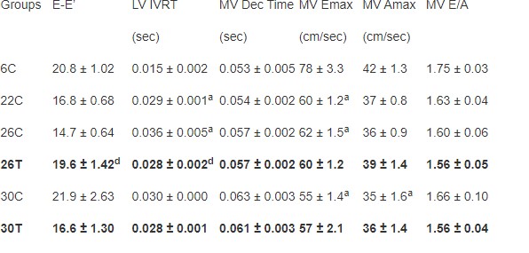
As described in Table 3, no age-associated changes in E-E’ ratio was found in the female F344xBN rats. APAP treatment increased the E-E’ ratio at 26-months (19.61 ± 1.42) compared to control (14.73 ± 0.64; P = 0.017). Left ventricular isovolumetric relaxation time (LV IVRT) was significantly increased at 22- (0.029 ± 0.001 sec) and 26-months (0.036 ± 0.005 sec) compared to 6-months (0.015 ± 0.002 sec; P < 0.05). Treatment with APAP at 26-months (0.028 ± 0.002 sec) significantly decreased LV IVRT compared to control (P < 0.001). There were no changes in mitral valve deceleration (MV Dec) time with age or APAP treatment. With age mitral valve peak velocity of the E wave (MV Emax) significantly decreased at 22- (59.8 ± 1.2 cm/sec), 26- (61.5 ± 1.5 cm/sec), and 30-months (55.4 ± 1.4 cm/sec) when compared to 6-months (78.0 ± 3.3 cm/sec; P < 0.05). Aging also significantly decreased peak velocity of the A wave (MV Amax) at 30-months (35.1 ± 1.6 cm/sec) when compared to 6-months (41.5 ± 1.3 cm/sec; P < 0.05). No change in MV Emax or Amax was found with APAP treatment. No changes were found in MV E/A ratio with age or APAP treatment.
Table 3. Echocardiographic evaluation of cardiac functional parameters in female F344xBN rats with and without APAP (mean ± SEM; n = ## rats/group).
6C – 6-month animal; 22C – 22-month animal; 26C – 26-month animal; 26T – 26-month APAP-treated animal; 30C – 30-month animal; 30T – 30-month APAP-treated animal; aP < 0.05 significant difference from 6-month control animal;bP < 0.05 significant difference from 22-month control animal; cP < 0.05 significant difference from 26-month control animal; dP < 0.05 age-matched control vs. treated animal.
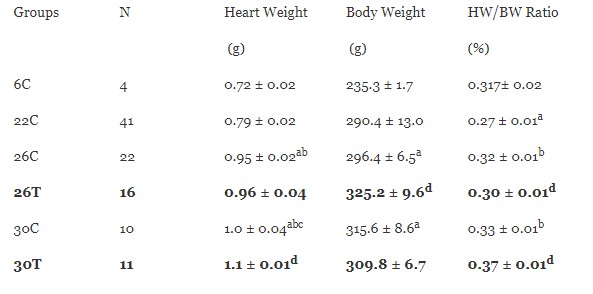
Table 4. Total body weight (BW) and heart weight (HW) in female F344xBN rats at 6-, 22-, 26-, and 30-months of age with or without APAP treatment (means ± SEM; n = ## rats/group).
6C – 6-month animal; 22C – 22-month animal; 26C – 26-month animal; 26T – 26-month APAP-treated animal; 30C – 30-month animal; 30T – 30-month APAP-treated animal; aP < 0.05 significant difference from 6-month control animal;bP < 0.05 significant difference from 22-month control animal; cP < 0.05 significant difference from 26-month control animal; dP < 0.05 age-matched control vs. treated animal.
Ejection Fraction (%)
Fractional Shortening (%)
0.082 ± 0.005a
0.579 ± 0.021d
0.084 ± 0.001d
0.029 ± 0.001a
0.036 ± 0.005a
0.028 ± 0.002d
Tricuspid valve dysfunction was higher in 22-, 26-, and 30-month (52.4%, 21.7%, 33.3%) female hearts relative to 6-month. APAP treatment at 26-months (33.3%) had a higher percentage of female rats with tricuspid dysfunction compared to 26-month control group. However, with APAP treatment at 30-months the percentage of female rats with tricuspid dysfunction (33.3%) was the same when relative to the 6-month group. The percentage of female rats with mitral dysfunction also was higher with age (22C: 19.0%; 26C: 17.4%; 30C: 13.3%) relative to 6-month rats. APAP treatment at 26-months had a similar percentage of mitral dysfunction compared to control; however, APAP treatment at 30-months had a lower percentage when compared to the 30-month control group. The presence of aortic dysfunction with age was only seen in the 30-month control group (20%). At 26-months with APAP treatment (26T: 16.7%; 30T: 15.0%) the percentage of female rats with aortic dysfunction was higher compared to control groups (26C: 0%; 30C: 20.0%). The percentage of aortic dysfunction in 30-month APAP rats was lower (20.0%) than the 30-month control rats (15%). Female rats with pulmonary valve dysfunction were higher but this percentage was similar or lower with APAP treatment at 26- and 30-months (26T: 77.8%; 30T: 45.0%) when compared to control groups (26C: 78.3%; 30C: 66.7%).
Electrocardiogram Analysis
The presence of arrhythmias was not found in the vast majority of female F344xBN rats, and therefore the incidence was not different between control and APAP treatment groups. In the 30-month APAP treated group one, premature ventricular contraction was observed (data not shown).
Heart Weight, Body Weight, and Heart Weight to Body Weight Ratio
Female F344xBN heart weights and body weights were compared in Table 4. Heart weight increased significantly with age at 26-months (0.954 ± 0.002 g) compared to 6- and 22-month groups (6C: 0.721 ± 0.002 g; 22C: 0.787 ± 0.02 g; P < 0.001). At 30-months (1.04 ± 0.04 g) heart weight significantly increased compared to all age groups (P < 0.05). APAP treatment significantly increased heart weight at 30-months when compared to control (P = 0.029). Body weight increased significantly at 26- (296.4 ± 6.5 g) and 30-months (315.6 ± 8.6 g) when compared to 6-month (235.3 ± 1.7 g; P < 0.05). When normalized to body weight, heart weights were significantly decreased at 22-months (0.272 ± 0.01%) compared to 6-months (0.307 ± 0.02%; P < 0.05). However, normalized heart weights at 26- (0.321 ± 0.01%) and 30-month (0.327 ± 0.01%) rats was significantly increased compared to 6-months (P < 0.05). APAP treatment significantly decreased heart weight after normalization to body weight at 26-months (0.295 ± 0.01%; P = 0.048) but significantly increased at 30-months (0.367 ± 0.01%; P = 0.002) compared to controls.
The present study was conducted to obtain the cardiac function reference values for the aging female F344xBN rats. The F344xBN rat, according to the National Institute of Aging, is the preferred animal model to study age-associated pathophysiological changes due to its longer life span and low occurrence of pathologies [22]. The 6-, 22-, 26-, and 30-month age groups were chosen based off survivability curves from the National Institute of Aging to represent females in the third, seventh, and eighth decade of life [22, 23]. These reference values are critical in order to compare the effects of aging on systolic and diastolic function in order to establish if the female F344XBN rat is an appropriate model for aging cardiac dysfunction.
Previous work in our laboratory has indicated that with aging there is an increase in oxidative-nitrosative stress which may responsible for the age-associated changes in cardiac structure and function [24-26]. This age-associated increase in oxidative-nitrosative stress is attenuated with chronic APAP treatment [20] due to its antioxidative properties. In order to investigate the role of age-associated oxidative-nitrosative stress on cardiac structure and function, APAP was administered to the treatment groups in order to see if it would attenuate these age-associated changes in cardiac function.
Increased risk of diastolic dysfunction with aging appears to occur more often in women compared to men [27]. These diastolic dysfunctions oftentimes precede the development of systolic dysfunction [27]. Diastolic dysfunction in humans is defined by prolonged deceleration time, a high A wave velocity, a low E wave velocity, and prolonged isovolumetric relaxation [27]. In the present study of the female F344xBN heart, we observed age-related increases in left ventricular relaxation time, decrease in E`, and a trend in mitral valve deceleration time. Nonetheless, we did not find any changes in the E/A ratio with age nor did APAP treatment produce a change (Table 3). In contrast, Boluyt et al. show that aged female F344 animals have increased LV IVRT, decreased E wave, and increased A wave velocity [28]. In our study, as in the Boluyt et al. study with age there was a longer left ventricular relaxation time, a reduced E’ wave velocity, a trend toward an increased mitral valve deceleration time, and no change in the E/A ratio [28]. Taken together, these data suggest that aging in female F344 and F344xBN rats, much like that seen in humans, is characterized by changes in diastolic function. APAP treatment had no effect on this age related change.
As previously observed in F344 rats, systolic function in female F344xBN rats did not appear to be significantly impaired with aging (Table 2) [28, 29]. Similar to the F344 rats, we found a slight increase in 26-month ejection fraction; this parameter, however, must be interpreted carefully as there was also increased valvular regurgitation at this measurement time [28]. Forman et al. find that F344 male rats have higher incidence of mitral regurgitation (MR) relative to female rats [29]. This may explain differences in age related functional changes between male and female rats. More investigation is needed to determine the mechanism of age related differences in cardiac function in male and female F344xBN rats. Nonetheless, our data are consistent with the notion that APAP significantly improved the ejection fraction (and therefore increased the EDV and reduced the ESV) at 26 months and there was trend towards improvement at 30 months (note that the ESV was significantly lower in treated compared to age matched controls at this time point).
Unlike our previous data regarding the presence of age associated arrhythmias in the male F334XBN [17], the aging female F344XBN failed to demonstrate age associated arrhythmias. However, APAP treatment attenuated the age associated incidents of valvular dysfunction.
These data suggest that aging in the female F344xBN rat heart is associated with changes in cardiac structure (Table 1) and function (Tables 2 and 3) and that chronic APAP treatment may diminish the incidence of valvular dysfunction in most situations, but increase the incidence in others (e.g., increased tricuspid dysfunction at 26 months). APAP appears to result in beneficial changes in ejection fraction (Table 2), but a variable effect on heart rate (i.e., increased at 26 months, but decreased at 30 months compared to control). This study provides reference values for cardiac structure and function for adult female F344XBN rats. Further investigation regarding other parameters of cardiac function is currently underway.
The American Heart Association estimates that 80 million Americans have one or more types of cardiovascular disease (CVD). It is estimated that 38.1 million of those afflicted are aged 60 or older. As a person ages he or she has a higher risk of developing myocardial infarction, stroke, atherosclerosis, peripheral occlusive disease, diabetes, hypertension [1-3].Thepotential bmechanism of increase risk of disease with age is that there is an increase of reactive oxygen species (ROS) which leads to oxidative stress [4]. Recently acetaminophen (APAP), an antipyretic and analgesic, has shown to be protective against oxidative stress in the heart and the vasculature of the brain due to its antioxidant properties [5-7].
Due to these antioxidant effects APAP is considered to be cardioprotective during ischemia/reperfusion, hypoxia/reoxygenation, exogenous peroxynitrite administration, and experimentally induced myocardial infarction due to its ability to block or reduce the development of mitochondrial permeability transition pores, mitochondrial swelling, cytochrome c release, late stage apoptosis, protein oxidation, production of hydroxyl radicals, arrhythmias, and peroxynitrite induced dysfunction in hearts [5, 8-16].
The Fischer 344/NNiaHSD x Brown Norway/BiNia F1 (F344xBN) has been recommended by the National Institute of Aging as a rat model of aging due to its increased life span and fewer age-associated pathologies compared to the standard model of aging – the Fischer 344 rat. Although several studies have investigated the age-associated changes in male and female F344 rats as well as male F344xBN rats [17-21], no study to our knowledge has examined the age-associated changes in structure and function in the female F344xBN heart using echocardiographic measures.
This information is crucial in determining whether or not the female F344xBN is an appropriate aging model to study the development of cardiovascular disease. Due to evidence that age-associated increase in levels of oxidative stress may cause cardiac dysfunction, we also determined the function and structure of the aged female F344xBN heart with chronic APAP treatment. The purpose of this study was to determine the age-associated changes in the female F344xBN cardiac structure and function and whether chronic APAP treatment would attenuate these effects.
All procedures were performed in accordance with the Guide for the Care and Use of Laboratory Animals as approved by the Council of the American Physiological Society and the Animal Use Review Board of Marshall University. All procedures were conducted in strict accordance with the Public Health Service animal welfare policy. Young (6-month), adult (22-month), aged (26-month), and very aged (30-month) female F344xBN rats were obtained from the National Institute for Aging were housed two per cage in an Association for Assessment and Accreditation of Laboratory Animal Care International (AAALAC) approved vivarium. Housing conditions consisted of a 12 h-12 h light-dark cycle with a temperature maintained at 22 ± 2˚C. Animals were provided with food and water ad libitum. Rats were allowed to recover from shipment for at least two weeks before experimentation. During this time the animals were carefully observed and weighed weekly. Any rat showing signs of failure to thrive, such as precipitous weight loss, disinterest in the environment, or unexpected gait alterations were excluded from the study. Six Female F344xBN rats were randomly assigned to each group (6C – 6-month animal; 22C – 22-month animal; 26C – 26-month animal; 26T – 26-month APAP-treated animal; 30C – 30-month animal; 30T – 30-month APAP-treated animal).
A therapeutic dose of APAP (30 mg/kg/day) was administered through the drinking water for treated animals at 22-months of age. Changes in body weight and water intake were monitored throughout the study to maintain a therapeutic dose. The 6-, 22-, 26-, and 30-month age-matched control groups received tap water and were maintained under the same conditions as the APAP treated groups.
Control (6-, 22-, 26-, and 30-month) and APAP treated (26- and 30-month) rats were anesthetized with an intraperitoneal (ip) injection of ketamine (40 mg/kg) and xyaline (10 kg/mg). The ventral thoraxes where shaved, and the rats were placed either on their backs or left side and covered with ultrasonic transmission gel for adequate sonic transference. A Phillips 5500 ECHO system with a 12 MHz transducer was used to take two-dimensional ECHO measurements, two-dimensional guided M-mode, Doppler M-mode, and other recordings from parasternal long- and short-axis views. Parasternal long- and short axis views were used to determine two-dimensional cardiac structural measurements. The echocardiographic views were then used to position the M-mode echocardiographic line. In order to determine the presence of valve dysfunctions, valvular blood flow velocities were evaluated using pulse wave Doppler with the probe toward the apex (x-axis) and the depth along the y-axis (long axis procedure). Ejection fraction and fractional shortening during systole was calculated using the evaluation of wall structure from short-axis procedures with the probe oriented toward the left ventricle and across the heart. M-mode displays were analyzed by a digital echocardiographic analysis system.
The following measurements were selected for each assessment of cardiac structure and function. The structural parameters included diastolic (IVSd) and systolic (IVSs) left ventricular septal thickness, diastolic (LVIDd) and systolic (LVIDs) left ventricular internal dimension, diastiolic (LVPWd) and systolic (LVPWs) left ventricular posterior wall thickness, and right ventricular diastolic internal dimension (RVd). Functional measurement included ejection fraction (EF) and left ventricular fractional shortening during systole (FS).
Additional echocardiographic measurements included mitral valve deceleration (MV decel time), left ventricular mass (LVM), end-systolic volume (ESV), end-diastolic volume (EDV), peak velocity of the A wave (Amax), and peak velocity of the E wave (Emax) were used to evaluate systolic function. Mitral valve deceleration, Emax, Amax, and MV E/A ratio were used to evaluate diastolic function.
After completion of treatment and echocardiographic procedures, animals were anesthetized with an ip injection of ketamine (40 mg/kg) and xyaline (10 mg/kg) and supplemented as necessary for reflexive responses. Before heart collection, electrocardiograms (EKGs) with leads I, II, and III were measured in age-matched control and treated animals using the Biopac Student Lab software (BIOPAC Systems, Inc., Microsoft). After completion of EKG measurements, the heart was removed after a midline laparotomy and was then placed in Krebs-Ringer bicarbonate buffer (KRB) containing the following: 118 mM NaCl, 4.7 mM KCl, 2.5 mM CaCl2, 1.2 mM KH2PO4, 1.2 mM MgSO4, 24.2 mM NaHCO3, and 10 mM a-D-glucose (pH 7.4) equilibrated with 5% CO2/95% O2 and maintained at 37° C. Isolated hearts were quickly massaged to remove any blood from the ventricles, cleaned of connective tissue, weighed, and immediately snap frozen in liquid nitrogen for further analysis.
Results were reported as mean ± SEM. Statistical analyses were performed using Sigma Stat 3.5 statistical software (Systat Software, Inc). Age comparisons between ECHO structural, functional parameters, and morphologic indices were evaluated by one-way ANOVA or Kruskal-Wallis one-way Analysis of Variance on Ranks with the Student-Newman-Keuls or Dunn's methods as the post hoc test, respectively. Differences between age-matched control and treated animals were determined by Student’s T-test. The level of significance accepted a priori was P < 0.05.
Echocardiographic evaluation of cardiac structural parameters in female F344xBN rats are compared in Table 1. Left ventricular septal thickness (IVS) during systole (lVSs) significantly increased at 26-months (0.253 ± 0.003 cm) compared to 6-months (0.193 ± 0.008 cm; P < 0.05); however, no age-associated changes during diastole (lVSd) were found. Left ventricular internal dimension during systole (LVIDs) significantly increased at 22- (0.340 ± 0.006 cm) and 26-months (0.331 ± 0.009 cm) compared to 6-month (0.378 ± 0.017 cm; P < 0.05). Treatment with APAP significantly decreased LVIDs compared to control animals at 30-months (0.344 ± 0.015 vs 0.356 ± 0.011 cm, respectively; P = 0.004). During diastole LVID did not change with age but significantly increased with APAP treatment at 26-months (0.630 ± 0.009 cm) when compared to control (0.593 ± 0.009 cm; P < 0.001). Left ventricular posterior wall thickness (LVPW) during systole (LVPWs) and diastole (LVPWd) was significantly increased at 22- (0.273 ± 0.004 and 0.183 ± 0.004 cm, respectively) and 30-months (0.288 ± 0.009 and 0.189 ± 0.008 cm, respectively) compared to 6-months (0.193 ± 0.002 and 0.153 ± 0.010 cm, respectively; P < 0.05). APAP treatment increased both LVPWs and LVPWd thickness at 26-months compared to control (0.314 ± 0.007 and 0.185 ± 0.003 cm, respectively; P = 0.002). No changes were found in right ventricular dimension during diastole or left ventricular mass with age or APAP treatment.

Echocardiographic evaluation of cardiac functional parameters in female F344xBN rats can be found in Table 2 and 3. As described in Table 2, ejection fraction was significantly increased at 26-months (82 ± 1.0%) compared to 6-months (74 ± 0.9%; P < 0.05). APAP treatment at 26-months (86 ± 0.8%) also significantly increased the ejection fraction compared to control (P = 0.009). Fractional shortening significantly decreased at 30-months (41.5 ± 1.7%) compared to 26-months (45.6 ± 1.0%; P < 0.05). APAP treatment at 26-months (49.8 ± 1.2%) significantly increased the fractional shortening when compared to control (P = 0.007). End systolic volume (ESV) was significantly decreased at 26-months (0.082 ± 0.005 mL) when compared to 6-months (0.143 ± 0.017 mL; P < 0.05). APAP treatment significantly decreased ESV at 30-months (0.084 ± 0.001 mL) when compared to control (0.114 ± 0.10 mL; P=0.034). APAP treatment at 26-months (0.579 ± 0.021 mL) significantly increased end diastolic volume when compared to control (0.492 ± 0.20 mL; P = 0.005). Heart rate did not change with age but significantly increased with APAP treatment at 26- (286.6 ± 5.9 bpm; P = 0.015) and decreased at 30-months (238.7 ± 14.8 bpm; P = 0.029) when compared to controls.
6C – 6-month animal; 22C – 22-month animal; 26C – 26-month animal; 26T – 26-month APAP-treated animal; 30C – 30-month animal; 30T – 30-month APAP-treated animal; aP < 0.05 significant difference from 6-month control animal;cP < 0.05 significant difference from 26-month control animal; dP < 0.05 age-matched control vs. treated animal; N.T. – not tested.
As described in Table 3, no age-associated changes in E-E’ ratio was found in the female F344xBN rats. APAP treatment increased the E-E’ ratio at 26-months (19.61 ± 1.42) compared to control (14.73 ± 0.64; P = 0.017). Left ventricular isovolumetric relaxation time (LV IVRT) was significantly increased at 22- (0.029 ± 0.001 sec) and 26-months (0.036 ± 0.005 sec) compared to 6-months (0.015 ± 0.002 sec; P < 0.05). Treatment with APAP at 26-months (0.028 ± 0.002 sec) significantly decreased LV IVRT compared to control (P < 0.001). There were no changes in mitral valve deceleration (MV Dec) time with age or APAP treatment. With age mitral valve peak velocity of the E wave (MV Emax) significantly decreased at 22- (59.8 ± 1.2 cm/sec), 26- (61.5 ± 1.5 cm/sec), and 30-months (55.4 ± 1.4 cm/sec) when compared to 6-months (78.0 ± 3.3 cm/sec; P < 0.05). Aging also significantly decreased peak velocity of the A wave (MV Amax) at 30-months (35.1 ± 1.6 cm/sec) when compared to 6-months (41.5 ± 1.3 cm/sec; P < 0.05). No change in MV Emax or Amax was found with APAP treatment. No changes were found in MV E/A ratio with age or APAP treatment.
6C – 6-month animal; 22C – 22-month animal; 26C – 26-month animal; 26T – 26-month APAP-treated animal; 30C – 30-month animal; 30T – 30-month APAP-treated animal; aP < 0.05 significant difference from 6-month control animal;bP < 0.05 significant difference from 22-month control animal; cP < 0.05 significant difference from 26-month control animal; dP < 0.05 age-matched control vs. treated animal.
Tricuspid valve dysfunction was higher in 22-, 26-, and 30-month (52.4%, 21.7%, 33.3%) female hearts relative to 6-month. APAP treatment at 26-months (33.3%) had a higher percentage of female rats with tricuspid dysfunction compared to 26-month control group. However, with APAP treatment at 30-months the percentage of female rats with tricuspid dysfunction (33.3%) was the same when relative to the 6-month group. The percentage of female rats with mitral dysfunction also was higher with age (22C: 19.0%; 26C: 17.4%; 30C: 13.3%) relative to 6-month rats. APAP treatment at 26-months had a similar percentage of mitral dysfunction compared to control; however, APAP treatment at 30-months had a lower percentage when compared to the 30-month control group. The presence of aortic dysfunction with age was only seen in the 30-month control group (20%). At 26-months with APAP treatment (26T: 16.7%; 30T: 15.0%) the percentage of female rats with aortic dysfunction was higher compared to control groups (26C: 0%; 30C: 20.0%). The percentage of aortic dysfunction in 30-month APAP rats was lower (20.0%) than the 30-month control rats (15%). Female rats with pulmonary valve dysfunction were higher but this percentage was similar or lower with APAP treatment at 26- and 30-months (26T: 77.8%; 30T: 45.0%) when compared to control groups (26C: 78.3%; 30C: 66.7%).
The presence of arrhythmias was not found in the vast majority of female F344xBN rats, and therefore the incidence was not different between control and APAP treatment groups. In the 30-month APAP treated group one, premature ventricular contraction was observed (data not shown).
Female F344xBN heart weights and body weights were compared in Table 4. Heart weight increased significantly with age at 26-months (0.954 ± 0.002 g) compared to 6- and 22-month groups (6C: 0.721 ± 0.002 g; 22C: 0.787 ± 0.02 g; P < 0.001). At 30-months (1.04 ± 0.04 g) heart weight significantly increased compared to all age groups (P < 0.05). APAP treatment significantly increased heart weight at 30-months when compared to control (P = 0.029). Body weight increased significantly at 26- (296.4 ± 6.5 g) and 30-months (315.6 ± 8.6 g) when compared to 6-month (235.3 ± 1.7 g; P < 0.05). When normalized to body weight, heart weights were significantly decreased at 22-months (0.272 ± 0.01%) compared to 6-months (0.307 ± 0.02%; P < 0.05). However, normalized heart weights at 26- (0.321 ± 0.01%) and 30-month (0.327 ± 0.01%) rats was significantly increased compared to 6-months (P < 0.05). APAP treatment significantly decreased heart weight after normalization to body weight at 26-months (0.295 ± 0.01%; P = 0.048) but significantly increased at 30-months (0.367 ± 0.01%; P = 0.002) compared to controls.
6C – 6-month animal; 22C – 22-month animal; 26C – 26-month animal; 26T – 26-month APAP-treated animal; 30C – 30-month animal; 30T – 30-month APAP-treated animal; aP < 0.05 significant difference from 6-month control animal;bP < 0.05 significant difference from 22-month control animal; cP < 0.05 significant difference from 26-month control animal; dP < 0.05 age-matched control vs. treated animal.
The present study was conducted to obtain the cardiac function reference values for the aging female F344xBN rats. The F344xBN rat, according to the National Institute of Aging, is the preferred animal model to study age-associated pathophysiological changes due to its longer life span and low occurrence of pathologies [22]. The 6-, 22-, 26-, and 30-month age groups were chosen based off survivability curves from the National Institute of Aging to represent females in the third, seventh, and eighth decade of life [22, 23]. These reference values are critical in order to compare the effects of aging on systolic and diastolic function in order to establish if the female F344XBN rat is an appropriate model for aging cardiac dysfunction.
Previous work in our laboratory has indicated that with aging there is an increase in oxidative-nitrosative stress which may responsible for the age-associated changes in cardiac structure and function [24-26]. This age-associated increase in oxidative-nitrosative stress is attenuated with chronic APAP treatment [20] due to its antioxidative properties. In order to investigate the role of age-associated oxidative-nitrosative stress on cardiac structure and function, APAP was administered to the treatment groups in order to see if it would attenuate these age-associated changes in cardiac function.
Increased risk of diastolic dysfunction with aging appears to occur more often in women compared to men [27]. These diastolic dysfunctions oftentimes precede the development of systolic dysfunction [27]. Diastolic dysfunction in humans is defined by prolonged deceleration time, a high A wave velocity, a low E wave velocity, and prolonged isovolumetric relaxation [27]. In the present study of the female F344xBN heart, we observed age-related increases in left ventricular relaxation time, decrease in E`, and a trend in mitral valve deceleration time. Nonetheless, we did not find any changes in the E/A ratio with age nor did APAP treatment produce a change (Table 3). In contrast, Boluyt et al. show that aged female F344 animals have increased LV IVRT, decreased E wave, and increased A wave velocity [28]. In our study, as in the Boluyt et al. study with age there was a longer left ventricular relaxation time, a reduced E’ wave velocity, a trend toward an increased mitral valve deceleration time, and no change in the E/A ratio [28]. Taken together, these data suggest that aging in female F344 and F344xBN rats, much like that seen in humans, is characterized by changes in diastolic function. APAP treatment had no effect on this age related change.
As previously observed in F344 rats, systolic function in female F344xBN rats did not appear to be significantly impaired with aging (Table 2) [28, 29]. Similar to the F344 rats, we found a slight increase in 26-month ejection fraction; this parameter, however, must be interpreted carefully as there was also increased valvular regurgitation at this measurement time [28]. Forman et al. find that F344 male rats have higher incidence of mitral regurgitation (MR) relative to female rats [29]. This may explain differences in age related functional changes between male and female rats. More investigation is needed to determine the mechanism of age related differences in cardiac function in male and female F344xBN rats. Nonetheless, our data are consistent with the notion that APAP significantly improved the ejection fraction (and therefore increased the EDV and reduced the ESV) at 26 months and there was trend towards improvement at 30 months (note that the ESV was significantly lower in treated compared to age matched controls at this time point).
Unlike our previous data regarding the presence of age associated arrhythmias in the male F334XBN [17], the aging female F344XBN failed to demonstrate age associated arrhythmias. However, APAP treatment attenuated the age associated incidents of valvular dysfunction.
These data suggest that aging in the female F344xBN rat heart is associated with changes in cardiac structure (Table 1) and function (Tables 2 and 3) and that chronic APAP treatment may diminish the incidence of valvular dysfunction in most situations, but increase the incidence in others (e.g., increased tricuspid dysfunction at 26 months). APAP appears to result in beneficial changes in ejection fraction (Table 2), but a variable effect on heart rate (i.e., increased at 26 months, but decreased at 30 months compared to control). This study provides reference values for cardiac structure and function for adult female F344XBN rats. Further investigation regarding other parameters of cardiac function is currently underway.
Clearly Auctoresonline and particularly Psychology and Mental Health Care Journal is dedicated to improving health care services for individuals and populations. The editorial boards' ability to efficiently recognize and share the global importance of health literacy with a variety of stakeholders. Auctoresonline publishing platform can be used to facilitate of optimal client-based services and should be added to health care professionals' repertoire of evidence-based health care resources.

Journal of Clinical Cardiology and Cardiovascular Intervention The submission and review process was adequate. However I think that the publication total value should have been enlightened in early fases. Thank you for all.

Journal of Women Health Care and Issues By the present mail, I want to say thank to you and tour colleagues for facilitating my published article. Specially thank you for the peer review process, support from the editorial office. I appreciate positively the quality of your journal.

Journal of Clinical Research and Reports I would be very delighted to submit my testimonial regarding the reviewer board and the editorial office. The reviewer board were accurate and helpful regarding any modifications for my manuscript. And the editorial office were very helpful and supportive in contacting and monitoring with any update and offering help. It was my pleasure to contribute with your promising Journal and I am looking forward for more collaboration.

We would like to thank the Journal of Thoracic Disease and Cardiothoracic Surgery because of the services they provided us for our articles. The peer-review process was done in a very excellent time manner, and the opinions of the reviewers helped us to improve our manuscript further. The editorial office had an outstanding correspondence with us and guided us in many ways. During a hard time of the pandemic that is affecting every one of us tremendously, the editorial office helped us make everything easier for publishing scientific work. Hope for a more scientific relationship with your Journal.

The peer-review process which consisted high quality queries on the paper. I did answer six reviewers’ questions and comments before the paper was accepted. The support from the editorial office is excellent.

Journal of Neuroscience and Neurological Surgery. I had the experience of publishing a research article recently. The whole process was simple from submission to publication. The reviewers made specific and valuable recommendations and corrections that improved the quality of my publication. I strongly recommend this Journal.

Dr. Katarzyna Byczkowska My testimonial covering: "The peer review process is quick and effective. The support from the editorial office is very professional and friendly. Quality of the Clinical Cardiology and Cardiovascular Interventions is scientific and publishes ground-breaking research on cardiology that is useful for other professionals in the field.

Thank you most sincerely, with regard to the support you have given in relation to the reviewing process and the processing of my article entitled "Large Cell Neuroendocrine Carcinoma of The Prostate Gland: A Review and Update" for publication in your esteemed Journal, Journal of Cancer Research and Cellular Therapeutics". The editorial team has been very supportive.

Testimony of Journal of Clinical Otorhinolaryngology: work with your Reviews has been a educational and constructive experience. The editorial office were very helpful and supportive. It was a pleasure to contribute to your Journal.

Dr. Bernard Terkimbi Utoo, I am happy to publish my scientific work in Journal of Women Health Care and Issues (JWHCI). The manuscript submission was seamless and peer review process was top notch. I was amazed that 4 reviewers worked on the manuscript which made it a highly technical, standard and excellent quality paper. I appreciate the format and consideration for the APC as well as the speed of publication. It is my pleasure to continue with this scientific relationship with the esteem JWHCI.

This is an acknowledgment for peer reviewers, editorial board of Journal of Clinical Research and Reports. They show a lot of consideration for us as publishers for our research article “Evaluation of the different factors associated with side effects of COVID-19 vaccination on medical students, Mutah university, Al-Karak, Jordan”, in a very professional and easy way. This journal is one of outstanding medical journal.

Dear Hao Jiang, to Journal of Nutrition and Food Processing We greatly appreciate the efficient, professional and rapid processing of our paper by your team. If there is anything else we should do, please do not hesitate to let us know. On behalf of my co-authors, we would like to express our great appreciation to editor and reviewers.

As an author who has recently published in the journal "Brain and Neurological Disorders". I am delighted to provide a testimonial on the peer review process, editorial office support, and the overall quality of the journal. The peer review process at Brain and Neurological Disorders is rigorous and meticulous, ensuring that only high-quality, evidence-based research is published. The reviewers are experts in their fields, and their comments and suggestions were constructive and helped improve the quality of my manuscript. The review process was timely and efficient, with clear communication from the editorial office at each stage. The support from the editorial office was exceptional throughout the entire process. The editorial staff was responsive, professional, and always willing to help. They provided valuable guidance on formatting, structure, and ethical considerations, making the submission process seamless. Moreover, they kept me informed about the status of my manuscript and provided timely updates, which made the process less stressful. The journal Brain and Neurological Disorders is of the highest quality, with a strong focus on publishing cutting-edge research in the field of neurology. The articles published in this journal are well-researched, rigorously peer-reviewed, and written by experts in the field. The journal maintains high standards, ensuring that readers are provided with the most up-to-date and reliable information on brain and neurological disorders. In conclusion, I had a wonderful experience publishing in Brain and Neurological Disorders. The peer review process was thorough, the editorial office provided exceptional support, and the journal's quality is second to none. I would highly recommend this journal to any researcher working in the field of neurology and brain disorders.
Dear Agrippa Hilda, Journal of Neuroscience and Neurological Surgery, Editorial Coordinator, I trust this message finds you well. I want to extend my appreciation for considering my article for publication in your esteemed journal. I am pleased to provide a testimonial regarding the peer review process and the support received from your editorial office. The peer review process for my paper was carried out in a highly professional and thorough manner. The feedback and comments provided by the authors were constructive and very useful in improving the quality of the manuscript. This rigorous assessment process undoubtedly contributes to the high standards maintained by your journal.

International Journal of Clinical Case Reports and Reviews. I strongly recommend to consider submitting your work to this high-quality journal. The support and availability of the Editorial staff is outstanding and the review process was both efficient and rigorous.

Thank you very much for publishing my Research Article titled “Comparing Treatment Outcome Of Allergic Rhinitis Patients After Using Fluticasone Nasal Spray And Nasal Douching" in the Journal of Clinical Otorhinolaryngology. As Medical Professionals we are immensely benefited from study of various informative Articles and Papers published in this high quality Journal. I look forward to enriching my knowledge by regular study of the Journal and contribute my future work in the field of ENT through the Journal for use by the medical fraternity. The support from the Editorial office was excellent and very prompt. I also welcome the comments received from the readers of my Research Article.

Dear Erica Kelsey, Editorial Coordinator of Cancer Research and Cellular Therapeutics Our team is very satisfied with the processing of our paper by your journal. That was fast, efficient, rigorous, but without unnecessary complications. We appreciated the very short time between the submission of the paper and its publication on line on your site.

I am very glad to say that the peer review process is very successful and fast and support from the Editorial Office. Therefore, I would like to continue our scientific relationship for a long time. And I especially thank you for your kindly attention towards my article. Have a good day!

"We recently published an article entitled “Influence of beta-Cyclodextrins upon the Degradation of Carbofuran Derivatives under Alkaline Conditions" in the Journal of “Pesticides and Biofertilizers” to show that the cyclodextrins protect the carbamates increasing their half-life time in the presence of basic conditions This will be very helpful to understand carbofuran behaviour in the analytical, agro-environmental and food areas. We greatly appreciated the interaction with the editor and the editorial team; we were particularly well accompanied during the course of the revision process, since all various steps towards publication were short and without delay".

I would like to express my gratitude towards you process of article review and submission. I found this to be very fair and expedient. Your follow up has been excellent. I have many publications in national and international journal and your process has been one of the best so far. Keep up the great work.

We are grateful for this opportunity to provide a glowing recommendation to the Journal of Psychiatry and Psychotherapy. We found that the editorial team were very supportive, helpful, kept us abreast of timelines and over all very professional in nature. The peer review process was rigorous, efficient and constructive that really enhanced our article submission. The experience with this journal remains one of our best ever and we look forward to providing future submissions in the near future.

I am very pleased to serve as EBM of the journal, I hope many years of my experience in stem cells can help the journal from one way or another. As we know, stem cells hold great potential for regenerative medicine, which are mostly used to promote the repair response of diseased, dysfunctional or injured tissue using stem cells or their derivatives. I think Stem Cell Research and Therapeutics International is a great platform to publish and share the understanding towards the biology and translational or clinical application of stem cells.

I would like to give my testimony in the support I have got by the peer review process and to support the editorial office where they were of asset to support young author like me to be encouraged to publish their work in your respected journal and globalize and share knowledge across the globe. I really give my great gratitude to your journal and the peer review including the editorial office.

I am delighted to publish our manuscript entitled "A Perspective on Cocaine Induced Stroke - Its Mechanisms and Management" in the Journal of Neuroscience and Neurological Surgery. The peer review process, support from the editorial office, and quality of the journal are excellent. The manuscripts published are of high quality and of excellent scientific value. I recommend this journal very much to colleagues.

Dr.Tania Muñoz, My experience as researcher and author of a review article in The Journal Clinical Cardiology and Interventions has been very enriching and stimulating. The editorial team is excellent, performs its work with absolute responsibility and delivery. They are proactive, dynamic and receptive to all proposals. Supporting at all times the vast universe of authors who choose them as an option for publication. The team of review specialists, members of the editorial board, are brilliant professionals, with remarkable performance in medical research and scientific methodology. Together they form a frontline team that consolidates the JCCI as a magnificent option for the publication and review of high-level medical articles and broad collective interest. I am honored to be able to share my review article and open to receive all your comments.

“The peer review process of JPMHC is quick and effective. Authors are benefited by good and professional reviewers with huge experience in the field of psychology and mental health. The support from the editorial office is very professional. People to contact to are friendly and happy to help and assist any query authors might have. Quality of the Journal is scientific and publishes ground-breaking research on mental health that is useful for other professionals in the field”.

Dear editorial department: On behalf of our team, I hereby certify the reliability and superiority of the International Journal of Clinical Case Reports and Reviews in the peer review process, editorial support, and journal quality. Firstly, the peer review process of the International Journal of Clinical Case Reports and Reviews is rigorous, fair, transparent, fast, and of high quality. The editorial department invites experts from relevant fields as anonymous reviewers to review all submitted manuscripts. These experts have rich academic backgrounds and experience, and can accurately evaluate the academic quality, originality, and suitability of manuscripts. The editorial department is committed to ensuring the rigor of the peer review process, while also making every effort to ensure a fast review cycle to meet the needs of authors and the academic community. Secondly, the editorial team of the International Journal of Clinical Case Reports and Reviews is composed of a group of senior scholars and professionals with rich experience and professional knowledge in related fields. The editorial department is committed to assisting authors in improving their manuscripts, ensuring their academic accuracy, clarity, and completeness. Editors actively collaborate with authors, providing useful suggestions and feedback to promote the improvement and development of the manuscript. We believe that the support of the editorial department is one of the key factors in ensuring the quality of the journal. Finally, the International Journal of Clinical Case Reports and Reviews is renowned for its high- quality articles and strict academic standards. The editorial department is committed to publishing innovative and academically valuable research results to promote the development and progress of related fields. The International Journal of Clinical Case Reports and Reviews is reasonably priced and ensures excellent service and quality ratio, allowing authors to obtain high-level academic publishing opportunities in an affordable manner. I hereby solemnly declare that the International Journal of Clinical Case Reports and Reviews has a high level of credibility and superiority in terms of peer review process, editorial support, reasonable fees, and journal quality. Sincerely, Rui Tao.

Clinical Cardiology and Cardiovascular Interventions I testity the covering of the peer review process, support from the editorial office, and quality of the journal.

Clinical Cardiology and Cardiovascular Interventions, we deeply appreciate the interest shown in our work and its publication. It has been a true pleasure to collaborate with you. The peer review process, as well as the support provided by the editorial office, have been exceptional, and the quality of the journal is very high, which was a determining factor in our decision to publish with you.

The peer reviewers process is quick and effective, the supports from editorial office is excellent, the quality of journal is high. I would like to collabroate with Internatioanl journal of Clinical Case Reports and Reviews journal clinically in the future time.

Clinical Cardiology and Cardiovascular Interventions, I would like to express my sincerest gratitude for the trust placed in our team for the publication in your journal. It has been a true pleasure to collaborate with you on this project. I am pleased to inform you that both the peer review process and the attention from the editorial coordination have been excellent. Your team has worked with dedication and professionalism to ensure that your publication meets the highest standards of quality. We are confident that this collaboration will result in mutual success, and we are eager to see the fruits of this shared effort.

Dear Dr. Jessica Magne, Editorial Coordinator 0f Clinical Cardiology and Cardiovascular Interventions, I hope this message finds you well. I want to express my utmost gratitude for your excellent work and for the dedication and speed in the publication process of my article titled "Navigating Innovation: Qualitative Insights on Using Technology for Health Education in Acute Coronary Syndrome Patients." I am very satisfied with the peer review process, the support from the editorial office, and the quality of the journal. I hope we can maintain our scientific relationship in the long term.

Dear Monica Gissare, - Editorial Coordinator of Nutrition and Food Processing. ¨My testimony with you is truly professional, with a positive response regarding the follow-up of the article and its review, you took into account my qualities and the importance of the topic¨.

Dear Dr. Jessica Magne, Editorial Coordinator 0f Clinical Cardiology and Cardiovascular Interventions, The review process for the article “The Handling of Anti-aggregants and Anticoagulants in the Oncologic Heart Patient Submitted to Surgery” was extremely rigorous and detailed. From the initial submission to the final acceptance, the editorial team at the “Journal of Clinical Cardiology and Cardiovascular Interventions” demonstrated a high level of professionalism and dedication. The reviewers provided constructive and detailed feedback, which was essential for improving the quality of our work. Communication was always clear and efficient, ensuring that all our questions were promptly addressed. The quality of the “Journal of Clinical Cardiology and Cardiovascular Interventions” is undeniable. It is a peer-reviewed, open-access publication dedicated exclusively to disseminating high-quality research in the field of clinical cardiology and cardiovascular interventions. The journal's impact factor is currently under evaluation, and it is indexed in reputable databases, which further reinforces its credibility and relevance in the scientific field. I highly recommend this journal to researchers looking for a reputable platform to publish their studies.

Dear Editorial Coordinator of the Journal of Nutrition and Food Processing! "I would like to thank the Journal of Nutrition and Food Processing for including and publishing my article. The peer review process was very quick, movement and precise. The Editorial Board has done an extremely conscientious job with much help, valuable comments and advices. I find the journal very valuable from a professional point of view, thank you very much for allowing me to be part of it and I would like to participate in the future!”

Dealing with The Journal of Neurology and Neurological Surgery was very smooth and comprehensive. The office staff took time to address my needs and the response from editors and the office was prompt and fair. I certainly hope to publish with this journal again.Their professionalism is apparent and more than satisfactory. Susan Weiner

My Testimonial Covering as fellowing: Lin-Show Chin. The peer reviewers process is quick and effective, the supports from editorial office is excellent, the quality of journal is high. I would like to collabroate with Internatioanl journal of Clinical Case Reports and Reviews.

My experience publishing in Psychology and Mental Health Care was exceptional. The peer review process was rigorous and constructive, with reviewers providing valuable insights that helped enhance the quality of our work. The editorial team was highly supportive and responsive, making the submission process smooth and efficient. The journal's commitment to high standards and academic rigor makes it a respected platform for quality research. I am grateful for the opportunity to publish in such a reputable journal.

My experience publishing in International Journal of Clinical Case Reports and Reviews was exceptional. I Come forth to Provide a Testimonial Covering the Peer Review Process and the editorial office for the Professional and Impartial Evaluation of the Manuscript.

I would like to offer my testimony in the support. I have received through the peer review process and support the editorial office where they are to support young authors like me, encourage them to publish their work in your esteemed journals, and globalize and share knowledge globally. I really appreciate your journal, peer review, and editorial office.

Dear Agrippa Hilda- Editorial Coordinator of Journal of Neuroscience and Neurological Surgery, "The peer review process was very quick and of high quality, which can also be seen in the articles in the journal. The collaboration with the editorial office was very good."

I would like to express my sincere gratitude for the support and efficiency provided by the editorial office throughout the publication process of my article, “Delayed Vulvar Metastases from Rectal Carcinoma: A Case Report.” I greatly appreciate the assistance and guidance I received from your team, which made the entire process smooth and efficient. The peer review process was thorough and constructive, contributing to the overall quality of the final article. I am very grateful for the high level of professionalism and commitment shown by the editorial staff, and I look forward to maintaining a long-term collaboration with the International Journal of Clinical Case Reports and Reviews.

To Dear Erin Aust, I would like to express my heartfelt appreciation for the opportunity to have my work published in this esteemed journal. The entire publication process was smooth and well-organized, and I am extremely satisfied with the final result. The Editorial Team demonstrated the utmost professionalism, providing prompt and insightful feedback throughout the review process. Their clear communication and constructive suggestions were invaluable in enhancing my manuscript, and their meticulous attention to detail and dedication to quality are truly commendable. Additionally, the support from the Editorial Office was exceptional. From the initial submission to the final publication, I was guided through every step of the process with great care and professionalism. The team's responsiveness and assistance made the entire experience both easy and stress-free. I am also deeply impressed by the quality and reputation of the journal. It is an honor to have my research featured in such a respected publication, and I am confident that it will make a meaningful contribution to the field.

"I am grateful for the opportunity of contributing to [International Journal of Clinical Case Reports and Reviews] and for the rigorous review process that enhances the quality of research published in your esteemed journal. I sincerely appreciate the time and effort of your team who have dedicatedly helped me in improvising changes and modifying my manuscript. The insightful comments and constructive feedback provided have been invaluable in refining and strengthening my work".

I thank the ‘Journal of Clinical Research and Reports’ for accepting this article for publication. This is a rigorously peer reviewed journal which is on all major global scientific data bases. I note the review process was prompt, thorough and professionally critical. It gave us an insight into a number of important scientific/statistical issues. The review prompted us to review the relevant literature again and look at the limitations of the study. The peer reviewers were open, clear in the instructions and the editorial team was very prompt in their communication. This journal certainly publishes quality research articles. I would recommend the journal for any future publications.

Dear Jessica Magne, with gratitude for the joint work. Fast process of receiving and processing the submitted scientific materials in “Clinical Cardiology and Cardiovascular Interventions”. High level of competence of the editors with clear and correct recommendations and ideas for enriching the article.

We found the peer review process quick and positive in its input. The support from the editorial officer has been very agile, always with the intention of improving the article and taking into account our subsequent corrections.

My article, titled 'No Way Out of the Smartphone Epidemic Without Considering the Insights of Brain Research,' has been republished in the International Journal of Clinical Case Reports and Reviews. The review process was seamless and professional, with the editors being both friendly and supportive. I am deeply grateful for their efforts.

To Dear Erin Aust – Editorial Coordinator of Journal of General Medicine and Clinical Practice! I declare that I am absolutely satisfied with your work carried out with great competence in following the manuscript during the various stages from its receipt, during the revision process to the final acceptance for publication. Thank Prof. Elvira Farina

Dear Jessica, and the super professional team of the ‘Clinical Cardiology and Cardiovascular Interventions’ I am sincerely grateful to the coordinated work of the journal team for the no problem with the submission of my manuscript: “Cardiometabolic Disorders in A Pregnant Woman with Severe Preeclampsia on the Background of Morbid Obesity (Case Report).” The review process by 5 experts was fast, and the comments were professional, which made it more specific and academic, and the process of publication and presentation of the article was excellent. I recommend that my colleagues publish articles in this journal, and I am interested in further scientific cooperation. Sincerely and best wishes, Dr. Oleg Golyanovskiy.

Dear Ashley Rosa, Editorial Coordinator of the journal - Psychology and Mental Health Care. " The process of obtaining publication of my article in the Psychology and Mental Health Journal was positive in all areas. The peer review process resulted in a number of valuable comments, the editorial process was collaborative and timely, and the quality of this journal has been quickly noticed, resulting in alternative journals contacting me to publish with them." Warm regards, Susan Anne Smith, PhD. Australian Breastfeeding Association.

Dear Jessica Magne, Editorial Coordinator, Clinical Cardiology and Cardiovascular Interventions, Auctores Publishing LLC. I appreciate the journal (JCCI) editorial office support, the entire team leads were always ready to help, not only on technical front but also on thorough process. Also, I should thank dear reviewers’ attention to detail and creative approach to teach me and bring new insights by their comments. Surely, more discussions and introduction of other hemodynamic devices would provide better prevention and management of shock states. Your efforts and dedication in presenting educational materials in this journal are commendable. Best wishes from, Farahnaz Fallahian.

Dear Maria Emerson, Editorial Coordinator, International Journal of Clinical Case Reports and Reviews, Auctores Publishing LLC. I am delighted to have published our manuscript, "Acute Colonic Pseudo-Obstruction (ACPO): A rare but serious complication following caesarean section." I want to thank the editorial team, especially Maria Emerson, for their prompt review of the manuscript, quick responses to queries, and overall support. Yours sincerely Dr. Victor Olagundoye.

Dear Ashley Rosa, Editorial Coordinator, International Journal of Clinical Case Reports and Reviews. Many thanks for publishing this manuscript after I lost confidence the editors were most helpful, more than other journals Best wishes from, Susan Anne Smith, PhD. Australian Breastfeeding Association.

Dear Agrippa Hilda, Editorial Coordinator, Journal of Neuroscience and Neurological Surgery. The entire process including article submission, review, revision, and publication was extremely easy. The journal editor was prompt and helpful, and the reviewers contributed to the quality of the paper. Thank you so much! Eric Nussbaum, MD

Dr Hala Al Shaikh This is to acknowledge that the peer review process for the article ’ A Novel Gnrh1 Gene Mutation in Four Omani Male Siblings, Presentation and Management ’ sent to the International Journal of Clinical Case Reports and Reviews was quick and smooth. The editorial office was prompt with easy communication.

Dear Erin Aust, Editorial Coordinator, Journal of General Medicine and Clinical Practice. We are pleased to share our experience with the “Journal of General Medicine and Clinical Practice”, following the successful publication of our article. The peer review process was thorough and constructive, helping to improve the clarity and quality of the manuscript. We are especially thankful to Ms. Erin Aust, the Editorial Coordinator, for her prompt communication and continuous support throughout the process. Her professionalism ensured a smooth and efficient publication experience. The journal upholds high editorial standards, and we highly recommend it to fellow researchers seeking a credible platform for their work. Best wishes By, Dr. Rakhi Mishra.

Dear Jessica Magne, Editorial Coordinator, Clinical Cardiology and Cardiovascular Interventions, Auctores Publishing LLC. The peer review process of the journal of Clinical Cardiology and Cardiovascular Interventions was excellent and fast, as was the support of the editorial office and the quality of the journal. Kind regards Walter F. Riesen Prof. Dr. Dr. h.c. Walter F. Riesen.

Dear Ashley Rosa, Editorial Coordinator, International Journal of Clinical Case Reports and Reviews, Auctores Publishing LLC. Thank you for publishing our article, Exploring Clozapine's Efficacy in Managing Aggression: A Multiple Single-Case Study in Forensic Psychiatry in the international journal of clinical case reports and reviews. We found the peer review process very professional and efficient. The comments were constructive, and the whole process was efficient. On behalf of the co-authors, I would like to thank you for publishing this article. With regards, Dr. Jelle R. Lettinga.

Dear Clarissa Eric, Editorial Coordinator, Journal of Clinical Case Reports and Studies, I would like to express my deep admiration for the exceptional professionalism demonstrated by your journal. I am thoroughly impressed by the speed of the editorial process, the substantive and insightful reviews, and the meticulous preparation of the manuscript for publication. Additionally, I greatly appreciate the courteous and immediate responses from your editorial office to all my inquiries. Best Regards, Dariusz Ziora

Dear Chrystine Mejia, Editorial Coordinator, Journal of Neurodegeneration and Neurorehabilitation, Auctores Publishing LLC, We would like to thank the editorial team for the smooth and high-quality communication leading up to the publication of our article in the Journal of Neurodegeneration and Neurorehabilitation. The reviewers have extensive knowledge in the field, and their relevant questions helped to add value to our publication. Kind regards, Dr. Ravi Shrivastava.

Dear Clarissa Eric, Editorial Coordinator, Journal of Clinical Case Reports and Studies, Auctores Publishing LLC, USA Office: +1-(302)-520-2644. I would like to express my sincere appreciation for the efficient and professional handling of my case report by the ‘Journal of Clinical Case Reports and Studies’. The peer review process was not only fast but also highly constructive—the reviewers’ comments were clear, relevant, and greatly helped me improve the quality and clarity of my manuscript. I also received excellent support from the editorial office throughout the process. Communication was smooth and timely, and I felt well guided at every stage, from submission to publication. The overall quality and rigor of the journal are truly commendable. I am pleased to have published my work with Journal of Clinical Case Reports and Studies, and I look forward to future opportunities for collaboration. Sincerely, Aline Tollet, UCLouvain.

Dear Ms. Mayra Duenas, Editorial Coordinator, International Journal of Clinical Case Reports and Reviews. “The International Journal of Clinical Case Reports and Reviews represented the “ideal house” to share with the research community a first experience with the use of the Simeox device for speech rehabilitation. High scientific reputation and attractive website communication were first determinants for the selection of this Journal, and the following submission process exceeded expectations: fast but highly professional peer review, great support by the editorial office, elegant graphic layout. Exactly what a dynamic research team - also composed by allied professionals - needs!" From, Chiara Beccaluva, PT - Italy.

Dear Maria Emerson, Editorial Coordinator, we have deeply appreciated the professionalism demonstrated by the International Journal of Clinical Case Reports and Reviews. The reviewers have extensive knowledge of our field and have been very efficient and fast in supporting the process. I am really looking forward to further collaboration. Thanks. Best regards, Dr. Claudio Ligresti
