AUCTORES
Globalize your Research

Research Article | DOI: https://doi.org/10.31579/2578-8965/155
1 MFM Unit, Department of Obstetrics and Gynecology, Security Forces Hospital, Riyadh, Saudi Arabia.
2 Obstetrics and Gynecology Department, Specialized medical center, Riyadh, Saudi Arabia.
*Corresponding Author: Elham Al Mardawi, MFM Unit, Department of Obstetrics and Gynecology, Security Forces Hospital, Riyadh, Saudi Arabia.
Citation: Elham A. Mardawi, Asrar Bajaba, Ahmad T. Chamsi, (2023), Period Prevalence of antenatally diagnosed congenital abnormalities; Single Center Study, J. Obstetrics Gynecology and Reproductive Sciences, 7(8) DOI:10.31579/2578-8965/155
Copyright: © 2023, Elham Al Mardawi. This is an open-access article distributed under the terms of The Creative Commons Attribution License, which permits unrestricted use, distribution, and reproduction in any medium, provided the original author and source are credited.
Received: 20 February 2023 | Accepted: 27 February 2023 | Published: 18 December 2023
Keywords: congenital; anomaly; abnormality; consanguinity; preconception
Objective: To find out the period prevalence of congenital malformation diagnosed in security forces hospital – Riyadh –kingdom of Saudi Arabia, during the study period from Jan 2012 till Dec 2014 and the possible associated risk factors.
Method: This is a retrospective chart review of all pregnant ladies who were following at security forces hospital – Riyadh – in the period between Jan 2012 till Dec 2014 in whom congenital abnormalities were diagnosed by ultrasound.
Results: out of 18748 scans done for 9374 patients during the study period, 283 cases of congenital abnormalities were diagnosed, which gives a period prevalence of 3.02 %. The majority -around 70% of these anomalies- involved one body system; out of them 31% were renal anomalies.
Conclusion: The period prevalence of congenital abnormalities in our study group is similar to that seen in other population worldwide. Strikingly enough, consanguinity in our population appears to play a major associated risk factor.
Congenital anomalies are also known as birth defects, congenital disorders, or congenital malformations (CM). Congenital anomalies can be defined as structural or functional anomalies (e.g., metabolic disorders) that occur during intrauterine life and can be identified prenatally, at birth or later in life [1].
A congenital anomaly can be defined also as: any abnormality of physical structure found at birth or during the first few weeks of life; or any irreversible condition existing before birth in which there is sufficient deviation in the usual number, size, shape, location of any part or organ to warrant its designation as abnormal [2,3]. Because congenital anomalies are considered among the most common causes of disability in developed and developing countries [3], it began to emerge as one of the major childhood health problems [4,5].
CM may be minor or major. A minor malformation is defined as a structural abnormality presenting at birth which has minimal effects on clinical function, but may have cosmetic effect e.g., pre auricular tag. On the other hand, a major malformation has a significant effect on function or on social acceptability e.g., ventricular septal defect and cleft lip.
Malformation can be categorized into three groups; single malformation, multiple malformations with recognizable patterns (syndromes) that are related by pathophysiology and result from a common etiology [6, 7,8].
Most of the congenital anomalies are of multifactorial causation. Purely genetic factors (chromosomes, single gene mutations) are believed to account for 15-20% of all congenital anomalies leaving up to 80% due to multifactorial inheritance or environmental exposures [9,10]. Risks factors like infectious agents, chemical compounds, radiation, use of medication, maternal metabolic diseases, multiple births, maternal life event stress, prematurity and occupational exposure are associated with higher risk of congenital disorders [6]. Furthermore, low schooling and low socioeconomic status in the population are other factors which are highly relevant [11]. Environmental exposure can have a preconception mutagenic action or a post-conception teratogenic action [12]. Deficiency of folic acid and other nutrients such as vitamin B1 in the periconceptional period are established risk factors for neural tube defects [13,14].
The danger of anomalies is increasing in old women pregnancies and in pregnancies which are not monitored. The abnormal intrauterine environment is regarded as another cause for impaired fetal development [14].
The role of environmental pollutants, drugs, and infectious agent in the causation of congenital defects is a major global concern. However, the underlying causes for most congenital anomalies remain obscure and multifactorial causation is believed to be the underlying cause for most congenital anomalies [15]. Among the most common preventable strategies is folate supplementation during periconceptional period and in the first trimester for the prevention of neural tube defects. In some developed countries food fortification with folic acid significantly reduced the incidence of neural tube defects. The incidence of congenital malformation is much higher in the low-birth-weight neonate and consanguineous than non-consanguineous marriages [16].
Consanguineous marriages are regarded an important factor contributing to increased congenital malformations, recessive gene may thus come to light for the first time in an in bred descendant after being hidden for generations. For this reason, consanguinity influences the incidence of some inherited diseases [17]. Because of high consanguinity rates within the Muslim population, the incidence of congenital abnormalities in some Islamic countries is between 10 -45 % [18]. The high rate of consanguinity marriages in Saudi Arabia compared to the developed countries necessitates recognition of the prevalence and pattern of these anomalies in order to take the appropriate steps in prevention and management, thereby reducing the disease in society [19].
The registration and monitoring of the type and number of congenital anomalies is vital to identify possible clusters and trends, and to address concerns about recognized environmental teratogens. Early prenatal diagnosis of congenital anomalies is crucial for early counseling, intervention and possible fetal therapy [20].
In dealing with such a concerning issue, some preventive measures are available which can be classified as primary and secondary. Primary prevention involves folic acid supplementation, maternal disease prevention by vaccination, especially against Rubella and chickenpox. Secondary prevention is targeted at early antenatal detection followed by termination of affected pregnancies, but this involves social, legal and religious issues [21]. Reported prevalence of major congenital malformation in different population around the world has shown considerable variation and ranges from less than 1% to up to 8 %. [22, 23, 24, 25, 26, 27, 28, 29, 30]. Among those, about 3% in Unites states, 2.4%in the India, and 2% to 3% United Kingdom [23]. The most prevalent conditions include congenital heart defects, orofacial defects, Down syndrome, and the neural tube defects. By routine prenatal ultrasound during the antenatal period, approximately half of the diagnosed fetal malformation were abnormalities related to the urinary system [24].
According to World health statistics 2008, about 260 000 neonatal deaths worldwide are caused by congenital anomalies. This figure represents about 7% of all neonatal deaths, but ranges from 5% in the South-East Asian region to more than 25% in the European region. Congenital anomalies are also considered a leading cause of fetal death and an increasing cause of neonatal mortality in countries undergoing the epidemiological transition (for example, China).
Although congenital anomalies account for a small percentage of deaths of neonates and infants aged 1–59 months in middle-income and low-income countries than in the wealthiest countries, more than 95% of all child deaths due to congenital anomalies occur in these settings, indicating that congenital anomalies affect all countries and represent a significant challenge to public health globally [31].
In Saudi Arabia, a study estimated the incidence of major and minor congenital malformation among live born infant to be 2.7%. And the highest incidence was for the cardiovascular (0.7%) and the musculoskeletal malformation (0.4%) (32). Another study found the incidence of congenital abnormalities to be 2.3 % [13], with the incidence of malformation of gastrointestinal tract of (0.13%), for the neural tube defects (NTD) (0.19%) and for Down syndrome (0.18%) [33]. According to another hospital-based study in 2008; the antenatal prevalence of congenital anomalies was 2.79% [34].
Although routine screening for fetal abnormalities is very successful, there are limitations to the abilities of both the technique and the operators to detect every anomaly.
There are several reasons for this: not all anomalies are evident at 20 weeks, when the routine ultrasound examination for anomalies is performed; there is wide variation in both expertise of staff and quality of equipment; and some fetuses are difficult to scan because of maternal body habitus, reduction in liquor volume or a persistent difficult fetal position. There are very few structural abnormalities for which the detection rate approaches 100%.
In our study, we tried to look at all structural congenital malformations in our sample which we think represents the community in Saudi Arabia. The primary outcome of interest was the point prevalence of congenital malformations in our hospital while the secondary outcomes were; the risk factors, the types of these anomalies distributed per body systems and the neonatal outcomes.
This is a retrospective chart review descriptive study conducted in the department of Obstetrics and Gynecology at Security Forces Hospital-Riyadh. This hospital serves all ministry of interior dependents all over the country of Saudi Arabia. The study was conducted over (36 months), from Jan 2012 till Dec 2014.
The total number of deliveries was (18,347) deliveries, and the total number of congenital malformations was (283) cases. Period prevalence is calculated as the proportion of a population that has the condition at some time during a given period.
All pregnant ladies who were followed during their pregnancies in the obstetrics and gynecology Department during the study period and had at least one ultrasound been included. All cases diagnosed to have any congenital malformation by the 1st scan done by the sonographers were referred to one of our perinatologists to confirm the diagnosis and to do full counseling. Some cases required invasive procedures where a specific genetic disease or aneuploidy is suspected. Fetal echocardiography was also done in some cases when indicated.
Major congenital anomalies were classified according to the systems involved (renal, cardiac, skeletal, etc.). The cases were also categorized according to the number of systems involved into: either isolated anomalies (only one system involved) or complex anomalies (two or more systems involved).
A miscarriage was considered if pregnancy loss occurred before 24 weeks of gestational age while still birth was defined as fetal loss at a gestational age of 24 weeks and above.
The data was collected from the files of patients and presented in tables and figures using Microsoft Excel Software.
The management of each pregnancy was not modified by the study, so it was exempted from IRB approval. Department Approval was obtained prior to data collection process.
Out of 283 (3%) abnormal cases diagnosed in our hospital during the study period, 227 cases delivered in our hospital and 56 patients (24.6%), lost their fallow up. Among the 227 cases who were followed, 7 cases (3.08%) ended with spontaneous miscarriage , 34cases (14.97%) had termination of pregnancy due to the diagnosed anomalies , after discussion in multidisciplinary perinatal committee meetings, number of terminated cases to be 34 cases ( 12 cases before 24 week., 22 cases more than 24week. ) , 16 (7.04%) pregnancies ended with intrauterine fetal death ,and 170 cases (74.88%) ended with delivery of live born, out of them 40 babies died in the early neonatal period ENND. Figure (1)

Figure 1: 40 babies died in the early neonatal period ENND.
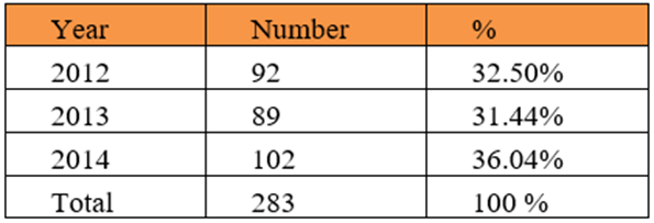
Table 1: distribution of the cases by year.
Regarding the maternal characteristics, the mean maternal age was 30.6 years, 90 (31.8%) of the cases were detected in mothers between (25-39) years of age, followed by women in age group between (30-34) years.
59 cases accounting for (20.84 %)1, and the last 7 cases (2.47 %) were detected at maternal age of 44 years or more. Table (2)
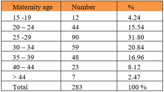
Table 2: Maternal age.
As for parity, the mean parity was 4, where 82 cases (28.77%) were diagnosed in mothers who were Para 4 and more. Table (3). Among our study population, more than half and exactly (56.5 %) had consanguineous marriages, which was counted as a risk factor, while (30%) had no risk factors what so ever. Interestingly (22.6%) had history of a previous baby with an abnormality. Table (4)
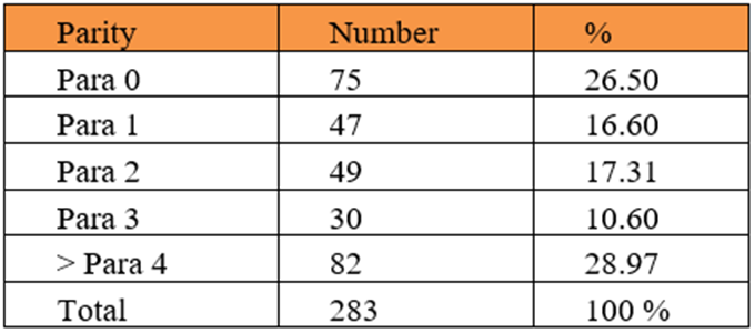
Table 3: Parity.
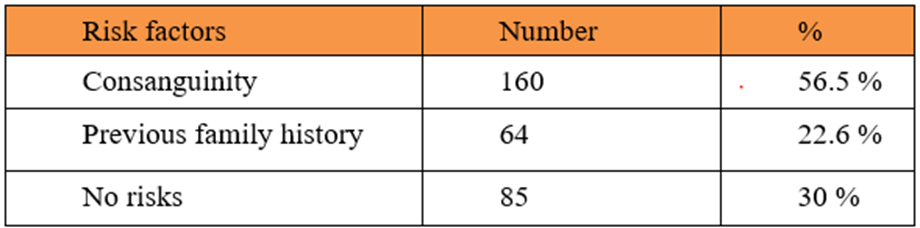
Table 4: risk factors.
When we looked at preconception folic acid intake, only 35 cases (12.36%) were taking it. Table (5)

Table 5: preconception use of folic acid.
As for the gestational age at which these abnormalities were detected, we found that 157 cases (55.47%) were diagnosed between (21-28 weeks) and the least were detected at gestational age of less than 20 weeks only 23 cases (8.12%). Table (6)
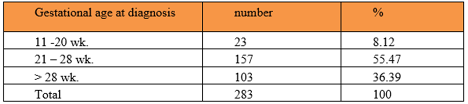
Table 6: Gestational age at diagnosis.
Regarding the nature of the abnormalities detected, whether isolated or multiple, most of them (70.3 %) were isolated. Table (7)

Table 7: type of anomalies.
By classifying the anomalies according to the system involved, we found that, renal anomalies were the most commonly detected ones, in 62 patient which accounts for (31.1%),
followed by central nervous system (CNS) anomalies 41 cases (20.6%), while the facial anomalies were the least commonly detected, only 5 cases (2.51%). Table (8)

Table 8: Isolated anomalies according to the system involved.
Looking at more details about the anomalies of these systems; in the renal system anomalies, we found the hydronephrosis accounting for the majority of the anomalies 21 cases (33.87%), followed with posterior urethral valve 20 cases (20.9%), while renal agenesis was found in 9 cases (9.67%). With regard to central nerve system anomalies; spina bifida was the most commonly diagnosed 13 cases (31.70%) through our study period, followed by isolated hydrocephalus 7 cases (17.07%). In cardiac system ventricular septum defect was found in most of the cases 10 cases (37.03%), followed by multiple cardiac anomalies 7 cases (25.92), 5 of our cases with cardiac abnormalities were confirmed to have hypo plastic left heart syndrome. As for the gastrointestinal system; the most commonly diagnosed anomaly was as follows: esophageal atresia, anorectal atresia, duodenal atresia accounting for 9 cases (45%), 7 cases (35%), and 4 cases (20%) respectively.
Looking at skeletal system; the most serious anomalies were skeletal dysplasia 9 cases (47.30%), 2 cases only with limb reduction (10.52%). As for the 13 cases of thoracic anomalies, 7 case (53.84%) had CPAM (congenital pulmonary airway malformation) and 6 cases (46.15%) had diaphragmatic hernia. Lastly cleft lip with or without cleft palate was seen in only 5 cases. Table (9)

Table 9: distribution according to type of congenital anomalies.
Fetal echocardiography either to confirm a cardiac abnormality in cases of isolated cardio vascular system (CVS) anomalies or to rule out an associated cardiac abnormality as a part of multi system involvement was done for 68 of our cases and yielded 40 abnormal results (58.8%). Table (10)
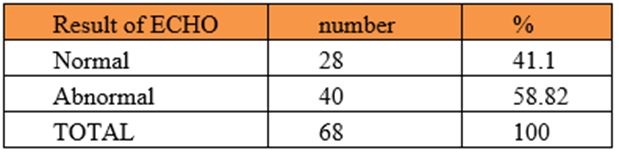
Table 10: Fetal echocardiography result among 68 cases referred to ECHO.
Invasive procedures for possible prenatal diagnosis of chromosomal/ genetic abnormalities were done for 56 out of all cases and 20 of them (35.7%) were having abnormal results. Table (11).
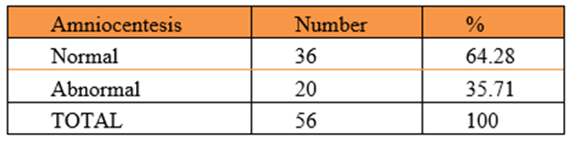
Table 11: Amniocentesis result among 56 cases requested invasive procedure exclude those refuse to do.
Regarding gestational age at delivery, 150 (72.1%) pregnancies ended by delivery at term. Table (12). Among the 34 cases who had termination of pregnancy, 12 cases were before viability, so a total of 208 cases carried their pregnancies beyond viability (24 week and above). As for the mode of delivery around two thirds 138 cases (66.45%) had vaginal delivery while (33.6%) had delivery by cesarean section. Table (13).
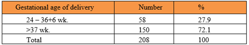
Table 12: Gestational age of delivery
(Excluding 7 case miscarriage, 12 case of termination of pregnancy before 24 weeks. among 227 cases delivered in our hospital)
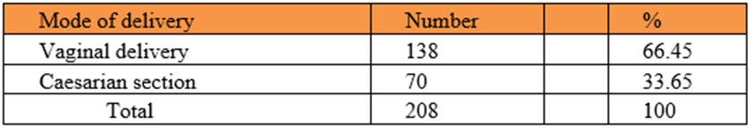
Table 13: Mode of delivery.
(Exclude 7 case miscarriage, 12 case of termination of pregnancy before 24 weeks. among 227 cases delivered in our hospital)
The outcome according to the birth weight is as shown in table (14), where 148 (65%) of born babies were weighing more 2500 gm, followed by 32
(14.09%) cases with birth weight between 2000-2500 gm. 117 cases (51.5%) of the abnormalities detected were among males, and (44.05%) happened in females, and we had 10 cases which accounted for (4.4%) where the gender was not determined. Table (15).
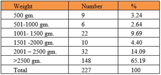
Table 14: Birth weight of the outcome among those delivered in the hospital.

Table 15: Outcome according to the gender among those completed fallow up in the hospital.
Table (16) is showing the number and percentages of babies who required NICU admission, and as expected most of them required so, 106 cases (62.36%). Among the outcome 40 babies (23.52%) ended with early neonatal death. Table (17).

Table 16: NICU admission among 170 delivered alive.

Table 17: the outcome among birth.
Most children, born with congenital anomalies and survive infancy are affected physically, mentally or socially and can be at increased risk of morbidity due to various health disorders [36].
Prevalence studies of congenital anomalies are useful to establish baseline rates, to document changes over time, and to identify clues to etiology. They are also important for planning and evaluating antenatal screening for congenital anomalies, particularly in high-risk populations [37]. The overall prevalence of major congenital malformation in this study during the study period was 3.01%, accounting for perinatal mortality rate of (47/10,000) live births [22].
In this study, congenital anomalies of the renal system were the most commonly encountered and accounted for 31% of all isolated anomalies. This was followed by malformation of central nerves system CNS (20.6%) and cardio vascular system CVS (13.56%). A similar study from Saudi Arabia reported that major congenital anomalies among all live births were mostly observed in the cardiovascular system (CVS), followed by musculoskeletal [32].
Maternal age is an important parameter in the birth of a congenitally malformed fetus. For this reason, females who are older than 35 years of age need to be examined more carefully since the risk of having a congenitally malformed fetus is increased [2]. In the present study, the median maternal age at diagnosis was 30 years which is close to the age reported by other authors as Sellout [5], who indicated that the median maternal age was 27.5 years. Also, they observed direct relation between advancing maternal age and the increasing incidence of congenital anomalies -lower incidence with age of <20> 35 years) to be the most frequent risk factor for birth defects in Brazil [25].
In this study, most of the congenital anomalies detected were seen in women who were para four or more 82 case (28.97%) followed by primai gravidas 75 case (26.50%).
An additional observation in this study was that the mean gestational age at delivery was 37 weeks and the time of diagnosis by ultrasound was 21-28 week. This outcome was similar to the observations of Khaskheli and Michels [38, 39].
Among the other risk factors studies, we found that consanguinity and having a positive previous history of congenital malformation were associated with higher risk. We also found that the mode of delivery was not influenced by having some of the abnormalities diagnosed, where the percentage of cesarean deliveries was very close to the rate for the general population in our unit. The late gestational age at diagnosis is a major factor affecting proper antenatal diagnosis and outcome. Only 56 cases had invasive prenatal diagnosis done, this can be explained by the late gestational age at diagnosis duo to late booking or late referral, in addition, some refused invasive testing.
A major limitation of this study was the retrospective nature of it, as we depended on data collected from patient medical records, and sometimes some information was missing, so we needed to make telephone calls to try to get as much of the information as possible. Also, the prevalence may have been underreported in this study as we only looked at cases with structural abnormalities.
Another factor that may have also resulted in lower rate of detection of malformation among the stillbirths in general is the lack of routine autopsy in these cases. Also, we included those malformation diagnosed by antenatal ultrasound, while still there are some others which were detected postnatally, or the mothers were un booked during their pregnancies and we had no records of their follow up.
The period prevalence of congenital abnormalities in our study is similar to that reported in other population worldwide. Strikingly enough, consanguinity in our population appears to play a major associated risk factor. Still, we think that the prevalence is under reported as only cases of structural anomalies were included, so we recommend a larger study to address this issue.
None
Acknowledgment
None
Authors’ contributions
AlMardawi drafted the manuscript. All Authors contributed to study conception and studydesign. Almardawi and Bajabaa contributed to literature review and data collection. All
Authors contributed to data analysis and data presentation in tables and figures. All
Authors reviewed manuscript for editorial and intellectual contents. All authors have read
and approved the final draft of manuscript.
Clearly Auctoresonline and particularly Psychology and Mental Health Care Journal is dedicated to improving health care services for individuals and populations. The editorial boards' ability to efficiently recognize and share the global importance of health literacy with a variety of stakeholders. Auctoresonline publishing platform can be used to facilitate of optimal client-based services and should be added to health care professionals' repertoire of evidence-based health care resources.

Journal of Clinical Cardiology and Cardiovascular Intervention The submission and review process was adequate. However I think that the publication total value should have been enlightened in early fases. Thank you for all.

Journal of Women Health Care and Issues By the present mail, I want to say thank to you and tour colleagues for facilitating my published article. Specially thank you for the peer review process, support from the editorial office. I appreciate positively the quality of your journal.

Journal of Clinical Research and Reports I would be very delighted to submit my testimonial regarding the reviewer board and the editorial office. The reviewer board were accurate and helpful regarding any modifications for my manuscript. And the editorial office were very helpful and supportive in contacting and monitoring with any update and offering help. It was my pleasure to contribute with your promising Journal and I am looking forward for more collaboration.

We would like to thank the Journal of Thoracic Disease and Cardiothoracic Surgery because of the services they provided us for our articles. The peer-review process was done in a very excellent time manner, and the opinions of the reviewers helped us to improve our manuscript further. The editorial office had an outstanding correspondence with us and guided us in many ways. During a hard time of the pandemic that is affecting every one of us tremendously, the editorial office helped us make everything easier for publishing scientific work. Hope for a more scientific relationship with your Journal.

The peer-review process which consisted high quality queries on the paper. I did answer six reviewers’ questions and comments before the paper was accepted. The support from the editorial office is excellent.

Journal of Neuroscience and Neurological Surgery. I had the experience of publishing a research article recently. The whole process was simple from submission to publication. The reviewers made specific and valuable recommendations and corrections that improved the quality of my publication. I strongly recommend this Journal.

Dr. Katarzyna Byczkowska My testimonial covering: "The peer review process is quick and effective. The support from the editorial office is very professional and friendly. Quality of the Clinical Cardiology and Cardiovascular Interventions is scientific and publishes ground-breaking research on cardiology that is useful for other professionals in the field.

Thank you most sincerely, with regard to the support you have given in relation to the reviewing process and the processing of my article entitled "Large Cell Neuroendocrine Carcinoma of The Prostate Gland: A Review and Update" for publication in your esteemed Journal, Journal of Cancer Research and Cellular Therapeutics". The editorial team has been very supportive.

Testimony of Journal of Clinical Otorhinolaryngology: work with your Reviews has been a educational and constructive experience. The editorial office were very helpful and supportive. It was a pleasure to contribute to your Journal.

Dr. Bernard Terkimbi Utoo, I am happy to publish my scientific work in Journal of Women Health Care and Issues (JWHCI). The manuscript submission was seamless and peer review process was top notch. I was amazed that 4 reviewers worked on the manuscript which made it a highly technical, standard and excellent quality paper. I appreciate the format and consideration for the APC as well as the speed of publication. It is my pleasure to continue with this scientific relationship with the esteem JWHCI.

This is an acknowledgment for peer reviewers, editorial board of Journal of Clinical Research and Reports. They show a lot of consideration for us as publishers for our research article “Evaluation of the different factors associated with side effects of COVID-19 vaccination on medical students, Mutah university, Al-Karak, Jordan”, in a very professional and easy way. This journal is one of outstanding medical journal.

Dear Hao Jiang, to Journal of Nutrition and Food Processing We greatly appreciate the efficient, professional and rapid processing of our paper by your team. If there is anything else we should do, please do not hesitate to let us know. On behalf of my co-authors, we would like to express our great appreciation to editor and reviewers.

As an author who has recently published in the journal "Brain and Neurological Disorders". I am delighted to provide a testimonial on the peer review process, editorial office support, and the overall quality of the journal. The peer review process at Brain and Neurological Disorders is rigorous and meticulous, ensuring that only high-quality, evidence-based research is published. The reviewers are experts in their fields, and their comments and suggestions were constructive and helped improve the quality of my manuscript. The review process was timely and efficient, with clear communication from the editorial office at each stage. The support from the editorial office was exceptional throughout the entire process. The editorial staff was responsive, professional, and always willing to help. They provided valuable guidance on formatting, structure, and ethical considerations, making the submission process seamless. Moreover, they kept me informed about the status of my manuscript and provided timely updates, which made the process less stressful. The journal Brain and Neurological Disorders is of the highest quality, with a strong focus on publishing cutting-edge research in the field of neurology. The articles published in this journal are well-researched, rigorously peer-reviewed, and written by experts in the field. The journal maintains high standards, ensuring that readers are provided with the most up-to-date and reliable information on brain and neurological disorders. In conclusion, I had a wonderful experience publishing in Brain and Neurological Disorders. The peer review process was thorough, the editorial office provided exceptional support, and the journal's quality is second to none. I would highly recommend this journal to any researcher working in the field of neurology and brain disorders.
Dear Agrippa Hilda, Journal of Neuroscience and Neurological Surgery, Editorial Coordinator, I trust this message finds you well. I want to extend my appreciation for considering my article for publication in your esteemed journal. I am pleased to provide a testimonial regarding the peer review process and the support received from your editorial office. The peer review process for my paper was carried out in a highly professional and thorough manner. The feedback and comments provided by the authors were constructive and very useful in improving the quality of the manuscript. This rigorous assessment process undoubtedly contributes to the high standards maintained by your journal.

International Journal of Clinical Case Reports and Reviews. I strongly recommend to consider submitting your work to this high-quality journal. The support and availability of the Editorial staff is outstanding and the review process was both efficient and rigorous.

Thank you very much for publishing my Research Article titled “Comparing Treatment Outcome Of Allergic Rhinitis Patients After Using Fluticasone Nasal Spray And Nasal Douching" in the Journal of Clinical Otorhinolaryngology. As Medical Professionals we are immensely benefited from study of various informative Articles and Papers published in this high quality Journal. I look forward to enriching my knowledge by regular study of the Journal and contribute my future work in the field of ENT through the Journal for use by the medical fraternity. The support from the Editorial office was excellent and very prompt. I also welcome the comments received from the readers of my Research Article.

Dear Erica Kelsey, Editorial Coordinator of Cancer Research and Cellular Therapeutics Our team is very satisfied with the processing of our paper by your journal. That was fast, efficient, rigorous, but without unnecessary complications. We appreciated the very short time between the submission of the paper and its publication on line on your site.

I am very glad to say that the peer review process is very successful and fast and support from the Editorial Office. Therefore, I would like to continue our scientific relationship for a long time. And I especially thank you for your kindly attention towards my article. Have a good day!

"We recently published an article entitled “Influence of beta-Cyclodextrins upon the Degradation of Carbofuran Derivatives under Alkaline Conditions" in the Journal of “Pesticides and Biofertilizers” to show that the cyclodextrins protect the carbamates increasing their half-life time in the presence of basic conditions This will be very helpful to understand carbofuran behaviour in the analytical, agro-environmental and food areas. We greatly appreciated the interaction with the editor and the editorial team; we were particularly well accompanied during the course of the revision process, since all various steps towards publication were short and without delay".

I would like to express my gratitude towards you process of article review and submission. I found this to be very fair and expedient. Your follow up has been excellent. I have many publications in national and international journal and your process has been one of the best so far. Keep up the great work.

We are grateful for this opportunity to provide a glowing recommendation to the Journal of Psychiatry and Psychotherapy. We found that the editorial team were very supportive, helpful, kept us abreast of timelines and over all very professional in nature. The peer review process was rigorous, efficient and constructive that really enhanced our article submission. The experience with this journal remains one of our best ever and we look forward to providing future submissions in the near future.

I am very pleased to serve as EBM of the journal, I hope many years of my experience in stem cells can help the journal from one way or another. As we know, stem cells hold great potential for regenerative medicine, which are mostly used to promote the repair response of diseased, dysfunctional or injured tissue using stem cells or their derivatives. I think Stem Cell Research and Therapeutics International is a great platform to publish and share the understanding towards the biology and translational or clinical application of stem cells.

I would like to give my testimony in the support I have got by the peer review process and to support the editorial office where they were of asset to support young author like me to be encouraged to publish their work in your respected journal and globalize and share knowledge across the globe. I really give my great gratitude to your journal and the peer review including the editorial office.

I am delighted to publish our manuscript entitled "A Perspective on Cocaine Induced Stroke - Its Mechanisms and Management" in the Journal of Neuroscience and Neurological Surgery. The peer review process, support from the editorial office, and quality of the journal are excellent. The manuscripts published are of high quality and of excellent scientific value. I recommend this journal very much to colleagues.

Dr.Tania Muñoz, My experience as researcher and author of a review article in The Journal Clinical Cardiology and Interventions has been very enriching and stimulating. The editorial team is excellent, performs its work with absolute responsibility and delivery. They are proactive, dynamic and receptive to all proposals. Supporting at all times the vast universe of authors who choose them as an option for publication. The team of review specialists, members of the editorial board, are brilliant professionals, with remarkable performance in medical research and scientific methodology. Together they form a frontline team that consolidates the JCCI as a magnificent option for the publication and review of high-level medical articles and broad collective interest. I am honored to be able to share my review article and open to receive all your comments.

“The peer review process of JPMHC is quick and effective. Authors are benefited by good and professional reviewers with huge experience in the field of psychology and mental health. The support from the editorial office is very professional. People to contact to are friendly and happy to help and assist any query authors might have. Quality of the Journal is scientific and publishes ground-breaking research on mental health that is useful for other professionals in the field”.

Dear editorial department: On behalf of our team, I hereby certify the reliability and superiority of the International Journal of Clinical Case Reports and Reviews in the peer review process, editorial support, and journal quality. Firstly, the peer review process of the International Journal of Clinical Case Reports and Reviews is rigorous, fair, transparent, fast, and of high quality. The editorial department invites experts from relevant fields as anonymous reviewers to review all submitted manuscripts. These experts have rich academic backgrounds and experience, and can accurately evaluate the academic quality, originality, and suitability of manuscripts. The editorial department is committed to ensuring the rigor of the peer review process, while also making every effort to ensure a fast review cycle to meet the needs of authors and the academic community. Secondly, the editorial team of the International Journal of Clinical Case Reports and Reviews is composed of a group of senior scholars and professionals with rich experience and professional knowledge in related fields. The editorial department is committed to assisting authors in improving their manuscripts, ensuring their academic accuracy, clarity, and completeness. Editors actively collaborate with authors, providing useful suggestions and feedback to promote the improvement and development of the manuscript. We believe that the support of the editorial department is one of the key factors in ensuring the quality of the journal. Finally, the International Journal of Clinical Case Reports and Reviews is renowned for its high- quality articles and strict academic standards. The editorial department is committed to publishing innovative and academically valuable research results to promote the development and progress of related fields. The International Journal of Clinical Case Reports and Reviews is reasonably priced and ensures excellent service and quality ratio, allowing authors to obtain high-level academic publishing opportunities in an affordable manner. I hereby solemnly declare that the International Journal of Clinical Case Reports and Reviews has a high level of credibility and superiority in terms of peer review process, editorial support, reasonable fees, and journal quality. Sincerely, Rui Tao.

Clinical Cardiology and Cardiovascular Interventions I testity the covering of the peer review process, support from the editorial office, and quality of the journal.

Clinical Cardiology and Cardiovascular Interventions, we deeply appreciate the interest shown in our work and its publication. It has been a true pleasure to collaborate with you. The peer review process, as well as the support provided by the editorial office, have been exceptional, and the quality of the journal is very high, which was a determining factor in our decision to publish with you.

The peer reviewers process is quick and effective, the supports from editorial office is excellent, the quality of journal is high. I would like to collabroate with Internatioanl journal of Clinical Case Reports and Reviews journal clinically in the future time.

Clinical Cardiology and Cardiovascular Interventions, I would like to express my sincerest gratitude for the trust placed in our team for the publication in your journal. It has been a true pleasure to collaborate with you on this project. I am pleased to inform you that both the peer review process and the attention from the editorial coordination have been excellent. Your team has worked with dedication and professionalism to ensure that your publication meets the highest standards of quality. We are confident that this collaboration will result in mutual success, and we are eager to see the fruits of this shared effort.

Dear Dr. Jessica Magne, Editorial Coordinator 0f Clinical Cardiology and Cardiovascular Interventions, I hope this message finds you well. I want to express my utmost gratitude for your excellent work and for the dedication and speed in the publication process of my article titled "Navigating Innovation: Qualitative Insights on Using Technology for Health Education in Acute Coronary Syndrome Patients." I am very satisfied with the peer review process, the support from the editorial office, and the quality of the journal. I hope we can maintain our scientific relationship in the long term.

Dear Monica Gissare, - Editorial Coordinator of Nutrition and Food Processing. ¨My testimony with you is truly professional, with a positive response regarding the follow-up of the article and its review, you took into account my qualities and the importance of the topic¨.

Dear Dr. Jessica Magne, Editorial Coordinator 0f Clinical Cardiology and Cardiovascular Interventions, The review process for the article “The Handling of Anti-aggregants and Anticoagulants in the Oncologic Heart Patient Submitted to Surgery” was extremely rigorous and detailed. From the initial submission to the final acceptance, the editorial team at the “Journal of Clinical Cardiology and Cardiovascular Interventions” demonstrated a high level of professionalism and dedication. The reviewers provided constructive and detailed feedback, which was essential for improving the quality of our work. Communication was always clear and efficient, ensuring that all our questions were promptly addressed. The quality of the “Journal of Clinical Cardiology and Cardiovascular Interventions” is undeniable. It is a peer-reviewed, open-access publication dedicated exclusively to disseminating high-quality research in the field of clinical cardiology and cardiovascular interventions. The journal's impact factor is currently under evaluation, and it is indexed in reputable databases, which further reinforces its credibility and relevance in the scientific field. I highly recommend this journal to researchers looking for a reputable platform to publish their studies.

Dear Editorial Coordinator of the Journal of Nutrition and Food Processing! "I would like to thank the Journal of Nutrition and Food Processing for including and publishing my article. The peer review process was very quick, movement and precise. The Editorial Board has done an extremely conscientious job with much help, valuable comments and advices. I find the journal very valuable from a professional point of view, thank you very much for allowing me to be part of it and I would like to participate in the future!”

Dealing with The Journal of Neurology and Neurological Surgery was very smooth and comprehensive. The office staff took time to address my needs and the response from editors and the office was prompt and fair. I certainly hope to publish with this journal again.Their professionalism is apparent and more than satisfactory. Susan Weiner

My Testimonial Covering as fellowing: Lin-Show Chin. The peer reviewers process is quick and effective, the supports from editorial office is excellent, the quality of journal is high. I would like to collabroate with Internatioanl journal of Clinical Case Reports and Reviews.

My experience publishing in Psychology and Mental Health Care was exceptional. The peer review process was rigorous and constructive, with reviewers providing valuable insights that helped enhance the quality of our work. The editorial team was highly supportive and responsive, making the submission process smooth and efficient. The journal's commitment to high standards and academic rigor makes it a respected platform for quality research. I am grateful for the opportunity to publish in such a reputable journal.

My experience publishing in International Journal of Clinical Case Reports and Reviews was exceptional. I Come forth to Provide a Testimonial Covering the Peer Review Process and the editorial office for the Professional and Impartial Evaluation of the Manuscript.

I would like to offer my testimony in the support. I have received through the peer review process and support the editorial office where they are to support young authors like me, encourage them to publish their work in your esteemed journals, and globalize and share knowledge globally. I really appreciate your journal, peer review, and editorial office.

Dear Agrippa Hilda- Editorial Coordinator of Journal of Neuroscience and Neurological Surgery, "The peer review process was very quick and of high quality, which can also be seen in the articles in the journal. The collaboration with the editorial office was very good."

I would like to express my sincere gratitude for the support and efficiency provided by the editorial office throughout the publication process of my article, “Delayed Vulvar Metastases from Rectal Carcinoma: A Case Report.” I greatly appreciate the assistance and guidance I received from your team, which made the entire process smooth and efficient. The peer review process was thorough and constructive, contributing to the overall quality of the final article. I am very grateful for the high level of professionalism and commitment shown by the editorial staff, and I look forward to maintaining a long-term collaboration with the International Journal of Clinical Case Reports and Reviews.

To Dear Erin Aust, I would like to express my heartfelt appreciation for the opportunity to have my work published in this esteemed journal. The entire publication process was smooth and well-organized, and I am extremely satisfied with the final result. The Editorial Team demonstrated the utmost professionalism, providing prompt and insightful feedback throughout the review process. Their clear communication and constructive suggestions were invaluable in enhancing my manuscript, and their meticulous attention to detail and dedication to quality are truly commendable. Additionally, the support from the Editorial Office was exceptional. From the initial submission to the final publication, I was guided through every step of the process with great care and professionalism. The team's responsiveness and assistance made the entire experience both easy and stress-free. I am also deeply impressed by the quality and reputation of the journal. It is an honor to have my research featured in such a respected publication, and I am confident that it will make a meaningful contribution to the field.

"I am grateful for the opportunity of contributing to [International Journal of Clinical Case Reports and Reviews] and for the rigorous review process that enhances the quality of research published in your esteemed journal. I sincerely appreciate the time and effort of your team who have dedicatedly helped me in improvising changes and modifying my manuscript. The insightful comments and constructive feedback provided have been invaluable in refining and strengthening my work".

I thank the ‘Journal of Clinical Research and Reports’ for accepting this article for publication. This is a rigorously peer reviewed journal which is on all major global scientific data bases. I note the review process was prompt, thorough and professionally critical. It gave us an insight into a number of important scientific/statistical issues. The review prompted us to review the relevant literature again and look at the limitations of the study. The peer reviewers were open, clear in the instructions and the editorial team was very prompt in their communication. This journal certainly publishes quality research articles. I would recommend the journal for any future publications.

Dear Jessica Magne, with gratitude for the joint work. Fast process of receiving and processing the submitted scientific materials in “Clinical Cardiology and Cardiovascular Interventions”. High level of competence of the editors with clear and correct recommendations and ideas for enriching the article.

We found the peer review process quick and positive in its input. The support from the editorial officer has been very agile, always with the intention of improving the article and taking into account our subsequent corrections.

My article, titled 'No Way Out of the Smartphone Epidemic Without Considering the Insights of Brain Research,' has been republished in the International Journal of Clinical Case Reports and Reviews. The review process was seamless and professional, with the editors being both friendly and supportive. I am deeply grateful for their efforts.

To Dear Erin Aust – Editorial Coordinator of Journal of General Medicine and Clinical Practice! I declare that I am absolutely satisfied with your work carried out with great competence in following the manuscript during the various stages from its receipt, during the revision process to the final acceptance for publication. Thank Prof. Elvira Farina

Dear Jessica, and the super professional team of the ‘Clinical Cardiology and Cardiovascular Interventions’ I am sincerely grateful to the coordinated work of the journal team for the no problem with the submission of my manuscript: “Cardiometabolic Disorders in A Pregnant Woman with Severe Preeclampsia on the Background of Morbid Obesity (Case Report).” The review process by 5 experts was fast, and the comments were professional, which made it more specific and academic, and the process of publication and presentation of the article was excellent. I recommend that my colleagues publish articles in this journal, and I am interested in further scientific cooperation. Sincerely and best wishes, Dr. Oleg Golyanovskiy.

Dear Ashley Rosa, Editorial Coordinator of the journal - Psychology and Mental Health Care. " The process of obtaining publication of my article in the Psychology and Mental Health Journal was positive in all areas. The peer review process resulted in a number of valuable comments, the editorial process was collaborative and timely, and the quality of this journal has been quickly noticed, resulting in alternative journals contacting me to publish with them." Warm regards, Susan Anne Smith, PhD. Australian Breastfeeding Association.

Dear Jessica Magne, Editorial Coordinator, Clinical Cardiology and Cardiovascular Interventions, Auctores Publishing LLC. I appreciate the journal (JCCI) editorial office support, the entire team leads were always ready to help, not only on technical front but also on thorough process. Also, I should thank dear reviewers’ attention to detail and creative approach to teach me and bring new insights by their comments. Surely, more discussions and introduction of other hemodynamic devices would provide better prevention and management of shock states. Your efforts and dedication in presenting educational materials in this journal are commendable. Best wishes from, Farahnaz Fallahian.

Dear Maria Emerson, Editorial Coordinator, International Journal of Clinical Case Reports and Reviews, Auctores Publishing LLC. I am delighted to have published our manuscript, "Acute Colonic Pseudo-Obstruction (ACPO): A rare but serious complication following caesarean section." I want to thank the editorial team, especially Maria Emerson, for their prompt review of the manuscript, quick responses to queries, and overall support. Yours sincerely Dr. Victor Olagundoye.

Dear Ashley Rosa, Editorial Coordinator, International Journal of Clinical Case Reports and Reviews. Many thanks for publishing this manuscript after I lost confidence the editors were most helpful, more than other journals Best wishes from, Susan Anne Smith, PhD. Australian Breastfeeding Association.

Dear Agrippa Hilda, Editorial Coordinator, Journal of Neuroscience and Neurological Surgery. The entire process including article submission, review, revision, and publication was extremely easy. The journal editor was prompt and helpful, and the reviewers contributed to the quality of the paper. Thank you so much! Eric Nussbaum, MD

Dr Hala Al Shaikh This is to acknowledge that the peer review process for the article ’ A Novel Gnrh1 Gene Mutation in Four Omani Male Siblings, Presentation and Management ’ sent to the International Journal of Clinical Case Reports and Reviews was quick and smooth. The editorial office was prompt with easy communication.

Dear Erin Aust, Editorial Coordinator, Journal of General Medicine and Clinical Practice. We are pleased to share our experience with the “Journal of General Medicine and Clinical Practice”, following the successful publication of our article. The peer review process was thorough and constructive, helping to improve the clarity and quality of the manuscript. We are especially thankful to Ms. Erin Aust, the Editorial Coordinator, for her prompt communication and continuous support throughout the process. Her professionalism ensured a smooth and efficient publication experience. The journal upholds high editorial standards, and we highly recommend it to fellow researchers seeking a credible platform for their work. Best wishes By, Dr. Rakhi Mishra.

Dear Jessica Magne, Editorial Coordinator, Clinical Cardiology and Cardiovascular Interventions, Auctores Publishing LLC. The peer review process of the journal of Clinical Cardiology and Cardiovascular Interventions was excellent and fast, as was the support of the editorial office and the quality of the journal. Kind regards Walter F. Riesen Prof. Dr. Dr. h.c. Walter F. Riesen.

Dear Ashley Rosa, Editorial Coordinator, International Journal of Clinical Case Reports and Reviews, Auctores Publishing LLC. Thank you for publishing our article, Exploring Clozapine's Efficacy in Managing Aggression: A Multiple Single-Case Study in Forensic Psychiatry in the international journal of clinical case reports and reviews. We found the peer review process very professional and efficient. The comments were constructive, and the whole process was efficient. On behalf of the co-authors, I would like to thank you for publishing this article. With regards, Dr. Jelle R. Lettinga.

Dear Clarissa Eric, Editorial Coordinator, Journal of Clinical Case Reports and Studies, I would like to express my deep admiration for the exceptional professionalism demonstrated by your journal. I am thoroughly impressed by the speed of the editorial process, the substantive and insightful reviews, and the meticulous preparation of the manuscript for publication. Additionally, I greatly appreciate the courteous and immediate responses from your editorial office to all my inquiries. Best Regards, Dariusz Ziora

Dear Chrystine Mejia, Editorial Coordinator, Journal of Neurodegeneration and Neurorehabilitation, Auctores Publishing LLC, We would like to thank the editorial team for the smooth and high-quality communication leading up to the publication of our article in the Journal of Neurodegeneration and Neurorehabilitation. The reviewers have extensive knowledge in the field, and their relevant questions helped to add value to our publication. Kind regards, Dr. Ravi Shrivastava.

Dear Clarissa Eric, Editorial Coordinator, Journal of Clinical Case Reports and Studies, Auctores Publishing LLC, USA Office: +1-(302)-520-2644. I would like to express my sincere appreciation for the efficient and professional handling of my case report by the ‘Journal of Clinical Case Reports and Studies’. The peer review process was not only fast but also highly constructive—the reviewers’ comments were clear, relevant, and greatly helped me improve the quality and clarity of my manuscript. I also received excellent support from the editorial office throughout the process. Communication was smooth and timely, and I felt well guided at every stage, from submission to publication. The overall quality and rigor of the journal are truly commendable. I am pleased to have published my work with Journal of Clinical Case Reports and Studies, and I look forward to future opportunities for collaboration. Sincerely, Aline Tollet, UCLouvain.

Dear Ms. Mayra Duenas, Editorial Coordinator, International Journal of Clinical Case Reports and Reviews. “The International Journal of Clinical Case Reports and Reviews represented the “ideal house” to share with the research community a first experience with the use of the Simeox device for speech rehabilitation. High scientific reputation and attractive website communication were first determinants for the selection of this Journal, and the following submission process exceeded expectations: fast but highly professional peer review, great support by the editorial office, elegant graphic layout. Exactly what a dynamic research team - also composed by allied professionals - needs!" From, Chiara Beccaluva, PT - Italy.

Dear Maria Emerson, Editorial Coordinator, we have deeply appreciated the professionalism demonstrated by the International Journal of Clinical Case Reports and Reviews. The reviewers have extensive knowledge of our field and have been very efficient and fast in supporting the process. I am really looking forward to further collaboration. Thanks. Best regards, Dr. Claudio Ligresti

Dear Chrystine Mejia, Editorial Coordinator, Journal of Neurodegeneration and Neurorehabilitation. “The peer review process was efficient and constructive, and the editorial office provided excellent communication and support throughout. The journal ensures scientific rigor and high editorial standards, while also offering a smooth and timely publication process. We sincerely appreciate the work of the editorial team in facilitating the dissemination of innovative approaches such as the Bonori Method.” Best regards, Dr. Giselle Pentón-Rol.
