AUCTORES
Globalize your Research

Research Article | DOI: https://doi.org/10.31579/2641-0419/103
1Division of Rheumatology, Tygerberg Academic Hospital, National Health Laboratory Services. Faculty of Health Sciences, Stellenbosch University and Tygerberg Academic Hospital, Cape Town, South Africa
2Department of Medicine, Stellenbosch University and Tygerberg hospital
3Division of Nephrology, Department of Medicine, Stellenbosch University and Tygerberg Hospital
*Corresponding Author: Amani Kaawan, Division of Rheumatology, Tygerberg Academic Hospital, National Health Laboratory Services. Faculty of Health Sciences, Stellenbosch University and Tygerberg Academic Hospital, Cape Town, South Africa.
Citation: Amani Kaawan., Mou Manie., Adel M Aboshakwa., Razeen Davids.,(2020) MMED PROJECT The Clinical Profile of Takayasu Disease at Tygerberg Academic Hospital – a retrospective study. J. Clinical Cardiology and Cardiovascular Interventions, 3(11); Doi:10.31579/2641-0419/103
Copyright: © 2020 Amani Kaawan, This is an open-access article distributed under the terms of the Creative Commons Attribution License, which permits unrestricted use, distribution, and reproduction in any medium, provided the original author and source are credited.
Received: 02 November 2020 | Accepted: 12 November 2020 | Published: 19 November 2020
Keywords: Takayasu’s arteritis
Takayasu’s arteritis (TA) is a disease of unknown etiology that causes chronic inflammation of the large blood vessels and usually involves the aorta and its branches. The inflammatory process initially leads to thickening of the arterial wall and may result in stenosis, occlusion, dilatation or aneurysm formation. Although TA occurs more commonly in certain regions such as Asia, it is a global disease. Currently, there is a dearth of information on this condition in African countries, including South Africa.
Results: We have identified 50 patients meeting the ACR criteria for the diagnosis of TA. Data was drawn from the records of the Division of Rheumatology as well as the angiographic records of TA. This included patient demographics, mode of presentation, classification, co-morbidities, complications, clinical and laboratory features, radiological findings, drug therapy as well as outcomes. The cohort comprised 34 of mixed race, 12 black and 4 white patients. The mean age at diagnosis was 16-56 (28.2) years. The most common presention was hypertension, followed by CVA, heart failure, and syncope. Angiography revealed Type V (most of aorta) and Type I (aortic arch branches) lesions to be the most common and that stenosis was much more common than aneurysm formation. Two patients had concomitant antiphospholipid syndrome. Another two had concomitant internal jugular vein thrombosis. Six patients had significant mediastinal lymphadenopathy, only one of whom had TB.
Corticosteroids and additional immunosuppressive therapy were used to control disease activity in most patients. Biological treatment was used in two patients. Surgical intervention consisted mainly of renal auto transplantation. Twelve percent of patients had repeated admissions, which included planned surgical intervention. We had two mortalities, both as a result of massive strokes. The mortality rate was 6%. Only 3 patients had active TB.
Conclusion: The study revealed gender and age characteristics similar to previous studies, confirming that TA affects mainly young females. The mortality rate of 6% in this cohort is significantly lower than that of previous series. The unusual findings in this study were:
Significant chest lymphadenopathy in 12% of the cases,the significantly lower percentage of active TB of only 6%, two cases of a rare finding of venous thrombosis as well as 2 cases of an uncommon finding in TA of antiphospholipid syndrome.
The diagnosis of TA may be difficult because of the non-specific nature of the illness in the early stages. Furthermore, TA exhibits different patterns of arterial involvement, disease expression and prognosis in different regions of the world. [1] The clinical presentation results from the arterial lesions and the systemic inflammatory response. At disease presentation or during relapses, TA patients may present with non-specific complaints such as fever, malaise, anorexia, weight loss, myalgia or arthralgia. [2] As arterial lesions progress, more characteristic features of TA may be found such as limb claudication, decreased or absent peripheral pulses, vascular bruits, hypertension and discrepancies in blood pressure between arms due to stenotic or occlusive lesions. [1] In severe cases, this may result in serious complications such as myocardial infarction, stroke and blindness. [3]
Imaging modalities are central to establishing the diagnosis of TA, determining the distribution of lesions and monitoring disease activity. [3] Conventional radiographic angiography [digital subtraction angiography (DSA)] is considered as the gold standard for the diagnosis of TA. However, it is invasive, poses some radiation risk and currently, has largely been replaced by computerized tomography angiography (CTA). Other non-invasive imaging methods including magnetic resonance angiography (MRA), color Doppler ultrasound (CDU), PET with 18F-fluorodeoxyglucose (18F-FDG) and 18F-FDG PET/CT have recently gained ground. In a study conducted in 2004, it was shown that non-invasive imaging methods, deoxyglucose positron emission tomography (PET) and magnetic resonance imaging (MRI)) provided important additional information about disease activity and progression of vessel wall thickening when compared with X ray angiography. [4]A meta-analysis done to assess the diagnostic value of 18-FDG-PET in estimating disease activity in TA concluded that 18-FDG-PET had moderate diagnosis value in assessing TA activity and may add additional value to the current diagnosis methods. [5]
The goal of treatment are to stop the acute inflammatory process and prevent further damage to the vessel wall. The mainstay of therapy is glucocorticoids (usually prednisone); however, other medications include cytotoxic drugs, such as methotrexate, which are often used to help control the disease and reduce the need for prednisone. Newer agents include mycophenolate leflunomide and biologics such as TNF alpha blockers (infliximab, etanercept), tocilizumab, abatacept and rituximab are also increasingly being used for treatment of resistant cases. [3] Treatment of symptomatic fibrotic lesions (stenosis or occlusions) requires either vascular interventional or surgical therapy. This can be achieved by angioplasty with or without stenting or in severe cases, by vascular resection and surgical interventions.
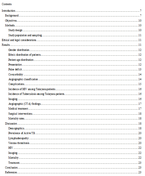
As shown in (table 1), currently there is a paucity of data on TA from the African continent including South Africa. A literature search has revealed only 3 publications on TA from South Africa. One important study was conducted at the Groote Schuur Hospital (GSH) in Cape Town (South Africa). [21] The study reported on 272 patients over a fifty-year period (1952-2002). Patients had a mean age of twenty-five years, of which 75% were female and comprised predominantly mixed race (68%).Some limitations of the study included a lack of information on non-vascular symptoms, the use of outdated imaging techniques, older treatment regimens as well as lack of information on co-morbidities such as HIV, diabetes and atherosclerosis. Additionally, the study reported the use of anti-TB therapy without definite confirmation of the TB diagnosis in some of the cases. Another study reporting on paediatric cases in Gauteng (South Africa) was conducted over a period of fifteen years and included thirty-one patients with a higher prevalence of TA among black patients. [3] This particular study was conducted at the Baragwanath Hospital (Johannesburg, South Africa). [3] The third study was conducted at the King Edward VIII Hospital in Durban (South Africa) and was a small series (11 patients) reporting mainly on the morphological and radiographic features of TA. [3]
A publication from Tunisia in 2010 reported on 27 patients with a mean age of diagnosis was 33 years and 88.9% were females. [3] A second Tunisian study included a limited number of 11 females and was conducted over a 12 year period, wherein the mean age at the time of diagnosis was 29.1 years. .[3] Furthermore, other published studies from the African continent was from Morocco with 47 patients.. [3]
Studies from other regions in the world revealed comparable data. A Turkish study included 248 patients from fifteen different rheumatology centres. Results indicated that the mean age was 40 years and the majority (228 out of 248) were females. [3] These findings are supported by a Brazilian study (Sao Paulo) wherein data was collected from three different public universities (UNIFESP, USP, UNICAMP). Seventy-three patients were recruited and showed a mean age of 27 years, with a predominantly female gender. [4]
In terms of its association with TB, tuberculosis has remained an important differential and possible etiological factor. However, tuberculous aortitis tends to cause erosion of the vessel wall with the formation of true or false aneurysms, particularly affecting the descending thoracic and abdominal aorta. Dissection and rupture are important complications rather than the stenosis typical of Takayasu arteritis. The incidence of rupture and bleeding complications of aneurysmal Takayasu arteritis is low. previous studies reported that the association ranged from 5.0 % to 48.0% . However, there was also a lack of information on whether the TB was active or not and, in some cases, the diagnosis of TB appeared questionable. The question of an association of TA with HIV has also not been addressed in previous studies from this region, even though HIV is known to cause thrombosis of large vessels. Information and previous reports on other features of the disease also appear scarce, with special reference to the systemic features such as fever, malaise, lymphadenopathy, weight loss, skin rash, synovitis and anemia.
Studies relating to the main affected arteries of TA in this country is limited. In terms of the most common subtype of TA in South Africa, reports appear varied due to the different demographic profile and regional differences. There is also very limited information on the use of the latest imaging techniques employed for the diagnosis of TA, the treatment regimes, responses and adverse effects to treatment.
Lastly, complications such as arterial aneurysm rupture, angioplasty and surgical bypass of vascular occlusive lesions which require surgical intervention have not been reported on in any significant detail in previous studies. It should be noted that indications for surgery such as revascularization have also been changed over the last few years.
This study attempts to shed light on some of the unresolved areas referred to above.
Objectives
The aims of the study were to determine and describe:
-demographics such as age, gender and race;
-prevalence of TA in the South African region;
-the type of association with active or previous TB or HIV;
- the presenting features and the prevalence and of other co-morbidities in TA patients;
-the affected arteries in TA patients;
-the imaging techniques employed for TA diagnosis;
-the treatment regimes, responses and outcomes treatment;
Methods
Study design
The current study was done at Tygerberg Academic Hospital, a tertiary referral center situated in the city of Cape Town (Western Cape Province, South Africa). Tygerberg Academic Hospital provides a tertiary service to a population of approximately 1.5 million people (the TBH drainage area). Most of the patients who visit the hospital are indigent and are of mixed-race ethnicity.
This was a retrospective observational study, conducted at the Division of Rheumatology at this hospital. Case records of patients with TA meeting the 1997 American College of Rheumatology (ACR) classification criteria were reviewed. Data collected were TA patient demographics, presentations, classification, co-morbidities, complications, clinical and laboratory features, radiological findings, drug therapy and outcomes. Data was accessed from the administrative databases of the Division of Rheumatology at Tygerberg Academic Hospital, allowing the investigator to review the in-patient and out-patient records of the TA patients.
The inclusion criteria were the positive diagnosis of TA according to ACR criteria, which requires that at least three of the six criteria are met (see Table 2 below). [2]

Ethical and legal considerations
The study was approved by the Ethics Committee of the Faculty of Health Sciences of the University of Stellenbosch (project number (S18/09/183) and the Research Committee of the Department of Medicine, Tygerberg Academic Hospital. The study was conducted in accordance to the Helsinki Declaration as well as MRC and ICH guidelines.
The names of the participants were not mentioned in the output of the research. Where the investigator only had access to the collected data in order to ensure the confidentiality and integrity of the data.
A total of 50 patients with Takayasu arteritis (TA) were identified.
Females represented the majority (82.35% n=42) of patients.
Approximately, a third of the population were of mixed ethnicity (68% n=34), almost a quarter (24% n=12) indicated were of black ethnicity and a smaller percentage of patients indicated white ethnicity.

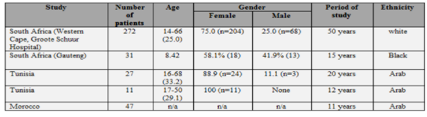
Almost half of the patients (44% n=22) were between the ages of 26-35 years, followed by less than a third (31% n=17) who were between the ages of 16-25 years. Only seven patients (14.0% n=7) belonged to the 36-45 years age group and four patients (8.0% n=4) were in the age group of 46 and over.
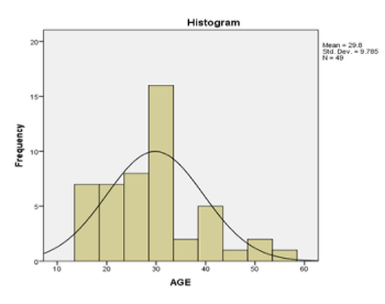
As shown in table (3), half of the patients (50% n=25) had hypertension (HPT) in their presentation, followed by eleven patients (24.0% n=12) who presented with cerebrovascular accident (CVA). One patient had both hypertension and CVA, and five patients (10.0% n=5) presented with heart failure (HF).
The above Table refers to the main methods of presentation, some patients had more than one presenting feature which represent 5 patient (10%) of total cohort. Of the three patients who presented with syncope, one had loss of vision; whereas one patient presented with constitutional symptoms and erythema nodosum. In the group of patients who presented with hypertension, one also had abdominal pain and BP discrepancy and another one had dry gangrene of the lower limb. Another patient also presented with lymphadenopathy. In the CVA group, one patient also had left side paresthesia and one patient also had palpitations.
Of two patients in whom we could not identify the mode of presentation, one of them was diagnosed at age of two years with no clear documentation of his presentation and the other had incomplete documentation.Significant lymphadenopathy was found in six patients (12% n=6) which refers to “pathological” lymph nodes of 2 cm or more, particularly in the hilar or mediastinal region. The regions in the chest where the lymph node was found were: mediastinal in one patient, hilar in another patient, one pretreacheal and two patients were subcarinal with cervical lymph nodes. Supraclavicular and pretreacheal and perivascular (scattered) lymph nodes - on PET scan was detected in one patient. Three patients had RVD tests all of which were negative, and three patients had transbronchial lymph node biopsies all of which were negative for TB. Only one patient showed evidence of TB (on bronchial washing).
As shown in the table below, the majority (72%) of our cohort had pulse deficits, and most of them involved the radial artery: on the right side (36.1%) and on the left side in (61.1%).
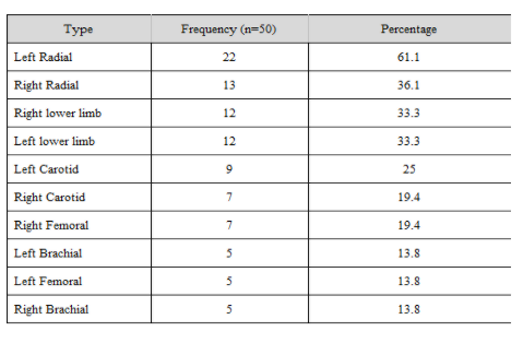
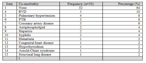
Table 5 shows the presence of co-morbidity in the cohort group. The majority of patients (66% n=32) had no co-morbidity.
There were 3 cases of active proven pulmonary tuberculosis (PTB) (6% n=3), two of whom were diagnosed on sputum testing and a third from bronchial washings. pulmonary hypertension was present in four patients. One patient had documented chronic thromboembolism and two had DCMO. The fourth patient had hypertensive heart disease.
One patient had interstitial lung disease but did not have pulmonary hypertension. Two patients had antiphospholipid antibody syndrome (APS), one of which had left carotid thrombosis and other one had cerebrovascular disease (2 strokes).
Five patients had retroviral disease (RVD). Two patients had chronic Hepatitis B, one patient had Arnold Chiari type1 with no syrinx; one patient had congenital heart disease; one patient had hypothyroidism, one had ureteric fistula, one patient had persistent haematuria (of unknown cause) and two patients had secondary syphilis.
In the current study, the new classification for TA was used as shown in Figure 2 below.
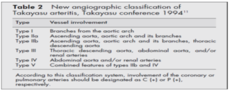
In our cohort, the majority of patients met Type V (38% n=19) of the ACR criteria. Type I was found in nine patients (18% n=9), Type III was found in seven patients (14% n=7), Type IIa and IIb were found in two patients, respectively.
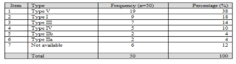
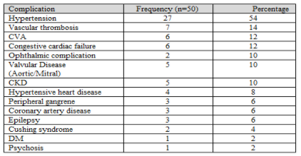
Table 9 shows complications related to disease and treatment. The majority of patients had HPT (54% n=27) which includes the earlier manifestation and subsequent complication with six patients reporting CVA (12% n=6).
Left Heart Failure was present in 6 patients. In four of these patients, this was assessed as being the result of severe hypertension and the other two was labeled “idiopathic”.
Three of the patients of our total cohort suffered a stroke subsequently .Chronic kidney disease was present in 5 patients (10%) and was thought to be mainly due to bilateral renal artery stenosis and severe hypertension.
Complication which occurred in 2 patients included optic nerve atrophy and glaucoma in another case. Valvular heart complications were present in 5 cases (10%) in the form of mitral regurgitation in two patients and aortic regurgitation in another 3 patients.
The vascular complications (arterial or venous thrombosis) occurred in seven patients (14% n=7). Pulmonary embolism was found in two patients with no obvious underlying venous thrombosis. Two of the patients had jugular vein thrombosis and the remaining were arterial in nature. Two patients had thromboembolic to the coronary vessels one had an intramural thrombus in the left ventricle (patient with DCMO).
Chronic kidney disease was found in (10%) of our patients where latest creatinine ranged from 140-180 umol/L.
Three patients (6% n=3) had coronary artery disease (CAD), the aetiology of which may be multifactorial. As mentioned, two patients had thromboemboli from the left ventricle and a third had diffuse atherosclerotic disease. A similar percentage of (6% n=3) was preset in patients who had peripheral vascular ischemia with evidence of gangrene in lower limbs and in the mesenteric vasculature. Steroid induced Cushing syndrome was present in two patients (4% n=2).
A total of 34 patients were tested for HIV (68%) in our cohort. Almost 15% (14.7%) of the patients who were tested were positive, thus the majority (85%) of the patients who were tested were negative.
Among our cohort, 20 patients (40%) were tested for TB. The majority (85%) of the tested population were negative, while 3 patients (15%) tested positive. It is important to note TB testing was based on clinical suspicion.
The majority (88% n=44) of the patients in this cohort had CTA and PET SCAN (88% n=44) each. PET SCAN is sometimes repeated as a follow up in detecting evidence of disease flare - 39 patients had both CTA and PET and 4 had only CTA). Half of patients (50% n=25) had Doppler, which is usually used as the initial diagnostic tool to confirm vasculature abnormality. In this cohort study, Doppler was frequently used in (50%) of patient to assess the renal and carotids. Only ten patients (20% n=10) had an MRI.

Angiographic (CTA) findings
The majority (58%) had stenosis, followed by stenosis and aneurysm in (32%) as shown in the table below.


Different types of options for treatment are available to TA patients depending on the course of the disease. The majority of patients (60% n=30) were treated with a combination of treatments including methotrexate or azathioprine with steroids.
Ten patients (20%) reported not being on treatment, due to “burnt-out” disease or patients did not have active disease at the time. Two patients received biologics; one received (Infliximab) and the other received tocilizumab.
Four patients (8% n=4) were treated with steroids only (Prednisone).
The efficacy of each treatment was not included in our study

Surgical interventions
Some patients had surgical intervention to treat complications arising from the disease. Twelve patients (24% n=12) had renal surgical intervention which included four of them undergoing autotransplantation. Five patients had renal artery graft, two patients had nephrectomy and one had kidney transplant. Furthermore, five patients (10% n=5) had aortic complications requiring surgical intervention, which included repair of AAA and arch of the aorta. Lastly, one patient case required repair of root of the aorta, which included aortic valve replacement. Two of patients required amputation, one had one toe amputated and the other two left fingers amputated.

Two patients (4%) died which both as a result of massive strokes.
Demographics
In terms of age distribution, our findings report a mean age of 28.2 years (16-56) years which is consistent with the Brazilian study (27 years) [19], the one Tunisian study (29.1) [15] and Groote Schuur series (25 years). [4] Higher mean ages were noted in the other Tunisian study (33.2 years) [16] and Turkish series (40 years). [18] In terms of ethnic distribution, our findings show that TA was predominantly found among the mixed-race group, which is similar to that of the GSH study. [21] The reason is that the Western Cape has a predominantly mixed-race population which explains the difference to the Gauteng series, where the majority of the population are of African ethnicity (black). [12] Gender distribution in this study among TA patients is supported by similar studies confirming the predominant female involvement (82%) and is consistent with other series by GSH (75%) [21], Turkish study (91.9%) [18], Tunisian study (88.9%) [15] and the Brazilian study [19]
Hypertension
Hypertension was the most common presentation in TA patients. Over half of patients in this series (54%) presented with hypertension and is consistent with the with GSH series (77%) [21], Turkish series (43%) [18] and Greek series (78.0%). [4] The pathogenesis of the hypertension is postulated as being due to renal artery stenosis in most cases or “coarctation of the aorta” in other cases. It should be noted that the majority of the patients had category Type V (38%) which affects the ascending aorta, aortic arch and its branches, and thoracic descending aorta as well as abdominal aorta and/or renal artery.
Theoretically, patients with TA have suppressed immunity. This is thought to be due to the immune mediated nature of the disease as well as the treatment with steroids and other immunosuppressive therapy.
In our cohort, 10% of our patients tested positive for TB. However, although all patients were subjected to an appropriate history-taking and had a chest radiograph, extensive testing for TB was not conducted on those patients assessed as not likely to have TB. Thus, the TB status of the majority (60%) was not known. Definitive exclusion of TB was also not conducted in other studies. However, a low prevalence of TB of only 6% (n=3) is unexpectedly lower in comparison to other studies. This is of particular significance considering the fact that patients in our cohort hail from a predominantly poor and disadvantaged communities where there is a pandemic of TB.
A more direct comparison can be made to the GSH study where the proportion of TA patients diagnosed with TB was as high as 20%. [21] A potential problem with the diagnosis of TB in this series was that a strongly positive Mantoux was also regarded as evidence of “active” TB. Currently routine Mantoux screening as evidence of TB is not the norm. In terms of TB prevalence, the Korean series reported prevalence of 17%, Turkish series (6.4%) [1] and the Brazilian study 41.0%.[19] Generally a high percentage of TB (up to 90%) has been reported in pediatric series including the South African study.
Although lymphadenopathy is a well-documented finding in patients with TA, the presence of large mediastinal nodes mimicking TB is rare. Current literature reports isolated cases of “massive” lymphadenopathy mimicking TB. In our cohort, six patients (12% n=6) had significant lymphadenopathy. In these cases; however, it is mandatory to exclude TB. Although one patient clearly had TB, evidence of TB for the remainder of the population was not available. It may be suggested that previous cases of severe lymphadenopathy due to TA, may have been regarded as being due to TB and in some cases, were subjected to anti-TB treatment. This is the first study that documents significant lymphadenopathy in 10% of the cases of TA.
Two patients in this study had internal jugular vein thrombosis and two had the CTA proven pulmonary embolism. However, there was no obvious site of venous thrombosis evident in the patients with pulmonary embolism. As TA is a disease affecting large arteries, venous thrombosis did not form part of the clinical features of this condition. Internal jugular vein thrombosis is also very rare, but 2 cases were evidenced in our cohort. Furthermore, isolated case reports relating to venous thrombosis and one reporting on internal jugular vein thrombosis have been noted. [5]
Two female patients of our cohort had antiphospholipid syndrome increasing the risk of venous and arterial thrombosis. The rarity is clear because the literature search revealed the association of TA with antiphospholipid syndrome as 2 case reports, one from Korea in which an elderly female was affected [5], and in the other a young Japanese man (17 years old). [6]
Cardiomyopathy:
Although 6 patients presented with heart failure, two of the cases did not have a clear cause such as severe hypertension or ischaemic heart disease. We feel that it is reasonable to assume that these cases were due to the rare entity of TA associated cardiomyopathy. Furthermore, there was no data exploring this topic, only a case report of TA presented with heart failure. [6]
In this study only 34 of the 50 patients were tested for HIV. HIV testing was not routinely done but reserved for those with underlying clinical suspicion of this condition. Thus, just under 15% of those tested were positive for HIV making it very difficult to draw any meaningful conclusions on the effect of this condition. From the limited numbers who were HIV positive, it does not appear that this condition presented differently in this sub group.
In our patients the commonest vessel affected was the subclavian artery (42%) and is consistent the Gauteng pediatric study (52.2%) and the Tunisian series. In the GSH series, 70% involved the entire aorta. [21] Our study showed that most TA patients had Type V (32%), which mirrors the findings of the Brazilian series (52%). [19]
CT angiogram has been and remains the imaging method of choice for the diagnosis of TA. Most of the patients had Type V disease, which was also the case in the GSH study. PET CT scanning has been shown to be a good adjunctive method of imaging in cases where there may be uncertainty on whether there is active vasculitis in the patient or not. The radiographic findings characteristic on CTA, namely stenosis and aneurysm formation, do not necessarily imply active disease. In this study, 80% of the patients had PET-CT scanning done, some of whom had more than one. This number is much larger than that which have reported previously and it appears that this may become a routine part of the follow up if there is a need to determine “active” TA.
In this study only 2 patients (4%) died. This represents a low mortality compared to previous reports and is a much lower than the mortality than that reported from the GSH of 20%.[21] This may reflect earlier and improved diagnosis of milder cases which may have been missed in the past or, may be due to alternative therapies which were not available previously (see below). [17]
Largely because of limitation of state resources, only two patients received biologic therapy. One patient received infliximab and the other patient received infliximab which caused reactivation of TB. She subsequently received tocilizumab and is currently still on this treatment.
Conclusion
In summary this study revealed gender and age characteristics similar to previous studies, confirming that TA affects mainly young females. The mortality rate of 6% in this cohort is significantly lower than that reported in previous series.
Some of the unexpected findings in this study was the significantly lower percentage of active TB. This study also revealed 6 cases of “massive” chest lymphadenopathy only one of which was due to TB. It may well be that some of the TB cases reported in previous studies may have been over-diagnosed.
This study also revealed two (2) unusual cases of venous thrombosis - supporting isolated previous reports of venous thrombosis.
Another previously documented but rare finding in TA was that there were 2 cases of cardiomyopathy in our cohort.
A prospective study involving larger numbers from different institutions may shed some light on these intriguing findings.
Clearly Auctoresonline and particularly Psychology and Mental Health Care Journal is dedicated to improving health care services for individuals and populations. The editorial boards' ability to efficiently recognize and share the global importance of health literacy with a variety of stakeholders. Auctoresonline publishing platform can be used to facilitate of optimal client-based services and should be added to health care professionals' repertoire of evidence-based health care resources.

Journal of Clinical Cardiology and Cardiovascular Intervention The submission and review process was adequate. However I think that the publication total value should have been enlightened in early fases. Thank you for all.

Journal of Women Health Care and Issues By the present mail, I want to say thank to you and tour colleagues for facilitating my published article. Specially thank you for the peer review process, support from the editorial office. I appreciate positively the quality of your journal.

Journal of Clinical Research and Reports I would be very delighted to submit my testimonial regarding the reviewer board and the editorial office. The reviewer board were accurate and helpful regarding any modifications for my manuscript. And the editorial office were very helpful and supportive in contacting and monitoring with any update and offering help. It was my pleasure to contribute with your promising Journal and I am looking forward for more collaboration.

We would like to thank the Journal of Thoracic Disease and Cardiothoracic Surgery because of the services they provided us for our articles. The peer-review process was done in a very excellent time manner, and the opinions of the reviewers helped us to improve our manuscript further. The editorial office had an outstanding correspondence with us and guided us in many ways. During a hard time of the pandemic that is affecting every one of us tremendously, the editorial office helped us make everything easier for publishing scientific work. Hope for a more scientific relationship with your Journal.

The peer-review process which consisted high quality queries on the paper. I did answer six reviewers’ questions and comments before the paper was accepted. The support from the editorial office is excellent.

Journal of Neuroscience and Neurological Surgery. I had the experience of publishing a research article recently. The whole process was simple from submission to publication. The reviewers made specific and valuable recommendations and corrections that improved the quality of my publication. I strongly recommend this Journal.

Dr. Katarzyna Byczkowska My testimonial covering: "The peer review process is quick and effective. The support from the editorial office is very professional and friendly. Quality of the Clinical Cardiology and Cardiovascular Interventions is scientific and publishes ground-breaking research on cardiology that is useful for other professionals in the field.

Thank you most sincerely, with regard to the support you have given in relation to the reviewing process and the processing of my article entitled "Large Cell Neuroendocrine Carcinoma of The Prostate Gland: A Review and Update" for publication in your esteemed Journal, Journal of Cancer Research and Cellular Therapeutics". The editorial team has been very supportive.

Testimony of Journal of Clinical Otorhinolaryngology: work with your Reviews has been a educational and constructive experience. The editorial office were very helpful and supportive. It was a pleasure to contribute to your Journal.

Dr. Bernard Terkimbi Utoo, I am happy to publish my scientific work in Journal of Women Health Care and Issues (JWHCI). The manuscript submission was seamless and peer review process was top notch. I was amazed that 4 reviewers worked on the manuscript which made it a highly technical, standard and excellent quality paper. I appreciate the format and consideration for the APC as well as the speed of publication. It is my pleasure to continue with this scientific relationship with the esteem JWHCI.

This is an acknowledgment for peer reviewers, editorial board of Journal of Clinical Research and Reports. They show a lot of consideration for us as publishers for our research article “Evaluation of the different factors associated with side effects of COVID-19 vaccination on medical students, Mutah university, Al-Karak, Jordan”, in a very professional and easy way. This journal is one of outstanding medical journal.

Dear Hao Jiang, to Journal of Nutrition and Food Processing We greatly appreciate the efficient, professional and rapid processing of our paper by your team. If there is anything else we should do, please do not hesitate to let us know. On behalf of my co-authors, we would like to express our great appreciation to editor and reviewers.

As an author who has recently published in the journal "Brain and Neurological Disorders". I am delighted to provide a testimonial on the peer review process, editorial office support, and the overall quality of the journal. The peer review process at Brain and Neurological Disorders is rigorous and meticulous, ensuring that only high-quality, evidence-based research is published. The reviewers are experts in their fields, and their comments and suggestions were constructive and helped improve the quality of my manuscript. The review process was timely and efficient, with clear communication from the editorial office at each stage. The support from the editorial office was exceptional throughout the entire process. The editorial staff was responsive, professional, and always willing to help. They provided valuable guidance on formatting, structure, and ethical considerations, making the submission process seamless. Moreover, they kept me informed about the status of my manuscript and provided timely updates, which made the process less stressful. The journal Brain and Neurological Disorders is of the highest quality, with a strong focus on publishing cutting-edge research in the field of neurology. The articles published in this journal are well-researched, rigorously peer-reviewed, and written by experts in the field. The journal maintains high standards, ensuring that readers are provided with the most up-to-date and reliable information on brain and neurological disorders. In conclusion, I had a wonderful experience publishing in Brain and Neurological Disorders. The peer review process was thorough, the editorial office provided exceptional support, and the journal's quality is second to none. I would highly recommend this journal to any researcher working in the field of neurology and brain disorders.
Dear Agrippa Hilda, Journal of Neuroscience and Neurological Surgery, Editorial Coordinator, I trust this message finds you well. I want to extend my appreciation for considering my article for publication in your esteemed journal. I am pleased to provide a testimonial regarding the peer review process and the support received from your editorial office. The peer review process for my paper was carried out in a highly professional and thorough manner. The feedback and comments provided by the authors were constructive and very useful in improving the quality of the manuscript. This rigorous assessment process undoubtedly contributes to the high standards maintained by your journal.

International Journal of Clinical Case Reports and Reviews. I strongly recommend to consider submitting your work to this high-quality journal. The support and availability of the Editorial staff is outstanding and the review process was both efficient and rigorous.

Thank you very much for publishing my Research Article titled “Comparing Treatment Outcome Of Allergic Rhinitis Patients After Using Fluticasone Nasal Spray And Nasal Douching" in the Journal of Clinical Otorhinolaryngology. As Medical Professionals we are immensely benefited from study of various informative Articles and Papers published in this high quality Journal. I look forward to enriching my knowledge by regular study of the Journal and contribute my future work in the field of ENT through the Journal for use by the medical fraternity. The support from the Editorial office was excellent and very prompt. I also welcome the comments received from the readers of my Research Article.

Dear Erica Kelsey, Editorial Coordinator of Cancer Research and Cellular Therapeutics Our team is very satisfied with the processing of our paper by your journal. That was fast, efficient, rigorous, but without unnecessary complications. We appreciated the very short time between the submission of the paper and its publication on line on your site.

I am very glad to say that the peer review process is very successful and fast and support from the Editorial Office. Therefore, I would like to continue our scientific relationship for a long time. And I especially thank you for your kindly attention towards my article. Have a good day!

"We recently published an article entitled “Influence of beta-Cyclodextrins upon the Degradation of Carbofuran Derivatives under Alkaline Conditions" in the Journal of “Pesticides and Biofertilizers” to show that the cyclodextrins protect the carbamates increasing their half-life time in the presence of basic conditions This will be very helpful to understand carbofuran behaviour in the analytical, agro-environmental and food areas. We greatly appreciated the interaction with the editor and the editorial team; we were particularly well accompanied during the course of the revision process, since all various steps towards publication were short and without delay".

I would like to express my gratitude towards you process of article review and submission. I found this to be very fair and expedient. Your follow up has been excellent. I have many publications in national and international journal and your process has been one of the best so far. Keep up the great work.

We are grateful for this opportunity to provide a glowing recommendation to the Journal of Psychiatry and Psychotherapy. We found that the editorial team were very supportive, helpful, kept us abreast of timelines and over all very professional in nature. The peer review process was rigorous, efficient and constructive that really enhanced our article submission. The experience with this journal remains one of our best ever and we look forward to providing future submissions in the near future.

I am very pleased to serve as EBM of the journal, I hope many years of my experience in stem cells can help the journal from one way or another. As we know, stem cells hold great potential for regenerative medicine, which are mostly used to promote the repair response of diseased, dysfunctional or injured tissue using stem cells or their derivatives. I think Stem Cell Research and Therapeutics International is a great platform to publish and share the understanding towards the biology and translational or clinical application of stem cells.

I would like to give my testimony in the support I have got by the peer review process and to support the editorial office where they were of asset to support young author like me to be encouraged to publish their work in your respected journal and globalize and share knowledge across the globe. I really give my great gratitude to your journal and the peer review including the editorial office.

I am delighted to publish our manuscript entitled "A Perspective on Cocaine Induced Stroke - Its Mechanisms and Management" in the Journal of Neuroscience and Neurological Surgery. The peer review process, support from the editorial office, and quality of the journal are excellent. The manuscripts published are of high quality and of excellent scientific value. I recommend this journal very much to colleagues.

Dr.Tania Muñoz, My experience as researcher and author of a review article in The Journal Clinical Cardiology and Interventions has been very enriching and stimulating. The editorial team is excellent, performs its work with absolute responsibility and delivery. They are proactive, dynamic and receptive to all proposals. Supporting at all times the vast universe of authors who choose them as an option for publication. The team of review specialists, members of the editorial board, are brilliant professionals, with remarkable performance in medical research and scientific methodology. Together they form a frontline team that consolidates the JCCI as a magnificent option for the publication and review of high-level medical articles and broad collective interest. I am honored to be able to share my review article and open to receive all your comments.

“The peer review process of JPMHC is quick and effective. Authors are benefited by good and professional reviewers with huge experience in the field of psychology and mental health. The support from the editorial office is very professional. People to contact to are friendly and happy to help and assist any query authors might have. Quality of the Journal is scientific and publishes ground-breaking research on mental health that is useful for other professionals in the field”.

Dear editorial department: On behalf of our team, I hereby certify the reliability and superiority of the International Journal of Clinical Case Reports and Reviews in the peer review process, editorial support, and journal quality. Firstly, the peer review process of the International Journal of Clinical Case Reports and Reviews is rigorous, fair, transparent, fast, and of high quality. The editorial department invites experts from relevant fields as anonymous reviewers to review all submitted manuscripts. These experts have rich academic backgrounds and experience, and can accurately evaluate the academic quality, originality, and suitability of manuscripts. The editorial department is committed to ensuring the rigor of the peer review process, while also making every effort to ensure a fast review cycle to meet the needs of authors and the academic community. Secondly, the editorial team of the International Journal of Clinical Case Reports and Reviews is composed of a group of senior scholars and professionals with rich experience and professional knowledge in related fields. The editorial department is committed to assisting authors in improving their manuscripts, ensuring their academic accuracy, clarity, and completeness. Editors actively collaborate with authors, providing useful suggestions and feedback to promote the improvement and development of the manuscript. We believe that the support of the editorial department is one of the key factors in ensuring the quality of the journal. Finally, the International Journal of Clinical Case Reports and Reviews is renowned for its high- quality articles and strict academic standards. The editorial department is committed to publishing innovative and academically valuable research results to promote the development and progress of related fields. The International Journal of Clinical Case Reports and Reviews is reasonably priced and ensures excellent service and quality ratio, allowing authors to obtain high-level academic publishing opportunities in an affordable manner. I hereby solemnly declare that the International Journal of Clinical Case Reports and Reviews has a high level of credibility and superiority in terms of peer review process, editorial support, reasonable fees, and journal quality. Sincerely, Rui Tao.

Clinical Cardiology and Cardiovascular Interventions I testity the covering of the peer review process, support from the editorial office, and quality of the journal.

Clinical Cardiology and Cardiovascular Interventions, we deeply appreciate the interest shown in our work and its publication. It has been a true pleasure to collaborate with you. The peer review process, as well as the support provided by the editorial office, have been exceptional, and the quality of the journal is very high, which was a determining factor in our decision to publish with you.

The peer reviewers process is quick and effective, the supports from editorial office is excellent, the quality of journal is high. I would like to collabroate with Internatioanl journal of Clinical Case Reports and Reviews journal clinically in the future time.

Clinical Cardiology and Cardiovascular Interventions, I would like to express my sincerest gratitude for the trust placed in our team for the publication in your journal. It has been a true pleasure to collaborate with you on this project. I am pleased to inform you that both the peer review process and the attention from the editorial coordination have been excellent. Your team has worked with dedication and professionalism to ensure that your publication meets the highest standards of quality. We are confident that this collaboration will result in mutual success, and we are eager to see the fruits of this shared effort.

Dear Dr. Jessica Magne, Editorial Coordinator 0f Clinical Cardiology and Cardiovascular Interventions, I hope this message finds you well. I want to express my utmost gratitude for your excellent work and for the dedication and speed in the publication process of my article titled "Navigating Innovation: Qualitative Insights on Using Technology for Health Education in Acute Coronary Syndrome Patients." I am very satisfied with the peer review process, the support from the editorial office, and the quality of the journal. I hope we can maintain our scientific relationship in the long term.

Dear Monica Gissare, - Editorial Coordinator of Nutrition and Food Processing. ¨My testimony with you is truly professional, with a positive response regarding the follow-up of the article and its review, you took into account my qualities and the importance of the topic¨.

Dear Dr. Jessica Magne, Editorial Coordinator 0f Clinical Cardiology and Cardiovascular Interventions, The review process for the article “The Handling of Anti-aggregants and Anticoagulants in the Oncologic Heart Patient Submitted to Surgery” was extremely rigorous and detailed. From the initial submission to the final acceptance, the editorial team at the “Journal of Clinical Cardiology and Cardiovascular Interventions” demonstrated a high level of professionalism and dedication. The reviewers provided constructive and detailed feedback, which was essential for improving the quality of our work. Communication was always clear and efficient, ensuring that all our questions were promptly addressed. The quality of the “Journal of Clinical Cardiology and Cardiovascular Interventions” is undeniable. It is a peer-reviewed, open-access publication dedicated exclusively to disseminating high-quality research in the field of clinical cardiology and cardiovascular interventions. The journal's impact factor is currently under evaluation, and it is indexed in reputable databases, which further reinforces its credibility and relevance in the scientific field. I highly recommend this journal to researchers looking for a reputable platform to publish their studies.

Dear Editorial Coordinator of the Journal of Nutrition and Food Processing! "I would like to thank the Journal of Nutrition and Food Processing for including and publishing my article. The peer review process was very quick, movement and precise. The Editorial Board has done an extremely conscientious job with much help, valuable comments and advices. I find the journal very valuable from a professional point of view, thank you very much for allowing me to be part of it and I would like to participate in the future!”

Dealing with The Journal of Neurology and Neurological Surgery was very smooth and comprehensive. The office staff took time to address my needs and the response from editors and the office was prompt and fair. I certainly hope to publish with this journal again.Their professionalism is apparent and more than satisfactory. Susan Weiner

My Testimonial Covering as fellowing: Lin-Show Chin. The peer reviewers process is quick and effective, the supports from editorial office is excellent, the quality of journal is high. I would like to collabroate with Internatioanl journal of Clinical Case Reports and Reviews.

My experience publishing in Psychology and Mental Health Care was exceptional. The peer review process was rigorous and constructive, with reviewers providing valuable insights that helped enhance the quality of our work. The editorial team was highly supportive and responsive, making the submission process smooth and efficient. The journal's commitment to high standards and academic rigor makes it a respected platform for quality research. I am grateful for the opportunity to publish in such a reputable journal.

My experience publishing in International Journal of Clinical Case Reports and Reviews was exceptional. I Come forth to Provide a Testimonial Covering the Peer Review Process and the editorial office for the Professional and Impartial Evaluation of the Manuscript.

I would like to offer my testimony in the support. I have received through the peer review process and support the editorial office where they are to support young authors like me, encourage them to publish their work in your esteemed journals, and globalize and share knowledge globally. I really appreciate your journal, peer review, and editorial office.

Dear Agrippa Hilda- Editorial Coordinator of Journal of Neuroscience and Neurological Surgery, "The peer review process was very quick and of high quality, which can also be seen in the articles in the journal. The collaboration with the editorial office was very good."

I would like to express my sincere gratitude for the support and efficiency provided by the editorial office throughout the publication process of my article, “Delayed Vulvar Metastases from Rectal Carcinoma: A Case Report.” I greatly appreciate the assistance and guidance I received from your team, which made the entire process smooth and efficient. The peer review process was thorough and constructive, contributing to the overall quality of the final article. I am very grateful for the high level of professionalism and commitment shown by the editorial staff, and I look forward to maintaining a long-term collaboration with the International Journal of Clinical Case Reports and Reviews.

To Dear Erin Aust, I would like to express my heartfelt appreciation for the opportunity to have my work published in this esteemed journal. The entire publication process was smooth and well-organized, and I am extremely satisfied with the final result. The Editorial Team demonstrated the utmost professionalism, providing prompt and insightful feedback throughout the review process. Their clear communication and constructive suggestions were invaluable in enhancing my manuscript, and their meticulous attention to detail and dedication to quality are truly commendable. Additionally, the support from the Editorial Office was exceptional. From the initial submission to the final publication, I was guided through every step of the process with great care and professionalism. The team's responsiveness and assistance made the entire experience both easy and stress-free. I am also deeply impressed by the quality and reputation of the journal. It is an honor to have my research featured in such a respected publication, and I am confident that it will make a meaningful contribution to the field.

"I am grateful for the opportunity of contributing to [International Journal of Clinical Case Reports and Reviews] and for the rigorous review process that enhances the quality of research published in your esteemed journal. I sincerely appreciate the time and effort of your team who have dedicatedly helped me in improvising changes and modifying my manuscript. The insightful comments and constructive feedback provided have been invaluable in refining and strengthening my work".

I thank the ‘Journal of Clinical Research and Reports’ for accepting this article for publication. This is a rigorously peer reviewed journal which is on all major global scientific data bases. I note the review process was prompt, thorough and professionally critical. It gave us an insight into a number of important scientific/statistical issues. The review prompted us to review the relevant literature again and look at the limitations of the study. The peer reviewers were open, clear in the instructions and the editorial team was very prompt in their communication. This journal certainly publishes quality research articles. I would recommend the journal for any future publications.

Dear Jessica Magne, with gratitude for the joint work. Fast process of receiving and processing the submitted scientific materials in “Clinical Cardiology and Cardiovascular Interventions”. High level of competence of the editors with clear and correct recommendations and ideas for enriching the article.

We found the peer review process quick and positive in its input. The support from the editorial officer has been very agile, always with the intention of improving the article and taking into account our subsequent corrections.

My article, titled 'No Way Out of the Smartphone Epidemic Without Considering the Insights of Brain Research,' has been republished in the International Journal of Clinical Case Reports and Reviews. The review process was seamless and professional, with the editors being both friendly and supportive. I am deeply grateful for their efforts.

To Dear Erin Aust – Editorial Coordinator of Journal of General Medicine and Clinical Practice! I declare that I am absolutely satisfied with your work carried out with great competence in following the manuscript during the various stages from its receipt, during the revision process to the final acceptance for publication. Thank Prof. Elvira Farina

Dear Jessica, and the super professional team of the ‘Clinical Cardiology and Cardiovascular Interventions’ I am sincerely grateful to the coordinated work of the journal team for the no problem with the submission of my manuscript: “Cardiometabolic Disorders in A Pregnant Woman with Severe Preeclampsia on the Background of Morbid Obesity (Case Report).” The review process by 5 experts was fast, and the comments were professional, which made it more specific and academic, and the process of publication and presentation of the article was excellent. I recommend that my colleagues publish articles in this journal, and I am interested in further scientific cooperation. Sincerely and best wishes, Dr. Oleg Golyanovskiy.

Dear Ashley Rosa, Editorial Coordinator of the journal - Psychology and Mental Health Care. " The process of obtaining publication of my article in the Psychology and Mental Health Journal was positive in all areas. The peer review process resulted in a number of valuable comments, the editorial process was collaborative and timely, and the quality of this journal has been quickly noticed, resulting in alternative journals contacting me to publish with them." Warm regards, Susan Anne Smith, PhD. Australian Breastfeeding Association.

Dear Jessica Magne, Editorial Coordinator, Clinical Cardiology and Cardiovascular Interventions, Auctores Publishing LLC. I appreciate the journal (JCCI) editorial office support, the entire team leads were always ready to help, not only on technical front but also on thorough process. Also, I should thank dear reviewers’ attention to detail and creative approach to teach me and bring new insights by their comments. Surely, more discussions and introduction of other hemodynamic devices would provide better prevention and management of shock states. Your efforts and dedication in presenting educational materials in this journal are commendable. Best wishes from, Farahnaz Fallahian.

Dear Maria Emerson, Editorial Coordinator, International Journal of Clinical Case Reports and Reviews, Auctores Publishing LLC. I am delighted to have published our manuscript, "Acute Colonic Pseudo-Obstruction (ACPO): A rare but serious complication following caesarean section." I want to thank the editorial team, especially Maria Emerson, for their prompt review of the manuscript, quick responses to queries, and overall support. Yours sincerely Dr. Victor Olagundoye.

Dear Ashley Rosa, Editorial Coordinator, International Journal of Clinical Case Reports and Reviews. Many thanks for publishing this manuscript after I lost confidence the editors were most helpful, more than other journals Best wishes from, Susan Anne Smith, PhD. Australian Breastfeeding Association.

Dear Agrippa Hilda, Editorial Coordinator, Journal of Neuroscience and Neurological Surgery. The entire process including article submission, review, revision, and publication was extremely easy. The journal editor was prompt and helpful, and the reviewers contributed to the quality of the paper. Thank you so much! Eric Nussbaum, MD

Dr Hala Al Shaikh This is to acknowledge that the peer review process for the article ’ A Novel Gnrh1 Gene Mutation in Four Omani Male Siblings, Presentation and Management ’ sent to the International Journal of Clinical Case Reports and Reviews was quick and smooth. The editorial office was prompt with easy communication.

Dear Erin Aust, Editorial Coordinator, Journal of General Medicine and Clinical Practice. We are pleased to share our experience with the “Journal of General Medicine and Clinical Practice”, following the successful publication of our article. The peer review process was thorough and constructive, helping to improve the clarity and quality of the manuscript. We are especially thankful to Ms. Erin Aust, the Editorial Coordinator, for her prompt communication and continuous support throughout the process. Her professionalism ensured a smooth and efficient publication experience. The journal upholds high editorial standards, and we highly recommend it to fellow researchers seeking a credible platform for their work. Best wishes By, Dr. Rakhi Mishra.

Dear Jessica Magne, Editorial Coordinator, Clinical Cardiology and Cardiovascular Interventions, Auctores Publishing LLC. The peer review process of the journal of Clinical Cardiology and Cardiovascular Interventions was excellent and fast, as was the support of the editorial office and the quality of the journal. Kind regards Walter F. Riesen Prof. Dr. Dr. h.c. Walter F. Riesen.

Dear Ashley Rosa, Editorial Coordinator, International Journal of Clinical Case Reports and Reviews, Auctores Publishing LLC. Thank you for publishing our article, Exploring Clozapine's Efficacy in Managing Aggression: A Multiple Single-Case Study in Forensic Psychiatry in the international journal of clinical case reports and reviews. We found the peer review process very professional and efficient. The comments were constructive, and the whole process was efficient. On behalf of the co-authors, I would like to thank you for publishing this article. With regards, Dr. Jelle R. Lettinga.

Dear Clarissa Eric, Editorial Coordinator, Journal of Clinical Case Reports and Studies, I would like to express my deep admiration for the exceptional professionalism demonstrated by your journal. I am thoroughly impressed by the speed of the editorial process, the substantive and insightful reviews, and the meticulous preparation of the manuscript for publication. Additionally, I greatly appreciate the courteous and immediate responses from your editorial office to all my inquiries. Best Regards, Dariusz Ziora

Dear Chrystine Mejia, Editorial Coordinator, Journal of Neurodegeneration and Neurorehabilitation, Auctores Publishing LLC, We would like to thank the editorial team for the smooth and high-quality communication leading up to the publication of our article in the Journal of Neurodegeneration and Neurorehabilitation. The reviewers have extensive knowledge in the field, and their relevant questions helped to add value to our publication. Kind regards, Dr. Ravi Shrivastava.

Dear Clarissa Eric, Editorial Coordinator, Journal of Clinical Case Reports and Studies, Auctores Publishing LLC, USA Office: +1-(302)-520-2644. I would like to express my sincere appreciation for the efficient and professional handling of my case report by the ‘Journal of Clinical Case Reports and Studies’. The peer review process was not only fast but also highly constructive—the reviewers’ comments were clear, relevant, and greatly helped me improve the quality and clarity of my manuscript. I also received excellent support from the editorial office throughout the process. Communication was smooth and timely, and I felt well guided at every stage, from submission to publication. The overall quality and rigor of the journal are truly commendable. I am pleased to have published my work with Journal of Clinical Case Reports and Studies, and I look forward to future opportunities for collaboration. Sincerely, Aline Tollet, UCLouvain.

Dear Ms. Mayra Duenas, Editorial Coordinator, International Journal of Clinical Case Reports and Reviews. “The International Journal of Clinical Case Reports and Reviews represented the “ideal house” to share with the research community a first experience with the use of the Simeox device for speech rehabilitation. High scientific reputation and attractive website communication were first determinants for the selection of this Journal, and the following submission process exceeded expectations: fast but highly professional peer review, great support by the editorial office, elegant graphic layout. Exactly what a dynamic research team - also composed by allied professionals - needs!" From, Chiara Beccaluva, PT - Italy.

Dear Maria Emerson, Editorial Coordinator, we have deeply appreciated the professionalism demonstrated by the International Journal of Clinical Case Reports and Reviews. The reviewers have extensive knowledge of our field and have been very efficient and fast in supporting the process. I am really looking forward to further collaboration. Thanks. Best regards, Dr. Claudio Ligresti

Dear Chrystine Mejia, Editorial Coordinator, Journal of Neurodegeneration and Neurorehabilitation. “The peer review process was efficient and constructive, and the editorial office provided excellent communication and support throughout. The journal ensures scientific rigor and high editorial standards, while also offering a smooth and timely publication process. We sincerely appreciate the work of the editorial team in facilitating the dissemination of innovative approaches such as the Bonori Method.” Best regards, Dr. Giselle Pentón-Rol.
