AUCTORES
Globalize your Research

Research Article | DOI: https://doi.org/10.31579/2641-0419/138
1Na Homolce Hospital, Prague, Czech Republic,
2Medtronic Minneapolis USA,
3 Icahn School of Medicine at Mount Sinai, New York, New York, USA
*Corresponding Author: Kentaro Minami, Na Homolce Hospital, Roentgenova 37/2, 15030, Praha 5, Czech Republic.
Citation: Kentaro Minami, Jan Petrů, Milan Chovanec, Jan Sorf, Pavel Hála, (2021) Late Retrieval of the Leadless Micra Transcatheter Pacemaker System. J. Clinical Cardiology and Cardiovascular Interventions, 4(6); Doi:10.31579/2641-0419/138
Copyright: © 2021 Kentaro Minami, This is an open-access article distributed under the terms of the Creative Commons Attribution License, which permits unrestricted use, distribution, and reproduction in any medium, provided the original author and source are credited.
Received: 02 February 2021 | Accepted: 11 March 2021 | Published: 17 March 2021
Keywords: leadless cardiac pacemaker; pacemaker retrieval; micra transcatheter pacing system
Aim: We report our single-center experience with the retrieval and replacement of the chronically implanted Micra transcatheter pacing system (TPS).
Material and Methods: We included 6 patients with an implanted the Micra TPSs who subsequently underwent transvenous method of retrieval at our institution. The indication for device retrieval was pacemaker syndrome in two patients, battery depletion in three patients, and need for upgrade to biventricular pacing in one patient.
Results: After an implantation duration of 555 ± 373 days, the overall retrieval success rate was 83.3%, 5 of 6 patients. No procedure-related adverse device events occurred. In the single patient with unsuccessful retrieval, intracardiac echocardiography revealed that the Micra TPS was embedded within the cardiac wall and surrounding tissue. After retrieval, four patients received a new Micra TPS.
Conclusions: Late retrieval of an implanted Micra TPS was safe and feasible, which indicates the possibility for their safe and elective replacement with a new leadless pacing device.
Short title: Micra Transcatheter Pacemaker Retrieval
ICE = intracardiac echocardiography
LCP = leadless cardiac pacemaker
RAO = right anterior oblique
Leadless cardiac pacemakers (LCPs) are safe, effective alternatives to conventional transvenous pacemakers for patients who require single-chamber ventricular pacing [1,2]. The Micra transcatheter pacing system (TPS) (Medtronic, Minneapolis, MN, USA), which is currently the only commercially available LCP for clinical practice, exhibited a high implantation success rate and stable pacing performance in global prospective studies [3].Despite these promising results, the ability to retrieve a long-standing implanted TPS may be a useful management strategy in certain scenarios. With an alternative LCP of different size and shape, it has been demonstrated that retrievability is possible even after implant durations of up to 6 years, with success rates ranging between 80-90% [4-6]. However, that LCP was removed from clinical use because of a battery dysfunction, and its longer and thinner profile (than the TPS device) precludes one from extrapolating from this experience to the feasibility, safety or efficacy of TPS retrievability. Although some successful retrieval experiences in the acute phase have been reported for the TPS device, retrieval of a long-standing implanted TPS devices has not been well-studied [7]. Therefore, we report our single-center clinical experiences regarding the retrieval of chronically implanted Micra TPS.
Materials and Methods
The present study included six patients (2 females) who underwent active fixation of the Micra TPS to the right ventricle (RV) in our institution between September 2014 and June 2018. The Micra TPS implant technique has been previously described. [1] Demographic data were obtained from the medical records. The study protocol was approved by the Institution Research and Ethics Committee of Na Homolce Hospital and was carried out in accordance with the Declaration of Helsinki. The basic principle regarding the possibility of retrieval or abandon of the Micra TPS was clearly explained to all the patients. Only the patients who strongly desired to be performed the retrieval of the implanted old Micra TPS were enrolled in this study. Written informed consent was obtained from each patient.
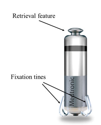
It has four self-expanding tines which are designed to provide a robust holding force within the cardiac tissue of right ventricular (RV). The tines are separate from the pacing electrode, which allows stable pacing performance and avoids cardiac tissue injury. The device was designed for acute retrieval capability: the proximal portion of the TPS contains a retrieve feature that can be snared, and the operators must engage the proximal retrieve feature via the snare.
All retrieval procedures were performed by two experienced operators. The retrieval catheter system was inserted via the right femoral vein accessed using the 23 Fr sheath employed for TPS insertion. Since there is no dedicated system for TPS retrieval, we used the regular delivery catheter system for TPS implantation. Prior to retrieval, a contrast agent was injected through a pigtail catheter to determine the LCP position. An intracardiac echocardiography (ICE) catheter was advanced to the right atrium using a left femoral venous approaching during all retrieval procedures to identify LCP movement, tricuspid valve damage, and the presence of pericardial effusion. For pacemaker-dependent patients, a temporary pacing lead was placed within the RV through the left femoral vein during the retrieval procedure.
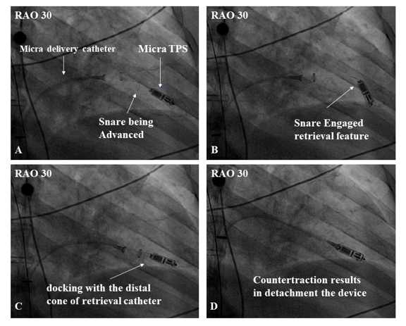
A single-loop 7 mm snare wire (Amplatz Goose Neck Microsnare, ev3Inc., Plymouth, MN, USA) was inserted through the central lumen of the TPS delivery catheter, which in turn was subsequently introduced through the 23 Fr sheath. This “retrieval system” was advanced under fluoroscopy and ICE to the junction of the inferior vena cava and right atrium. The system’s distal cone was manoeuvred into the RV to the proximal aspect of the device. The snare was advanced and deployed around the proximal retrieval feature of the TPS. After confirming coaxial alignment between the snare and the retrieval feature with multi-plane fluoroscopy, the snare was closed and locked around the retrieval feature. The snare loop was then tightened to hold the device firmly, and tension was applied, along with countertraction force from the distal cone, to release the tines from the myocardium, thereby allowing withdrawal of the device into the distal cone. The delivery catheter and Micra TPS were then withdrawn into the introducer sheath and removed from the body (Figure 2, Supplementary Online Videos A B, and C).
All patients received a new pacing device immediately after the retrieval procedure. When a new TPS was implanted after retrieval, it was generally positioned at the apical septum, slightly above the initial location of the explanted TPS, to avoid reimplantation into any fibrous tissue relate to the old device.
Patient characteristics are shown in Table 1.

The mean duration time from implantation to retrieval was 554 ± 373 days (range, 280 to 1219 days). The indication for device retrieval included pacemaker syndrome in two patients, battery depletion due to high pacing threshold and complete dependence of ventricular pacing in three patients, and need for upgrade to biventricular pacing due to the development of low left ventricular ejection fraction from continuous ventricular pacing in one patient.
Successful retrieval, defined as complete removal of the LCP, was achieved in 5 patients (83%). The mean procedural fluoroscopy time was 19 ± 16 min. In all five successful retrieval cases, the proximal retrieval feature could be snared and connected tightly with the delivery catheter. After careful introduction of the distal cone of the delivery catheter over the LCP capsule to cover the full body of the implanted TPS, continuous contraction allowed the tines to be released from the cardiac tissue, and the LCP was pulled back into the distal cone. We carefully inspected all retrieved devices for cardiac tissue remnants, but only blood coagulum was present. No procedure-related adverse device events occurred.
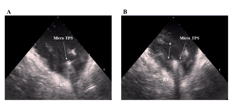
Device retrieval was unsuccessful in one patient. In this patient, the microsnare successfully engaged the proximal retrieval feature of the device; however, even after tight fixation of the snare, we were unable to advance the distal cone over the device. We did not attempt to pull the engaged device because of concern that insufficient countertraction might tear the myocardial tissue. Intraprocedural ICE showed that the Micra TPS was almost embedded within the cardiac wall (Figure 2).
After retrieval, four patients each received a new Micra TPS immediately. All reimplantations were successful and satisfactory electrical parameters were recorded (pacing threshold, 0.57±0.33 V, sensing threshold at 12.4±4.5 mV; and impedance, 551±78 Ω). The remaining two patients received conventional transvenous pacemakers – either dual-chamber or biventricular devices. No procedure-related adverse events occurred during reimplantation. And specifically, no pericardial effusion or tricuspid valve damage was observed on intraoperative ICE.
The primary findings of the present study are as follows: (A) retrieval of chronically-implanted TPS devices was performed safely with currently available endovascular tools, and (B) implantation of a new leadless cardiac pacing device immediately after retrieval of the old one could be a safe and feasible strategy. Although the manufacture recommends abandonment of the TPS at end-of-life, its retrieval may be preferred in specific scenarios. However, there are no detailed studies regarding the safety and feasibility for retrieval and replacement of chronically-implanted TPS devices. In the present study, retrieval was performed only after a clear discussion with patients regarding the basic principle of the options of retrieval or abandonment of the TPS. If the retrieval attempt was unsuccessful, it was planned to abandon the old TPS and implant the appropriate new cardiac pacing device.
First, although there is no dedicated system for the retrieval of a chronically implanted TPS devices, retrieval could be performed safely with currently available tools. However, there are several clinical implications including the possibility for elective reimplantation of a new LCP, reductions in the risk of potential device-to-device interactions and long-term risks of multiple devices implanted in the RV. Although acute retrievability has been reported, to the best of our knowledge, this is the first study to examine the safety and feasibility of retrieval of chronically-implanted TPS devices. According to a careful review of the literature (see Table 2), there are several case reports of TPS retrieval - totalling 29 retrieval patients with explantation performed within 100 days of the initial implantation procedure. Among the five retrieval cases which were performed after 100 days from initial implantation, three were retrieved successfully (60%). In contrast, in our study, all patients had the TPS implanted for at least 280 days and the longest implantation period was 1219 days.

In our experience reimplantation of a new TPS immediately after retrieval was safe and effective in patients who require replacement due to end of battery life or other reasons. In the four patients who received a new TPS immediately after retrieval, all reimplantation procedures were performed safely without any procedure-related complications and with excellent electrical measurements. Reimplantation of a new TPS after retrieval may be preferable to traditional transvenous device systems because an LCP can prevent transvenous lead- and pocket-related complications, and requires only one right femoral vein puncture for both retrieval and reimplantation.
Retrieval was unsuccessful in only one of six patients. In this patient, although the proximal retrieval feature of the TPS could be engaged by the snare, detaching the TPS from the myocardium was not possible. Notably, in this case, the ICE imaging revealed that the TPS was embedded into the ventricular myocardium (Figure 2). Reports describing adherent tissue surrounding an implanted TPS device are limited. One autopsy study of the patient with an implanted TPS showed device encapsulation [8]. Another report noted a TPS device totally covered with fibrous tissue at autopsy less than one year after implantation [9]. The severity of embeddedness within fibrous tissue might be associated with the difficulty of TPS retrieval. ICE imaging appears helpful not only for the retrieval manipulation of the snare and the early detection of pericardial effusion during retrieval but also for the assessment of the retrieval difficulty.
Two approaches can be utilized for retrieval of the TPS (Figure 3-A, 3-B).
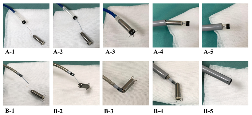
Both approaches require femoral venous access with insertion of a TPS introducer sheath, but then diverge with regard to the specific tool employed to engage the TPS during the next step: the snare is advanced through either i) the integrated protectable sleeve of the TPS delivery catheter (as employed in our case series), or ii) a steerable sheath [10]. In using a steerable sheath, a short sheath (11-16 Fr) is first inserted into the introducer sheath to prevent back bleeding, and a steerable sheath (for example, 8.5 Fr, Agilis NXT, Abbott Laboratories, Abbott Park, IL, USA) is inserted and advanced into the RV.
The differences between the two approaches are the acceptable snare size, and the ability to apply countertraction force to detach the TPS from the myocardium. That is, it is easier to snare the retrieval feature using a steerable sheath, because this sheath allows the use of a 20 mm loop diameter snare (or a tri-loop snare if desired). However, the steerable sheath is limited by the inability to withdraw the LP into the steerable sheath due to its smaller diameter. In contrast, the TPS delivery catheter only accommodates a 7 mm snare; this snare size is small, but still feasible to engage the proximal retrieval feature of the TPS. On the other hand, this approach allows the ability to provide true countertraction using the distal cone. Indeed, because of the chronic nature of the TPS implantations, leading to our expectation of encountering fibrous tissue intimately associated with the device, we used the TPS delivery system with the 7 mm loop snare for all of our retrieval cases. Finally, one practical limitation of this latter approach is the need for a TPS delivery catheter. This can be accomplished by first placing a new TPS device in the heart, and then using the same deliver catheter to retrieve the old LCP. Of course, it would be the most ideal if a dedicated retrieval kit was developed address some of the practical limitations of the current off-the-shelf tools approaches, as well as the fibrous tissue adhesions surrounding the device that can prevent successful retrieval.
Based on our experiences, retrieval of chronically implanted TPS devices can be performed safely, and immediate reimplantation of a new leadless cardiac pacing device is feasible. Using ICE during retrieval might play a critical role not only in the rapid detection of pericardial effusion but also in the assessment of the retrieval difficulty. Our findings may overcome the concern of potential multiple intracardiac devices in patients with long life expectancy and allow more extensive use of LPs. A worldwide registry and future studies regarding retrieval of chronically-implanted Micra TPS devices should be implemented to better elucidate its safety and efficacy.
Limitations: This study has several limitations. First, it is a non-randomized observational single-center experience with a small sample size. Second, the learning curve for the procedure may have influenced the fluoroscopy time and retrieval success rate. However, all retrieval procedures in this study were performed by two experienced operators. Third, it is important to recognize that the meantime from implantation-to-explantation was only ~2 tears, with the longest duration being just over three years. One cannot conclude from the experience that retrieval success will remain in the 80% range when even longer follow-up times. Finally, the difficulty of retrieving an infected TPS devices remains unclear, although based on clinical experience with lead extraction, if anything, infection should facilitate retrieval.
The retrieval of a long-standing implanted Micra TPS device was safely performed, which may indicate the possibility of their safe and elective replacement with a new leadless cardiac pacing device.
Supplemental Materials
Online Video A, B, C


Drs Neužil and Reddy were the leading operators for retrieval of the TPS device. Drs Neužil and Minami took part in data evaluation and writing this manuscript; Dr Reddy participated in critical revisions of the manuscript. All co-authors contributed fully terms of the design of the study, the evaluation of data, the actual manuscript preparation. The corresponding author confirms that all authors have seen and approved the final text.
Clearly Auctoresonline and particularly Psychology and Mental Health Care Journal is dedicated to improving health care services for individuals and populations. The editorial boards' ability to efficiently recognize and share the global importance of health literacy with a variety of stakeholders. Auctoresonline publishing platform can be used to facilitate of optimal client-based services and should be added to health care professionals' repertoire of evidence-based health care resources.

Journal of Clinical Cardiology and Cardiovascular Intervention The submission and review process was adequate. However I think that the publication total value should have been enlightened in early fases. Thank you for all.

Journal of Women Health Care and Issues By the present mail, I want to say thank to you and tour colleagues for facilitating my published article. Specially thank you for the peer review process, support from the editorial office. I appreciate positively the quality of your journal.

Journal of Clinical Research and Reports I would be very delighted to submit my testimonial regarding the reviewer board and the editorial office. The reviewer board were accurate and helpful regarding any modifications for my manuscript. And the editorial office were very helpful and supportive in contacting and monitoring with any update and offering help. It was my pleasure to contribute with your promising Journal and I am looking forward for more collaboration.

We would like to thank the Journal of Thoracic Disease and Cardiothoracic Surgery because of the services they provided us for our articles. The peer-review process was done in a very excellent time manner, and the opinions of the reviewers helped us to improve our manuscript further. The editorial office had an outstanding correspondence with us and guided us in many ways. During a hard time of the pandemic that is affecting every one of us tremendously, the editorial office helped us make everything easier for publishing scientific work. Hope for a more scientific relationship with your Journal.

The peer-review process which consisted high quality queries on the paper. I did answer six reviewers’ questions and comments before the paper was accepted. The support from the editorial office is excellent.

Journal of Neuroscience and Neurological Surgery. I had the experience of publishing a research article recently. The whole process was simple from submission to publication. The reviewers made specific and valuable recommendations and corrections that improved the quality of my publication. I strongly recommend this Journal.

Dr. Katarzyna Byczkowska My testimonial covering: "The peer review process is quick and effective. The support from the editorial office is very professional and friendly. Quality of the Clinical Cardiology and Cardiovascular Interventions is scientific and publishes ground-breaking research on cardiology that is useful for other professionals in the field.

Thank you most sincerely, with regard to the support you have given in relation to the reviewing process and the processing of my article entitled "Large Cell Neuroendocrine Carcinoma of The Prostate Gland: A Review and Update" for publication in your esteemed Journal, Journal of Cancer Research and Cellular Therapeutics". The editorial team has been very supportive.

Testimony of Journal of Clinical Otorhinolaryngology: work with your Reviews has been a educational and constructive experience. The editorial office were very helpful and supportive. It was a pleasure to contribute to your Journal.

Dr. Bernard Terkimbi Utoo, I am happy to publish my scientific work in Journal of Women Health Care and Issues (JWHCI). The manuscript submission was seamless and peer review process was top notch. I was amazed that 4 reviewers worked on the manuscript which made it a highly technical, standard and excellent quality paper. I appreciate the format and consideration for the APC as well as the speed of publication. It is my pleasure to continue with this scientific relationship with the esteem JWHCI.

This is an acknowledgment for peer reviewers, editorial board of Journal of Clinical Research and Reports. They show a lot of consideration for us as publishers for our research article “Evaluation of the different factors associated with side effects of COVID-19 vaccination on medical students, Mutah university, Al-Karak, Jordan”, in a very professional and easy way. This journal is one of outstanding medical journal.

Dear Hao Jiang, to Journal of Nutrition and Food Processing We greatly appreciate the efficient, professional and rapid processing of our paper by your team. If there is anything else we should do, please do not hesitate to let us know. On behalf of my co-authors, we would like to express our great appreciation to editor and reviewers.

As an author who has recently published in the journal "Brain and Neurological Disorders". I am delighted to provide a testimonial on the peer review process, editorial office support, and the overall quality of the journal. The peer review process at Brain and Neurological Disorders is rigorous and meticulous, ensuring that only high-quality, evidence-based research is published. The reviewers are experts in their fields, and their comments and suggestions were constructive and helped improve the quality of my manuscript. The review process was timely and efficient, with clear communication from the editorial office at each stage. The support from the editorial office was exceptional throughout the entire process. The editorial staff was responsive, professional, and always willing to help. They provided valuable guidance on formatting, structure, and ethical considerations, making the submission process seamless. Moreover, they kept me informed about the status of my manuscript and provided timely updates, which made the process less stressful. The journal Brain and Neurological Disorders is of the highest quality, with a strong focus on publishing cutting-edge research in the field of neurology. The articles published in this journal are well-researched, rigorously peer-reviewed, and written by experts in the field. The journal maintains high standards, ensuring that readers are provided with the most up-to-date and reliable information on brain and neurological disorders. In conclusion, I had a wonderful experience publishing in Brain and Neurological Disorders. The peer review process was thorough, the editorial office provided exceptional support, and the journal's quality is second to none. I would highly recommend this journal to any researcher working in the field of neurology and brain disorders.
Dear Agrippa Hilda, Journal of Neuroscience and Neurological Surgery, Editorial Coordinator, I trust this message finds you well. I want to extend my appreciation for considering my article for publication in your esteemed journal. I am pleased to provide a testimonial regarding the peer review process and the support received from your editorial office. The peer review process for my paper was carried out in a highly professional and thorough manner. The feedback and comments provided by the authors were constructive and very useful in improving the quality of the manuscript. This rigorous assessment process undoubtedly contributes to the high standards maintained by your journal.

International Journal of Clinical Case Reports and Reviews. I strongly recommend to consider submitting your work to this high-quality journal. The support and availability of the Editorial staff is outstanding and the review process was both efficient and rigorous.

Thank you very much for publishing my Research Article titled “Comparing Treatment Outcome Of Allergic Rhinitis Patients After Using Fluticasone Nasal Spray And Nasal Douching" in the Journal of Clinical Otorhinolaryngology. As Medical Professionals we are immensely benefited from study of various informative Articles and Papers published in this high quality Journal. I look forward to enriching my knowledge by regular study of the Journal and contribute my future work in the field of ENT through the Journal for use by the medical fraternity. The support from the Editorial office was excellent and very prompt. I also welcome the comments received from the readers of my Research Article.

Dear Erica Kelsey, Editorial Coordinator of Cancer Research and Cellular Therapeutics Our team is very satisfied with the processing of our paper by your journal. That was fast, efficient, rigorous, but without unnecessary complications. We appreciated the very short time between the submission of the paper and its publication on line on your site.

I am very glad to say that the peer review process is very successful and fast and support from the Editorial Office. Therefore, I would like to continue our scientific relationship for a long time. And I especially thank you for your kindly attention towards my article. Have a good day!

"We recently published an article entitled “Influence of beta-Cyclodextrins upon the Degradation of Carbofuran Derivatives under Alkaline Conditions" in the Journal of “Pesticides and Biofertilizers” to show that the cyclodextrins protect the carbamates increasing their half-life time in the presence of basic conditions This will be very helpful to understand carbofuran behaviour in the analytical, agro-environmental and food areas. We greatly appreciated the interaction with the editor and the editorial team; we were particularly well accompanied during the course of the revision process, since all various steps towards publication were short and without delay".

I would like to express my gratitude towards you process of article review and submission. I found this to be very fair and expedient. Your follow up has been excellent. I have many publications in national and international journal and your process has been one of the best so far. Keep up the great work.

We are grateful for this opportunity to provide a glowing recommendation to the Journal of Psychiatry and Psychotherapy. We found that the editorial team were very supportive, helpful, kept us abreast of timelines and over all very professional in nature. The peer review process was rigorous, efficient and constructive that really enhanced our article submission. The experience with this journal remains one of our best ever and we look forward to providing future submissions in the near future.

I am very pleased to serve as EBM of the journal, I hope many years of my experience in stem cells can help the journal from one way or another. As we know, stem cells hold great potential for regenerative medicine, which are mostly used to promote the repair response of diseased, dysfunctional or injured tissue using stem cells or their derivatives. I think Stem Cell Research and Therapeutics International is a great platform to publish and share the understanding towards the biology and translational or clinical application of stem cells.

I would like to give my testimony in the support I have got by the peer review process and to support the editorial office where they were of asset to support young author like me to be encouraged to publish their work in your respected journal and globalize and share knowledge across the globe. I really give my great gratitude to your journal and the peer review including the editorial office.

I am delighted to publish our manuscript entitled "A Perspective on Cocaine Induced Stroke - Its Mechanisms and Management" in the Journal of Neuroscience and Neurological Surgery. The peer review process, support from the editorial office, and quality of the journal are excellent. The manuscripts published are of high quality and of excellent scientific value. I recommend this journal very much to colleagues.

Dr.Tania Muñoz, My experience as researcher and author of a review article in The Journal Clinical Cardiology and Interventions has been very enriching and stimulating. The editorial team is excellent, performs its work with absolute responsibility and delivery. They are proactive, dynamic and receptive to all proposals. Supporting at all times the vast universe of authors who choose them as an option for publication. The team of review specialists, members of the editorial board, are brilliant professionals, with remarkable performance in medical research and scientific methodology. Together they form a frontline team that consolidates the JCCI as a magnificent option for the publication and review of high-level medical articles and broad collective interest. I am honored to be able to share my review article and open to receive all your comments.

“The peer review process of JPMHC is quick and effective. Authors are benefited by good and professional reviewers with huge experience in the field of psychology and mental health. The support from the editorial office is very professional. People to contact to are friendly and happy to help and assist any query authors might have. Quality of the Journal is scientific and publishes ground-breaking research on mental health that is useful for other professionals in the field”.

Dear editorial department: On behalf of our team, I hereby certify the reliability and superiority of the International Journal of Clinical Case Reports and Reviews in the peer review process, editorial support, and journal quality. Firstly, the peer review process of the International Journal of Clinical Case Reports and Reviews is rigorous, fair, transparent, fast, and of high quality. The editorial department invites experts from relevant fields as anonymous reviewers to review all submitted manuscripts. These experts have rich academic backgrounds and experience, and can accurately evaluate the academic quality, originality, and suitability of manuscripts. The editorial department is committed to ensuring the rigor of the peer review process, while also making every effort to ensure a fast review cycle to meet the needs of authors and the academic community. Secondly, the editorial team of the International Journal of Clinical Case Reports and Reviews is composed of a group of senior scholars and professionals with rich experience and professional knowledge in related fields. The editorial department is committed to assisting authors in improving their manuscripts, ensuring their academic accuracy, clarity, and completeness. Editors actively collaborate with authors, providing useful suggestions and feedback to promote the improvement and development of the manuscript. We believe that the support of the editorial department is one of the key factors in ensuring the quality of the journal. Finally, the International Journal of Clinical Case Reports and Reviews is renowned for its high- quality articles and strict academic standards. The editorial department is committed to publishing innovative and academically valuable research results to promote the development and progress of related fields. The International Journal of Clinical Case Reports and Reviews is reasonably priced and ensures excellent service and quality ratio, allowing authors to obtain high-level academic publishing opportunities in an affordable manner. I hereby solemnly declare that the International Journal of Clinical Case Reports and Reviews has a high level of credibility and superiority in terms of peer review process, editorial support, reasonable fees, and journal quality. Sincerely, Rui Tao.

Clinical Cardiology and Cardiovascular Interventions I testity the covering of the peer review process, support from the editorial office, and quality of the journal.

Clinical Cardiology and Cardiovascular Interventions, we deeply appreciate the interest shown in our work and its publication. It has been a true pleasure to collaborate with you. The peer review process, as well as the support provided by the editorial office, have been exceptional, and the quality of the journal is very high, which was a determining factor in our decision to publish with you.

The peer reviewers process is quick and effective, the supports from editorial office is excellent, the quality of journal is high. I would like to collabroate with Internatioanl journal of Clinical Case Reports and Reviews journal clinically in the future time.

Clinical Cardiology and Cardiovascular Interventions, I would like to express my sincerest gratitude for the trust placed in our team for the publication in your journal. It has been a true pleasure to collaborate with you on this project. I am pleased to inform you that both the peer review process and the attention from the editorial coordination have been excellent. Your team has worked with dedication and professionalism to ensure that your publication meets the highest standards of quality. We are confident that this collaboration will result in mutual success, and we are eager to see the fruits of this shared effort.

Dear Dr. Jessica Magne, Editorial Coordinator 0f Clinical Cardiology and Cardiovascular Interventions, I hope this message finds you well. I want to express my utmost gratitude for your excellent work and for the dedication and speed in the publication process of my article titled "Navigating Innovation: Qualitative Insights on Using Technology for Health Education in Acute Coronary Syndrome Patients." I am very satisfied with the peer review process, the support from the editorial office, and the quality of the journal. I hope we can maintain our scientific relationship in the long term.

Dear Monica Gissare, - Editorial Coordinator of Nutrition and Food Processing. ¨My testimony with you is truly professional, with a positive response regarding the follow-up of the article and its review, you took into account my qualities and the importance of the topic¨.

Dear Dr. Jessica Magne, Editorial Coordinator 0f Clinical Cardiology and Cardiovascular Interventions, The review process for the article “The Handling of Anti-aggregants and Anticoagulants in the Oncologic Heart Patient Submitted to Surgery” was extremely rigorous and detailed. From the initial submission to the final acceptance, the editorial team at the “Journal of Clinical Cardiology and Cardiovascular Interventions” demonstrated a high level of professionalism and dedication. The reviewers provided constructive and detailed feedback, which was essential for improving the quality of our work. Communication was always clear and efficient, ensuring that all our questions were promptly addressed. The quality of the “Journal of Clinical Cardiology and Cardiovascular Interventions” is undeniable. It is a peer-reviewed, open-access publication dedicated exclusively to disseminating high-quality research in the field of clinical cardiology and cardiovascular interventions. The journal's impact factor is currently under evaluation, and it is indexed in reputable databases, which further reinforces its credibility and relevance in the scientific field. I highly recommend this journal to researchers looking for a reputable platform to publish their studies.

Dear Editorial Coordinator of the Journal of Nutrition and Food Processing! "I would like to thank the Journal of Nutrition and Food Processing for including and publishing my article. The peer review process was very quick, movement and precise. The Editorial Board has done an extremely conscientious job with much help, valuable comments and advices. I find the journal very valuable from a professional point of view, thank you very much for allowing me to be part of it and I would like to participate in the future!”

Dealing with The Journal of Neurology and Neurological Surgery was very smooth and comprehensive. The office staff took time to address my needs and the response from editors and the office was prompt and fair. I certainly hope to publish with this journal again.Their professionalism is apparent and more than satisfactory. Susan Weiner

My Testimonial Covering as fellowing: Lin-Show Chin. The peer reviewers process is quick and effective, the supports from editorial office is excellent, the quality of journal is high. I would like to collabroate with Internatioanl journal of Clinical Case Reports and Reviews.

My experience publishing in Psychology and Mental Health Care was exceptional. The peer review process was rigorous and constructive, with reviewers providing valuable insights that helped enhance the quality of our work. The editorial team was highly supportive and responsive, making the submission process smooth and efficient. The journal's commitment to high standards and academic rigor makes it a respected platform for quality research. I am grateful for the opportunity to publish in such a reputable journal.

My experience publishing in International Journal of Clinical Case Reports and Reviews was exceptional. I Come forth to Provide a Testimonial Covering the Peer Review Process and the editorial office for the Professional and Impartial Evaluation of the Manuscript.

I would like to offer my testimony in the support. I have received through the peer review process and support the editorial office where they are to support young authors like me, encourage them to publish their work in your esteemed journals, and globalize and share knowledge globally. I really appreciate your journal, peer review, and editorial office.

Dear Agrippa Hilda- Editorial Coordinator of Journal of Neuroscience and Neurological Surgery, "The peer review process was very quick and of high quality, which can also be seen in the articles in the journal. The collaboration with the editorial office was very good."

I would like to express my sincere gratitude for the support and efficiency provided by the editorial office throughout the publication process of my article, “Delayed Vulvar Metastases from Rectal Carcinoma: A Case Report.” I greatly appreciate the assistance and guidance I received from your team, which made the entire process smooth and efficient. The peer review process was thorough and constructive, contributing to the overall quality of the final article. I am very grateful for the high level of professionalism and commitment shown by the editorial staff, and I look forward to maintaining a long-term collaboration with the International Journal of Clinical Case Reports and Reviews.

To Dear Erin Aust, I would like to express my heartfelt appreciation for the opportunity to have my work published in this esteemed journal. The entire publication process was smooth and well-organized, and I am extremely satisfied with the final result. The Editorial Team demonstrated the utmost professionalism, providing prompt and insightful feedback throughout the review process. Their clear communication and constructive suggestions were invaluable in enhancing my manuscript, and their meticulous attention to detail and dedication to quality are truly commendable. Additionally, the support from the Editorial Office was exceptional. From the initial submission to the final publication, I was guided through every step of the process with great care and professionalism. The team's responsiveness and assistance made the entire experience both easy and stress-free. I am also deeply impressed by the quality and reputation of the journal. It is an honor to have my research featured in such a respected publication, and I am confident that it will make a meaningful contribution to the field.

"I am grateful for the opportunity of contributing to [International Journal of Clinical Case Reports and Reviews] and for the rigorous review process that enhances the quality of research published in your esteemed journal. I sincerely appreciate the time and effort of your team who have dedicatedly helped me in improvising changes and modifying my manuscript. The insightful comments and constructive feedback provided have been invaluable in refining and strengthening my work".

I thank the ‘Journal of Clinical Research and Reports’ for accepting this article for publication. This is a rigorously peer reviewed journal which is on all major global scientific data bases. I note the review process was prompt, thorough and professionally critical. It gave us an insight into a number of important scientific/statistical issues. The review prompted us to review the relevant literature again and look at the limitations of the study. The peer reviewers were open, clear in the instructions and the editorial team was very prompt in their communication. This journal certainly publishes quality research articles. I would recommend the journal for any future publications.

Dear Jessica Magne, with gratitude for the joint work. Fast process of receiving and processing the submitted scientific materials in “Clinical Cardiology and Cardiovascular Interventions”. High level of competence of the editors with clear and correct recommendations and ideas for enriching the article.

We found the peer review process quick and positive in its input. The support from the editorial officer has been very agile, always with the intention of improving the article and taking into account our subsequent corrections.

My article, titled 'No Way Out of the Smartphone Epidemic Without Considering the Insights of Brain Research,' has been republished in the International Journal of Clinical Case Reports and Reviews. The review process was seamless and professional, with the editors being both friendly and supportive. I am deeply grateful for their efforts.

To Dear Erin Aust – Editorial Coordinator of Journal of General Medicine and Clinical Practice! I declare that I am absolutely satisfied with your work carried out with great competence in following the manuscript during the various stages from its receipt, during the revision process to the final acceptance for publication. Thank Prof. Elvira Farina

Dear Jessica, and the super professional team of the ‘Clinical Cardiology and Cardiovascular Interventions’ I am sincerely grateful to the coordinated work of the journal team for the no problem with the submission of my manuscript: “Cardiometabolic Disorders in A Pregnant Woman with Severe Preeclampsia on the Background of Morbid Obesity (Case Report).” The review process by 5 experts was fast, and the comments were professional, which made it more specific and academic, and the process of publication and presentation of the article was excellent. I recommend that my colleagues publish articles in this journal, and I am interested in further scientific cooperation. Sincerely and best wishes, Dr. Oleg Golyanovskiy.

Dear Ashley Rosa, Editorial Coordinator of the journal - Psychology and Mental Health Care. " The process of obtaining publication of my article in the Psychology and Mental Health Journal was positive in all areas. The peer review process resulted in a number of valuable comments, the editorial process was collaborative and timely, and the quality of this journal has been quickly noticed, resulting in alternative journals contacting me to publish with them." Warm regards, Susan Anne Smith, PhD. Australian Breastfeeding Association.

Dear Jessica Magne, Editorial Coordinator, Clinical Cardiology and Cardiovascular Interventions, Auctores Publishing LLC. I appreciate the journal (JCCI) editorial office support, the entire team leads were always ready to help, not only on technical front but also on thorough process. Also, I should thank dear reviewers’ attention to detail and creative approach to teach me and bring new insights by their comments. Surely, more discussions and introduction of other hemodynamic devices would provide better prevention and management of shock states. Your efforts and dedication in presenting educational materials in this journal are commendable. Best wishes from, Farahnaz Fallahian.

Dear Maria Emerson, Editorial Coordinator, International Journal of Clinical Case Reports and Reviews, Auctores Publishing LLC. I am delighted to have published our manuscript, "Acute Colonic Pseudo-Obstruction (ACPO): A rare but serious complication following caesarean section." I want to thank the editorial team, especially Maria Emerson, for their prompt review of the manuscript, quick responses to queries, and overall support. Yours sincerely Dr. Victor Olagundoye.

Dear Ashley Rosa, Editorial Coordinator, International Journal of Clinical Case Reports and Reviews. Many thanks for publishing this manuscript after I lost confidence the editors were most helpful, more than other journals Best wishes from, Susan Anne Smith, PhD. Australian Breastfeeding Association.

Dear Agrippa Hilda, Editorial Coordinator, Journal of Neuroscience and Neurological Surgery. The entire process including article submission, review, revision, and publication was extremely easy. The journal editor was prompt and helpful, and the reviewers contributed to the quality of the paper. Thank you so much! Eric Nussbaum, MD

Dr Hala Al Shaikh This is to acknowledge that the peer review process for the article ’ A Novel Gnrh1 Gene Mutation in Four Omani Male Siblings, Presentation and Management ’ sent to the International Journal of Clinical Case Reports and Reviews was quick and smooth. The editorial office was prompt with easy communication.

Dear Erin Aust, Editorial Coordinator, Journal of General Medicine and Clinical Practice. We are pleased to share our experience with the “Journal of General Medicine and Clinical Practice”, following the successful publication of our article. The peer review process was thorough and constructive, helping to improve the clarity and quality of the manuscript. We are especially thankful to Ms. Erin Aust, the Editorial Coordinator, for her prompt communication and continuous support throughout the process. Her professionalism ensured a smooth and efficient publication experience. The journal upholds high editorial standards, and we highly recommend it to fellow researchers seeking a credible platform for their work. Best wishes By, Dr. Rakhi Mishra.

Dear Jessica Magne, Editorial Coordinator, Clinical Cardiology and Cardiovascular Interventions, Auctores Publishing LLC. The peer review process of the journal of Clinical Cardiology and Cardiovascular Interventions was excellent and fast, as was the support of the editorial office and the quality of the journal. Kind regards Walter F. Riesen Prof. Dr. Dr. h.c. Walter F. Riesen.

Dear Ashley Rosa, Editorial Coordinator, International Journal of Clinical Case Reports and Reviews, Auctores Publishing LLC. Thank you for publishing our article, Exploring Clozapine's Efficacy in Managing Aggression: A Multiple Single-Case Study in Forensic Psychiatry in the international journal of clinical case reports and reviews. We found the peer review process very professional and efficient. The comments were constructive, and the whole process was efficient. On behalf of the co-authors, I would like to thank you for publishing this article. With regards, Dr. Jelle R. Lettinga.

Dear Clarissa Eric, Editorial Coordinator, Journal of Clinical Case Reports and Studies, I would like to express my deep admiration for the exceptional professionalism demonstrated by your journal. I am thoroughly impressed by the speed of the editorial process, the substantive and insightful reviews, and the meticulous preparation of the manuscript for publication. Additionally, I greatly appreciate the courteous and immediate responses from your editorial office to all my inquiries. Best Regards, Dariusz Ziora

Dear Chrystine Mejia, Editorial Coordinator, Journal of Neurodegeneration and Neurorehabilitation, Auctores Publishing LLC, We would like to thank the editorial team for the smooth and high-quality communication leading up to the publication of our article in the Journal of Neurodegeneration and Neurorehabilitation. The reviewers have extensive knowledge in the field, and their relevant questions helped to add value to our publication. Kind regards, Dr. Ravi Shrivastava.

Dear Clarissa Eric, Editorial Coordinator, Journal of Clinical Case Reports and Studies, Auctores Publishing LLC, USA Office: +1-(302)-520-2644. I would like to express my sincere appreciation for the efficient and professional handling of my case report by the ‘Journal of Clinical Case Reports and Studies’. The peer review process was not only fast but also highly constructive—the reviewers’ comments were clear, relevant, and greatly helped me improve the quality and clarity of my manuscript. I also received excellent support from the editorial office throughout the process. Communication was smooth and timely, and I felt well guided at every stage, from submission to publication. The overall quality and rigor of the journal are truly commendable. I am pleased to have published my work with Journal of Clinical Case Reports and Studies, and I look forward to future opportunities for collaboration. Sincerely, Aline Tollet, UCLouvain.

Dear Ms. Mayra Duenas, Editorial Coordinator, International Journal of Clinical Case Reports and Reviews. “The International Journal of Clinical Case Reports and Reviews represented the “ideal house” to share with the research community a first experience with the use of the Simeox device for speech rehabilitation. High scientific reputation and attractive website communication were first determinants for the selection of this Journal, and the following submission process exceeded expectations: fast but highly professional peer review, great support by the editorial office, elegant graphic layout. Exactly what a dynamic research team - also composed by allied professionals - needs!" From, Chiara Beccaluva, PT - Italy.

Dear Maria Emerson, Editorial Coordinator, we have deeply appreciated the professionalism demonstrated by the International Journal of Clinical Case Reports and Reviews. The reviewers have extensive knowledge of our field and have been very efficient and fast in supporting the process. I am really looking forward to further collaboration. Thanks. Best regards, Dr. Claudio Ligresti

Dear Chrystine Mejia, Editorial Coordinator, Journal of Neurodegeneration and Neurorehabilitation. “The peer review process was efficient and constructive, and the editorial office provided excellent communication and support throughout. The journal ensures scientific rigor and high editorial standards, while also offering a smooth and timely publication process. We sincerely appreciate the work of the editorial team in facilitating the dissemination of innovative approaches such as the Bonori Method.” Best regards, Dr. Giselle Pentón-Rol.
