AUCTORES
Globalize your Research

Review Article | DOI: https://doi.org/10.31579/2641-5194/059
1 Istanbul Medipol University School of Medicine Department of Gastroenterology. Istanbul. Turkey
2 Istanbul Yeditepe University School of Medicine. Istanbul. Turkey
3 Istanbul Health and Technology University. Department of Gastroenterology. Istanbul. Turkey
*Corresponding Author: Vedat Goral, Istanbul Medipol University School of Medicine Department of Gastroenterology. Istanbul. Turkey.
Citation: Goral V. , Kerem M.Goral , Ormeci N. (2022), Follow-up Strategies in Focal Liver Lesions and Treatment methods. J. Gastroenterology Pancreatology and Hepatobilary Disorders, 6(2); DOI: 10.31579/2641-5194/059
Copyright: © 2022 Vedat Goral. This is an open access article distributed under the Creative Commons Attribution License, which permits unrestricted use, distribution, and reproduction in any medium, provided the original work is properly cited.
Received: 03 December 2021 | Accepted: 24 December 2021 | Published: 11 January 2022
Keywords: focal liver lesions; follw-up; treatment
Today, advances in cross-sectional imaging have led to the detection and early recognition of incidental/focal liver lesions (FCL). In approximately 17,000 cases of chest CT, incidental liver lesions were found in 6% [1]. In general, FCL consists of hepatocytes, biliary epithelium, mesenchymal tissue, connective tissue, or metastasized cells from distant sites. Most incidental lesions are benign, some may require careful management and treatment. In evaluating the lesion, the patient's clinical history, underlying disease and age factor should be considered. FCL can be detected at a rate of 10-30% in normal healthy and chronic liver disease patients, and even in oncology patients with malignancy, FCLs can be highly benign (50-80%)
Most incidental lesions are benign, some may require careful management and treatment. In evaluating the lesion, the patient's clinical history, underlying disease and age factor should be considered. Early diagnosis and treatment should be decided whether it is necessary or not. Factors affecting this decision are the presence of other diseases, laboratory and radiological data. The characteristics of the lesion (size, margin, growth, etc.) should be determined and follow-up should be planned.
Ultrasonography (USG): It is the most commonly used diagnostic method. It is a great advantage that it is non-invasive and easy to apply. However, the diagnosis of FCLs can sometimes be inaccurate, as it depends on the performer and the ultrasound device. Therefore, USG should be used as a screening test, and advanced tests such as CT, MR, Elastography, CEUS, and PET should also be used for the definition of the lesion when necessary [3, 4].
Computed Tomography: The most important disadvantages are that it contains X-rays (increased risk of malignancy due to ionizing radiation exposure) and requires iodinated contrast material (allergic reaction and contrast nephropathy). Radiation exposure limits its use in pregnant women and children. In multiphasic studies, patients who will benefit from these protocols should be carefully selected because the dose is increased. Renal function tests should be checked before contrast is given, and iodinated contrast material should not be used in patients with stage 3-4 CRF [4, 5].
MR Imaging: Does not include X-rays. It is contraindicated in patients with a pacemaker. Claustrophobic patients may not tolerate it. Gadolinium-containing contrast agents, which are more reliable than iodinated contrast agents, are used. However, the use of contrast material with gadolinium is contraindicated in patients with stage 4-5 CRF (Nephrogenic systemic fibrosis). Dynamic studies, especially with hepatocyte-specific contrast agents (Gadobenic acid/Gd-BOPTA, gadoxtic acid/Gd-EOB-DTPA) help in the diagnosis by showing the contrast enhancement patterns of the lesions.
CEUS (Sulfur Hexafluoride (SF6) Microbuble contrast-enhanced ultrasonography): It is stated that it can be the second examination in the differential diagnosis of benign lesions, especially after ultrasound [6-8].CT and MRI contrast agents are contraindicated, as are patients with renal impairment and those with known allergic reactions to CT/MRI contrast agents. CEUS will be used in more and more diagnoses in the future.
Elastography: When a focal liver lesion is detected in the liver, the clinician usually chooses the next examination, should be determined and determine the appropriate method. A wrong application also negatively affects the treatment and patient prognosis. In 2014, ACG (American College of Gastroenterology) published a guideline [9-11]. Apart from that, there are some guidelines. The differential diagnosis, especially between benign and malignant lesions, is extremely important and can often be particularly challenging. Contrast-applied radiological techniques and/or liver biopsy are often necessary for diagnosis, but they have various contraindications or complications. Due to the limitations of these methods, there is an urgent need to develop a first-line, non-invasive, and simple method to diagnose FCLs. Elastography is a USG-based imaging method that provides information about the physical parameter corresponding to tissue stiffness and can be considered as a virtual biopsy. Various elastographic approaches have been developed, such as different elastography methods, transient elastography, and 2D wave elastography. These tools are currently used in the evaluation of liver fibrosis and focal lesions in other organs such as the breast and thyroid gland. It is particularly useful in the ability to distinguish between benign and malignant lesions, hepatocellular carcinoma and liver metastases, and in follow-up after percutaneous therapy. In the future, elastography will be used more often.
Dual Energy CT (DECT): If the patient has an unclear lesion, it may be preferred over conventional CT MRI [12-14].
PET/CT and PET/MR: In lesions >1 cm, it may eliminate the need for biopsy [13].
When a focal lesion is detected in the liver, risk factors should be considered first. Risk factors can be low or high grade [3-7].These risk factors are very useful in the diagnosis and prognosis of the lesion (Scheme 1 and 2).
1) Low risk factors;
- Absence of malignancy
- Absence of hepatic disease (hepatitis, PSC, ACH, NASH, hemochromatosis, anabolic steroid use, genetic disease, etc.)
- Young age
- If there are no symptoms, the risk of malignancy is low.
2) High risk factors; If the person
- Cirrhosis of the Liver
- Presence of hepatic disease other than liver cirrhosis (NASH, alcoholism, viral, metabolic, anabolic steroid use, glycogen storage disease, PSK, hereditary disease)
- Having a known malignant disease
- Advanced age
- History of estrogen or other drug use
- If there are features such as travel history (parasitic diseases), the probability of the detected lesion being malignant increases.
Lesions detected in the liver can be benign or malignant.
1) Benign lesions in the liver
- Hepatic hemangioma
- FNH
- Hepatic adenoma
- Hepatic cysts
- Biliary hamartoma
- Abscess may be in the form of Mesenchymal hamartoma.
2) Malignant lesions; HCC can be in the form of Cholangiocarcinoma, other liver malignancies and metastatic lesions.
When a lesion is detected in the liver;
- Does it pose a risk for the patient in the future?
- Is it possible to differentiate between benign and malignant?
- Does the lesion cause complications (bleeding, etc.)? parameters such as
If the lesion is <5>1 cm, the lesion should be investigated.
Hepatic hemangiomas (HH): It is the most common/common, benign liver lesion. It can be diagnosed at any age; Most of these lesions (up to 80%) are between 30-50 years of age, more common in women (3: 1) and mostly solitary, however, sometimes more than one hemangioma may be present in the liver [1, 10-12]. Small hemangiomas usually appear homogeneous, but larger hemangiomas (>4 cm) may appear heterogeneous. They are generally asymptomatic and have a good prognosis. Massive hemangiomas can sometimes cause abdominal pain and discomfort with pressure on neighboring organs [13-14].
In small HH suspicious lesions less than 3 cm, follow-up should be done after 6 months. For lesions larger than 3 cm, annual or biennial follow-up is recommended. If Kasabach-Merritt syndrome and symptomatic hemangioma are present, treatment should be prompt.
In hemangioma, growth of <3>3 mm per year. If it is stable in 6-12 months, there is no need for follow-up. However, if the growth is >3 mm per year, the council (gastroenterologist/hepatologist, hepatobiliary surgeon) is evaluated for surgery. Only in symptoms that tend to grow more than 3 cm per year or lesions greater than 10 cm in diameter should intervention be considered.
Indications for surgery are: It is performed in cases such as a) rupture with intraperitoneal bleeding, b) massive hemangiomas causing symptoms, and c) inability to exclude malignancy on imaging.
Small hepatic hemangiomas are less likely to develop complications during pregnancy or oral contraceptive drug (OCA) use. Conservative monitoring in pregnancy is recommended for patients with large tumors, but the presence of hemangioma is not a contraindication for OCA. We do not recommend contraception in asymptomatic female patients who wish to become pregnant. During pregnancy, routine liver ultrasound is not recommended. Estrogen may affect lesion growth, but the risk of lesion rupture is similar for pregnant and non-pregnant women.
In these patients, acute abdominal pain; may indicate thrombosis or intratumoral hemorrhage. By stretching the Glisson's capsule; in acute thrombosis; There may be fever and changes in LFT. Rarely, there may be secondary hemobilia due to the opening of the biliary tract.
Treatment methods,
- surgical enucleation that preserves the parenchyma,
- intra-arterial embolization or radioactive irradiation,
- It is in the form of liver transplantation.
In a series of 1185 cases, complications in enucleation were found to be quite low [14]. If a giant hemangioma (> 10 cm) and/or bleeding is present, transcatheter arterial embolization can be performed to reduce the lesion size before elective surgery.
The prognosis is generally quite good, with most lesions remaining asymptomatic and without complications. In a study of 76 asymptomatic patients, none of the patients developed symptoms or complications during a mean follow-up of 92 months [12] . Rupture risk is very rare and there is no relationship between hemangioma size and rupture risk.
Focal nodular hyperplasia (FNH) is a benign lesion with a central scar and a proliferation of surrounding hyperplastic hepatocytes. It is seen in the 2nd frequency among the benign lesions of the liver. FNH occurs in intrahepatic arteriovenous malformation as a local hyperplastic response to increased blood flow. Angiopoietin genes (ANGPT1 and ANGPT2) are implicated in etiopathogenesis. Typically, FNH is solitary and is more common in women. It is divided into two as inflammatory and telangiectatic [15-16].
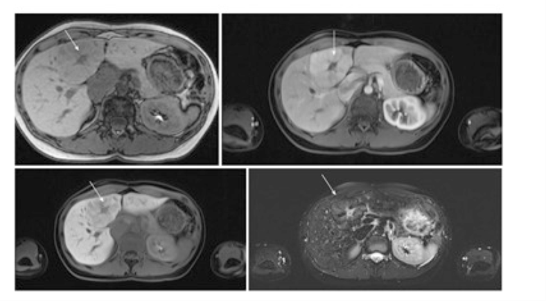
In a large series of patients referred for ultrasound or contrast-enhanced CT, the prevalence of FNH was found to be 0.2–1.6% ( ). Routine follow-up imaging is not recommended for asymptomatic patients with FNH because of low/slow growth risk or low complications. CEUS (Contrast Enhanced Ultrasonography), CT or MR can diagnose FNH almost 100% with typical imaging (Picture 1) [10, 11]. For FNH, follow-up is not necessary unless there is underlying vascular liver disease.
According to hepatocellular adenoma, they can be symptomatic in 40%. In general, it should be followed every 6-12 months. A biopsy is not required for diagnosis. If the appearance on CT is questionable, a biopsy may be required. In the study involving 30 FNH patients (34 lesions) monitored by ultrasound, 33 lesions (97%) either remained stable or decreased in size at a mean follow-up of 42 months [15].
If the diagnosis is uncertain and the person has a history of cancer, even if the lesion is small, surgical treatment is performed. If 0.5 cm of growth per year and the lesion diameter is >3-4 cm, surgical treatment is indicated. Laparoscopic / robotic liver resection has advantages such as less operative blood loss, less postoperative pain, and shorter hospital stay.
Embolization and radiofrequency ablation are not primary treatments. If the patient does not want surgery, these may come up.
The prognosis is excellent, the lesion is mostly stable or may regress over time, complications (eg bleeding) are very rare. Malignant transformation has not been reported.
Discontinuation of oral contraceptives and other estrogen-containing drugs should not be insisted upon. Pregnancy is not contraindicated in these patients. Women with FNH who continue to take these drugs should have follow-up imaging every 6-12 months. In enlarged and symptomatic cases, embolization and resection are performed. If there is no growth and no symptoms, no treatment is required.
Hepatic adenomas (HA); It is an uncommon, solid, benign liver lesion. Hepatic adenomas consist of hepatocytes, do not contain the portal vein, central vein and bile duct, and are distinguished from FNH with this feature. In young women, it is associated with the use of estrogen-containing drugs. Patients with glycogen storage disease or metabolic syndrome are at higher risk of developing HA [13].
There are 4 subtypes of FNH:
A) Hepatocyte nuclear factor – 1α (HNF-1α) inactivated hepatic adenomas (30-40%)
B) Inflammatory hepatic adenomas (40-55%)
C) β-catenin activated hepatic adenomas (10-20%)
D) Unclassifiable (5-10%). They do not have the typical clinical or imaging appearances.
Inflammatory adenomas should be followed up because of the risk of bleeding. The risk of malignancy is higher in β-catenin-activated adenomas. Inflammatory hepatic adenomas appear strongly hyperintense on T2-weighted MRI, which may be diffuse or margin-like (Atoll sign) at the periphery of the lesion (12). Normally, a follow-up of 2 years at 6 months intervals is recommended.
Mutations of catenin β1 (CTNNB1) in Exon 3 (coding for β-catenin) occur in 10-15% of hepatic adenomas. These are associated with a higher risk of malignant transformation. In contrast, in a subset of HA (5-10%), two hot spots in exons 7 and 8 are associated with CTNNB1 mutations and do not increase the risk of malignancy. These variants of hepatic adenoma do not have typical imaging features and may therefore be difficult to distinguish from HCC or FNH. Hepatic adenomas with catenin β1 mutations may also show contrast enhancement in the hepatobiliary phase of MRI using liver-specific contrast media.
Treatment decisions depend on gender, size, and progression. In addition to weight loss, lifestyle changes such as discontinuation of OCA should be recommended. Resection is recommended in men, regardless of size and in the presence of proven β‐catenin mutation. In women, after lifestyle change, 6 months of observation is recommended, and for nodules ≥ 5 cm and those that continue to grow, resection is indicated. Lesions <5>
Haemorrhagic HA that is hemodynamically unstable should be embolized and any remaining lesion on follow-up imaging is an indication for resection. In multiple HA, liver transplantation is not recommended, but may be considered in people with underlying liver disease.
Simple Liver Cyst: They are benign lesions that are not associated with the biliary tract. It is asymptomatic and detected incidentally on USG [12]. Its incidence in the community varies between 5-14% [16, 17]. They can be single or multiple. Cysts should not show mural thickening, nodularity, or increased contrast on USG, CT, or MRI. Cysts seen between the liver and the diaphragm are different from simple hepatic cysts and are diagnosed as diaphragmatic mesothelial cysts. Typical localization and often bilobular appearance are important in the differential diagnosis. They are often asymptomatic and do not require treatment.
Treatment indications;
- symptomatic cysts
- evidence of septations
- calcification or
- if biliary cystadenoma and cystadenocarcinoma are suspected.
Surgical intervention; consists of fenestration, enucleation, aspiration, and sclerotherapy.
Hydatid Cyst in Liver: When small, they resemble simple cysts. In cysts larger than 5 cm, CT and MRI are applied in the follow-up. Laboratory tests are valuable in follow-up. Treatment is medical (Pharmaceutical, PAIR, Knitting method) or surgery [17].
Liver Abscess: Abscesses; It can be classified as pyogenic, amebic or fungal. In cases such as cholangitis, portal phlebitis, pathogens enter through the portal venous system or biliary tract. The possibility of occult colorectal neoplasia should be considered, especially in patients diagnosed with pyogenic liver abscess due to K. pneumoniae and in the absence of apparent underlying hepatobiliary disease [18, 19].
Peribiliary abscesses tend to be scattered, small and adjacent to the biliary tree; In appendicitis or diverticulitis, pathogens can cause larger lesions in the liver via the hepatic artery or portal vein (hematogenous).
Amoebic abscesses are nonspecific and their frequency has decreased considerably today. USG and MRI guide the diagnosis. Treatment can be medical or surgical.
Pyogenic abscess is treated with drainage or surgery. In Nepal, in 102 patients with pyogenic liver abscess who did not have abscess drainage, the mean time to ultrasonographic resolution of abscesses <10> 10 cm was 22 weeks [19]. For patients with persistent clinical symptoms with evidence of persistent abscess following drainage intervention and antibiotic therapy, reassessment for re-drainage is required. If this is not possible, surgical intervention is indicated.
Bile duct hamartomas are congenital malformations of the ductal plate that are not connected to the bile ducts. They are usually discovered incidentally on abdominal imaging (20). Although not of clinical significance, they may mimic disseminated small liver metastases in the cancer patient. Biliary hamartomas are typically small (5-10 mm in size) and are usually widely distributed in both lobes of the liver. On ultrasound, they appear as small hyperechoic or hypoechoic lesions and may show artifacts (comet appearance). On CT, they appear as round, oval, or irregularly shaped small cystic lesions without contrast enhancement, but sometimes thin rim enhancement may be present and therefore mimic hypovascular liver metastases.
In general; It is symptomatic and diagnosed incidentally. It is a rare, benign malformation of the intrahepatic biliary tract (15). It is usually seen as small (<15>
Bile duct hamartomas may rarely be very large, up to 20 cm, and may be symptomatic due to internal bleeding or pressure on adjacent structures. Among the differential diagnoses of biliary hamartomas; peribiliary cysts (predominantly in the perihilar region in patients with liver parenchymal disease), polycystic disease, and Caroli's disease (cysts communicate with the bile ducts and are associated with bile duct abnormalities). They can also sometimes mimic liver abscesses.
Biliary cystadenoma and cystadenocarcinoma: Biliary cystadenoma is a rare, slow growing, multiloculated cystic benign tumor (21). They are slow growing neoplastic lesions originating from the bile ducts. Often – they show intrahepatic localization (85%). It is generally seen in middle-aged women and is considered premalignant. Although it develops slowly, it requires treatment with its precancerous feature. Therefore, early recognition is important. Although benign, these tumors tend to degenerate into malignant and any such tumor should be considered potentially malignant. In both cystadenoma and cystadenocarcinoma, coarse calcifications can be seen on USG and CT, but they are not a sign of benignity [16].
Bilioma: A collection of encapsulated bile of the biliary tree due to traumatic or iatrogenic causes. It appears as a collection showing unilocular, subcapsular or intraparenchymal fluid density (0-15 HU). It is localized in the gallbladder cavity or in the surrounding structures. Biliomas are treated with both percutaneous drainage and surgery.
Hepatic angiosarcoma is a rare tumor. As in patients with hemochromatosis, there is a strong association with prior exposure to carcinogens such as vinyl chloride and Thorotrast. However, in the majority, the tumor is idiopathic. Pathologically, angiosarcoma presents as large, solitary masses or multiple tumor nodules of varying size containing multiple vascular channels. Therefore, they should be followed regularly [12].
Epithelioid hemangioendothelioma (EHE) is a rare tumor of vascular origin, not to be confused with infantile hemangioendothelioma, which is a very different tumor. These hepatic tumors are characterized by multiple, peripherally based lesions that gradually become confluent masses. In addition to the unusual peripheral liver distribution, an important characteristic feature is the presence of capsular retraction due to fibrosis and scarring. Follow-up should be done with MRI or CT at regular intervals [12].
Regenerative nodules develop in response to liver injury, consist of proliferation of hepatocytes and surrounding stroma. Typically, they occur in liver cirrhosis.
Dysplastic nodules (DN): Differentiation of dysplastic nodules from HCC must be supported by radiological and several parameters (trabecular irregularity, increased nuclear/cytoplasmic ratio) and immunohistological markers. The DN is typically hypovascular or isovascular to the liver during the arterial phase and isoechoic to the liver in later phases. Better DN diagnosis can be obtained in patients with cirrhosis evaluated with Gd-EOB-DTPA MRI ( ). AASLD practice guidelines recommend repeat ultrasound examination after 3 months for new nodules <1>1 cm. Overall, there is still no definitive answer as to whether a much earlier diagnosis will mean a better outcome [12].
HCC develops against the background of chronic liver disease. It occurs frequently in Asian and Mediterranean countries, and develops on the background of chronic liver disease in Europe. AFP (AFP, AFP-L3, DCP) follow-up is important, but its sensitivity is 60% and specificity is 80%. Persistently rising AFP is important. AFP elevation is not specific for HCC, and may be elevated in acute/chronic viral hepatitis and decompensated liver diseases, pregnancy, ovarian Tm, and gastric cancer. In non-cirrhotic HCC, the diagnosis needs to be confirmed by biopsy. New biomarkers, e.g. MicroRNA panels or exosome-derived proteins may be promising in the future diagnosis of HCC [22-29].
In HCC, lesions <1> 1 cm. With FNAB, the correct diagnosis is made between 82 and 87%. In the absence of diagnostic uncertainty or cirrhosis, a biopsy is required to confirm preoperative HCC. AFP level is also important [22].
Control-follow-up is done with USG at 6-month intervals. 6 months interval is due to the doubling time of the tumor (mean 117 days, 29-398 days). If the lesion is <1>
Fibrolamellar HCC: Fibrolamellar HCC (FL-HCC) is a less aggressive tumor with a better prognosis than classical HCC. On CT, FL-HCC appears as a large, well-defined vascular mass with a lobulated surface and often a central scar and calcifications in 70% of cases ( ). On MR imaging, FL-HCC is typically hypointense on T1- and hyperintense on T2-weighted images, with central scar hypointense on both sequences. This is in contrast to FNH scar, which is most often hyperintense on T2-weighted images. The fibrous central regions of both FNH and FL-HCC, CT and extracellular gadolinium MRI show delayed retention of contrast agents. Compared with FNH, the contrast enhancement in FL-HCC is heterogeneous compared to the generally homogeneous contrast enhancement pattern of FNH. Follow-up should be like classic HCC.
Cholangiocarcinoma: It constitutes 20% of primary liver tumors (30). It arises from biliary epithelial cells. Its frequency has been increasing in recent years (31). Biopsy, MRCP, CT, ERCP, tumor markers guide the diagnosis. Since the symptoms are detected late, the diagnosis may also be late. It starts from the intrahepatic and spreads to the peri hiller and extraheaptic locations. The presence of primary sclerosing cholangitis, liver cirrhosis, choledochal cyst, cholelithiasis is a risk factor for CC.
Hepatic Lymphoma: Primary hepatic lymphoma (PHL) is a rare form of non-Hodgkin lymphoma (NHL) that mainly involves the liver, as opposed to a predominant lymph node or spleen involvement in other subtypes of NHL [32-33]. The liver is the major reticuloendothelial organ and liver involvement secondary to systemic NHL is common, such that 40% of patients with NHL have liver involvement. Most patients with PHL have vague symptoms such as nausea, vomiting, upper abdominal pain or discomfort, and about a third have structural symptoms such as fever, muscle pain, and weight loss. However, due to the low incidence of initially characteristically vague symptoms, patients with PHL often undergo extensive investigations before reaching a definitive diagnosis. The diagnosis of PHL depends on a liver biopsy, which should be compatible with lymphoma, and the absence of extrahepatic lymphoproliferative involvement.
Primary hepatic lymphoma can often be confused with other space-occupying liver lesions, namely hepatocellular carcinoma, hepatic adenoma, focal hyperplasia of the liver, and hepatic hemangioma. Sometimes a hepatologist and gastroenterologist should consider the rare possibility of PHL when approaching space-occupying lesions of the liver, with the exception of hepatocellular carcinoma, which is particularly common.
Metastatic lesions: The liver is a common site for metastasis from solid tumors, and patients with a history of malignancy are at higher risk for metastatic disease. When the lesion is detected, features such as its margin, echopattern, size, growth pattern are investigated. Metastatic lesions may appear as hypo, iso and hyperechoic [12].
Liver Biopsy should be performed for the differential diagnosis of primary or metastatic Liver Tm.
In the follow-up of the lesion:
a) The size of the lesion
b) The edge of the lesion
c) Development pattern of the lesion
d) Complex structure of the lesion according to its homogeneity
e) Diversity of the lesion
f) Localization of the lesion
g) Criteria such as lesion growth pattern should be examined.
The diagnosis rate in metastatic liver lesions has been increasing in recent years.
Conclusion: Early diagnosis and treatment should be decided whether it is necessary or not. Factors affecting this decision are the presence of other diseases, laboratory and radiological data. The characteristics of the lesion (size, margin, growth, etc.) should be determined and follow-up should be planned.
In the near future, liquid biopsy techniques may hold the key to a safe and definitive diagnosis of FLL. The rapid development of artificial intelligence (AI) technology will be useful in diagnosis, differential diagnosis and follow-up in the future.
Clearly Auctoresonline and particularly Psychology and Mental Health Care Journal is dedicated to improving health care services for individuals and populations. The editorial boards' ability to efficiently recognize and share the global importance of health literacy with a variety of stakeholders. Auctoresonline publishing platform can be used to facilitate of optimal client-based services and should be added to health care professionals' repertoire of evidence-based health care resources.

Journal of Clinical Cardiology and Cardiovascular Intervention The submission and review process was adequate. However I think that the publication total value should have been enlightened in early fases. Thank you for all.

Journal of Women Health Care and Issues By the present mail, I want to say thank to you and tour colleagues for facilitating my published article. Specially thank you for the peer review process, support from the editorial office. I appreciate positively the quality of your journal.

Journal of Clinical Research and Reports I would be very delighted to submit my testimonial regarding the reviewer board and the editorial office. The reviewer board were accurate and helpful regarding any modifications for my manuscript. And the editorial office were very helpful and supportive in contacting and monitoring with any update and offering help. It was my pleasure to contribute with your promising Journal and I am looking forward for more collaboration.

We would like to thank the Journal of Thoracic Disease and Cardiothoracic Surgery because of the services they provided us for our articles. The peer-review process was done in a very excellent time manner, and the opinions of the reviewers helped us to improve our manuscript further. The editorial office had an outstanding correspondence with us and guided us in many ways. During a hard time of the pandemic that is affecting every one of us tremendously, the editorial office helped us make everything easier for publishing scientific work. Hope for a more scientific relationship with your Journal.

The peer-review process which consisted high quality queries on the paper. I did answer six reviewers’ questions and comments before the paper was accepted. The support from the editorial office is excellent.

Journal of Neuroscience and Neurological Surgery. I had the experience of publishing a research article recently. The whole process was simple from submission to publication. The reviewers made specific and valuable recommendations and corrections that improved the quality of my publication. I strongly recommend this Journal.

Dr. Katarzyna Byczkowska My testimonial covering: "The peer review process is quick and effective. The support from the editorial office is very professional and friendly. Quality of the Clinical Cardiology and Cardiovascular Interventions is scientific and publishes ground-breaking research on cardiology that is useful for other professionals in the field.

Thank you most sincerely, with regard to the support you have given in relation to the reviewing process and the processing of my article entitled "Large Cell Neuroendocrine Carcinoma of The Prostate Gland: A Review and Update" for publication in your esteemed Journal, Journal of Cancer Research and Cellular Therapeutics". The editorial team has been very supportive.

Testimony of Journal of Clinical Otorhinolaryngology: work with your Reviews has been a educational and constructive experience. The editorial office were very helpful and supportive. It was a pleasure to contribute to your Journal.

Dr. Bernard Terkimbi Utoo, I am happy to publish my scientific work in Journal of Women Health Care and Issues (JWHCI). The manuscript submission was seamless and peer review process was top notch. I was amazed that 4 reviewers worked on the manuscript which made it a highly technical, standard and excellent quality paper. I appreciate the format and consideration for the APC as well as the speed of publication. It is my pleasure to continue with this scientific relationship with the esteem JWHCI.

This is an acknowledgment for peer reviewers, editorial board of Journal of Clinical Research and Reports. They show a lot of consideration for us as publishers for our research article “Evaluation of the different factors associated with side effects of COVID-19 vaccination on medical students, Mutah university, Al-Karak, Jordan”, in a very professional and easy way. This journal is one of outstanding medical journal.

Dear Hao Jiang, to Journal of Nutrition and Food Processing We greatly appreciate the efficient, professional and rapid processing of our paper by your team. If there is anything else we should do, please do not hesitate to let us know. On behalf of my co-authors, we would like to express our great appreciation to editor and reviewers.

As an author who has recently published in the journal "Brain and Neurological Disorders". I am delighted to provide a testimonial on the peer review process, editorial office support, and the overall quality of the journal. The peer review process at Brain and Neurological Disorders is rigorous and meticulous, ensuring that only high-quality, evidence-based research is published. The reviewers are experts in their fields, and their comments and suggestions were constructive and helped improve the quality of my manuscript. The review process was timely and efficient, with clear communication from the editorial office at each stage. The support from the editorial office was exceptional throughout the entire process. The editorial staff was responsive, professional, and always willing to help. They provided valuable guidance on formatting, structure, and ethical considerations, making the submission process seamless. Moreover, they kept me informed about the status of my manuscript and provided timely updates, which made the process less stressful. The journal Brain and Neurological Disorders is of the highest quality, with a strong focus on publishing cutting-edge research in the field of neurology. The articles published in this journal are well-researched, rigorously peer-reviewed, and written by experts in the field. The journal maintains high standards, ensuring that readers are provided with the most up-to-date and reliable information on brain and neurological disorders. In conclusion, I had a wonderful experience publishing in Brain and Neurological Disorders. The peer review process was thorough, the editorial office provided exceptional support, and the journal's quality is second to none. I would highly recommend this journal to any researcher working in the field of neurology and brain disorders.
Dear Agrippa Hilda, Journal of Neuroscience and Neurological Surgery, Editorial Coordinator, I trust this message finds you well. I want to extend my appreciation for considering my article for publication in your esteemed journal. I am pleased to provide a testimonial regarding the peer review process and the support received from your editorial office. The peer review process for my paper was carried out in a highly professional and thorough manner. The feedback and comments provided by the authors were constructive and very useful in improving the quality of the manuscript. This rigorous assessment process undoubtedly contributes to the high standards maintained by your journal.

International Journal of Clinical Case Reports and Reviews. I strongly recommend to consider submitting your work to this high-quality journal. The support and availability of the Editorial staff is outstanding and the review process was both efficient and rigorous.

Thank you very much for publishing my Research Article titled “Comparing Treatment Outcome Of Allergic Rhinitis Patients After Using Fluticasone Nasal Spray And Nasal Douching" in the Journal of Clinical Otorhinolaryngology. As Medical Professionals we are immensely benefited from study of various informative Articles and Papers published in this high quality Journal. I look forward to enriching my knowledge by regular study of the Journal and contribute my future work in the field of ENT through the Journal for use by the medical fraternity. The support from the Editorial office was excellent and very prompt. I also welcome the comments received from the readers of my Research Article.

Dear Erica Kelsey, Editorial Coordinator of Cancer Research and Cellular Therapeutics Our team is very satisfied with the processing of our paper by your journal. That was fast, efficient, rigorous, but without unnecessary complications. We appreciated the very short time between the submission of the paper and its publication on line on your site.

I am very glad to say that the peer review process is very successful and fast and support from the Editorial Office. Therefore, I would like to continue our scientific relationship for a long time. And I especially thank you for your kindly attention towards my article. Have a good day!

"We recently published an article entitled “Influence of beta-Cyclodextrins upon the Degradation of Carbofuran Derivatives under Alkaline Conditions" in the Journal of “Pesticides and Biofertilizers” to show that the cyclodextrins protect the carbamates increasing their half-life time in the presence of basic conditions This will be very helpful to understand carbofuran behaviour in the analytical, agro-environmental and food areas. We greatly appreciated the interaction with the editor and the editorial team; we were particularly well accompanied during the course of the revision process, since all various steps towards publication were short and without delay".

I would like to express my gratitude towards you process of article review and submission. I found this to be very fair and expedient. Your follow up has been excellent. I have many publications in national and international journal and your process has been one of the best so far. Keep up the great work.

We are grateful for this opportunity to provide a glowing recommendation to the Journal of Psychiatry and Psychotherapy. We found that the editorial team were very supportive, helpful, kept us abreast of timelines and over all very professional in nature. The peer review process was rigorous, efficient and constructive that really enhanced our article submission. The experience with this journal remains one of our best ever and we look forward to providing future submissions in the near future.

I am very pleased to serve as EBM of the journal, I hope many years of my experience in stem cells can help the journal from one way or another. As we know, stem cells hold great potential for regenerative medicine, which are mostly used to promote the repair response of diseased, dysfunctional or injured tissue using stem cells or their derivatives. I think Stem Cell Research and Therapeutics International is a great platform to publish and share the understanding towards the biology and translational or clinical application of stem cells.

I would like to give my testimony in the support I have got by the peer review process and to support the editorial office where they were of asset to support young author like me to be encouraged to publish their work in your respected journal and globalize and share knowledge across the globe. I really give my great gratitude to your journal and the peer review including the editorial office.

I am delighted to publish our manuscript entitled "A Perspective on Cocaine Induced Stroke - Its Mechanisms and Management" in the Journal of Neuroscience and Neurological Surgery. The peer review process, support from the editorial office, and quality of the journal are excellent. The manuscripts published are of high quality and of excellent scientific value. I recommend this journal very much to colleagues.

Dr.Tania Muñoz, My experience as researcher and author of a review article in The Journal Clinical Cardiology and Interventions has been very enriching and stimulating. The editorial team is excellent, performs its work with absolute responsibility and delivery. They are proactive, dynamic and receptive to all proposals. Supporting at all times the vast universe of authors who choose them as an option for publication. The team of review specialists, members of the editorial board, are brilliant professionals, with remarkable performance in medical research and scientific methodology. Together they form a frontline team that consolidates the JCCI as a magnificent option for the publication and review of high-level medical articles and broad collective interest. I am honored to be able to share my review article and open to receive all your comments.

“The peer review process of JPMHC is quick and effective. Authors are benefited by good and professional reviewers with huge experience in the field of psychology and mental health. The support from the editorial office is very professional. People to contact to are friendly and happy to help and assist any query authors might have. Quality of the Journal is scientific and publishes ground-breaking research on mental health that is useful for other professionals in the field”.

Dear editorial department: On behalf of our team, I hereby certify the reliability and superiority of the International Journal of Clinical Case Reports and Reviews in the peer review process, editorial support, and journal quality. Firstly, the peer review process of the International Journal of Clinical Case Reports and Reviews is rigorous, fair, transparent, fast, and of high quality. The editorial department invites experts from relevant fields as anonymous reviewers to review all submitted manuscripts. These experts have rich academic backgrounds and experience, and can accurately evaluate the academic quality, originality, and suitability of manuscripts. The editorial department is committed to ensuring the rigor of the peer review process, while also making every effort to ensure a fast review cycle to meet the needs of authors and the academic community. Secondly, the editorial team of the International Journal of Clinical Case Reports and Reviews is composed of a group of senior scholars and professionals with rich experience and professional knowledge in related fields. The editorial department is committed to assisting authors in improving their manuscripts, ensuring their academic accuracy, clarity, and completeness. Editors actively collaborate with authors, providing useful suggestions and feedback to promote the improvement and development of the manuscript. We believe that the support of the editorial department is one of the key factors in ensuring the quality of the journal. Finally, the International Journal of Clinical Case Reports and Reviews is renowned for its high- quality articles and strict academic standards. The editorial department is committed to publishing innovative and academically valuable research results to promote the development and progress of related fields. The International Journal of Clinical Case Reports and Reviews is reasonably priced and ensures excellent service and quality ratio, allowing authors to obtain high-level academic publishing opportunities in an affordable manner. I hereby solemnly declare that the International Journal of Clinical Case Reports and Reviews has a high level of credibility and superiority in terms of peer review process, editorial support, reasonable fees, and journal quality. Sincerely, Rui Tao.

Clinical Cardiology and Cardiovascular Interventions I testity the covering of the peer review process, support from the editorial office, and quality of the journal.

Clinical Cardiology and Cardiovascular Interventions, we deeply appreciate the interest shown in our work and its publication. It has been a true pleasure to collaborate with you. The peer review process, as well as the support provided by the editorial office, have been exceptional, and the quality of the journal is very high, which was a determining factor in our decision to publish with you.

The peer reviewers process is quick and effective, the supports from editorial office is excellent, the quality of journal is high. I would like to collabroate with Internatioanl journal of Clinical Case Reports and Reviews journal clinically in the future time.

Clinical Cardiology and Cardiovascular Interventions, I would like to express my sincerest gratitude for the trust placed in our team for the publication in your journal. It has been a true pleasure to collaborate with you on this project. I am pleased to inform you that both the peer review process and the attention from the editorial coordination have been excellent. Your team has worked with dedication and professionalism to ensure that your publication meets the highest standards of quality. We are confident that this collaboration will result in mutual success, and we are eager to see the fruits of this shared effort.

Dear Dr. Jessica Magne, Editorial Coordinator 0f Clinical Cardiology and Cardiovascular Interventions, I hope this message finds you well. I want to express my utmost gratitude for your excellent work and for the dedication and speed in the publication process of my article titled "Navigating Innovation: Qualitative Insights on Using Technology for Health Education in Acute Coronary Syndrome Patients." I am very satisfied with the peer review process, the support from the editorial office, and the quality of the journal. I hope we can maintain our scientific relationship in the long term.

Dear Monica Gissare, - Editorial Coordinator of Nutrition and Food Processing. ¨My testimony with you is truly professional, with a positive response regarding the follow-up of the article and its review, you took into account my qualities and the importance of the topic¨.

Dear Dr. Jessica Magne, Editorial Coordinator 0f Clinical Cardiology and Cardiovascular Interventions, The review process for the article “The Handling of Anti-aggregants and Anticoagulants in the Oncologic Heart Patient Submitted to Surgery” was extremely rigorous and detailed. From the initial submission to the final acceptance, the editorial team at the “Journal of Clinical Cardiology and Cardiovascular Interventions” demonstrated a high level of professionalism and dedication. The reviewers provided constructive and detailed feedback, which was essential for improving the quality of our work. Communication was always clear and efficient, ensuring that all our questions were promptly addressed. The quality of the “Journal of Clinical Cardiology and Cardiovascular Interventions” is undeniable. It is a peer-reviewed, open-access publication dedicated exclusively to disseminating high-quality research in the field of clinical cardiology and cardiovascular interventions. The journal's impact factor is currently under evaluation, and it is indexed in reputable databases, which further reinforces its credibility and relevance in the scientific field. I highly recommend this journal to researchers looking for a reputable platform to publish their studies.

Dear Editorial Coordinator of the Journal of Nutrition and Food Processing! "I would like to thank the Journal of Nutrition and Food Processing for including and publishing my article. The peer review process was very quick, movement and precise. The Editorial Board has done an extremely conscientious job with much help, valuable comments and advices. I find the journal very valuable from a professional point of view, thank you very much for allowing me to be part of it and I would like to participate in the future!”

Dealing with The Journal of Neurology and Neurological Surgery was very smooth and comprehensive. The office staff took time to address my needs and the response from editors and the office was prompt and fair. I certainly hope to publish with this journal again.Their professionalism is apparent and more than satisfactory. Susan Weiner

My Testimonial Covering as fellowing: Lin-Show Chin. The peer reviewers process is quick and effective, the supports from editorial office is excellent, the quality of journal is high. I would like to collabroate with Internatioanl journal of Clinical Case Reports and Reviews.

My experience publishing in Psychology and Mental Health Care was exceptional. The peer review process was rigorous and constructive, with reviewers providing valuable insights that helped enhance the quality of our work. The editorial team was highly supportive and responsive, making the submission process smooth and efficient. The journal's commitment to high standards and academic rigor makes it a respected platform for quality research. I am grateful for the opportunity to publish in such a reputable journal.

My experience publishing in International Journal of Clinical Case Reports and Reviews was exceptional. I Come forth to Provide a Testimonial Covering the Peer Review Process and the editorial office for the Professional and Impartial Evaluation of the Manuscript.

I would like to offer my testimony in the support. I have received through the peer review process and support the editorial office where they are to support young authors like me, encourage them to publish their work in your esteemed journals, and globalize and share knowledge globally. I really appreciate your journal, peer review, and editorial office.

Dear Agrippa Hilda- Editorial Coordinator of Journal of Neuroscience and Neurological Surgery, "The peer review process was very quick and of high quality, which can also be seen in the articles in the journal. The collaboration with the editorial office was very good."

I would like to express my sincere gratitude for the support and efficiency provided by the editorial office throughout the publication process of my article, “Delayed Vulvar Metastases from Rectal Carcinoma: A Case Report.” I greatly appreciate the assistance and guidance I received from your team, which made the entire process smooth and efficient. The peer review process was thorough and constructive, contributing to the overall quality of the final article. I am very grateful for the high level of professionalism and commitment shown by the editorial staff, and I look forward to maintaining a long-term collaboration with the International Journal of Clinical Case Reports and Reviews.

To Dear Erin Aust, I would like to express my heartfelt appreciation for the opportunity to have my work published in this esteemed journal. The entire publication process was smooth and well-organized, and I am extremely satisfied with the final result. The Editorial Team demonstrated the utmost professionalism, providing prompt and insightful feedback throughout the review process. Their clear communication and constructive suggestions were invaluable in enhancing my manuscript, and their meticulous attention to detail and dedication to quality are truly commendable. Additionally, the support from the Editorial Office was exceptional. From the initial submission to the final publication, I was guided through every step of the process with great care and professionalism. The team's responsiveness and assistance made the entire experience both easy and stress-free. I am also deeply impressed by the quality and reputation of the journal. It is an honor to have my research featured in such a respected publication, and I am confident that it will make a meaningful contribution to the field.

"I am grateful for the opportunity of contributing to [International Journal of Clinical Case Reports and Reviews] and for the rigorous review process that enhances the quality of research published in your esteemed journal. I sincerely appreciate the time and effort of your team who have dedicatedly helped me in improvising changes and modifying my manuscript. The insightful comments and constructive feedback provided have been invaluable in refining and strengthening my work".

I thank the ‘Journal of Clinical Research and Reports’ for accepting this article for publication. This is a rigorously peer reviewed journal which is on all major global scientific data bases. I note the review process was prompt, thorough and professionally critical. It gave us an insight into a number of important scientific/statistical issues. The review prompted us to review the relevant literature again and look at the limitations of the study. The peer reviewers were open, clear in the instructions and the editorial team was very prompt in their communication. This journal certainly publishes quality research articles. I would recommend the journal for any future publications.

Dear Jessica Magne, with gratitude for the joint work. Fast process of receiving and processing the submitted scientific materials in “Clinical Cardiology and Cardiovascular Interventions”. High level of competence of the editors with clear and correct recommendations and ideas for enriching the article.

We found the peer review process quick and positive in its input. The support from the editorial officer has been very agile, always with the intention of improving the article and taking into account our subsequent corrections.

My article, titled 'No Way Out of the Smartphone Epidemic Without Considering the Insights of Brain Research,' has been republished in the International Journal of Clinical Case Reports and Reviews. The review process was seamless and professional, with the editors being both friendly and supportive. I am deeply grateful for their efforts.

To Dear Erin Aust – Editorial Coordinator of Journal of General Medicine and Clinical Practice! I declare that I am absolutely satisfied with your work carried out with great competence in following the manuscript during the various stages from its receipt, during the revision process to the final acceptance for publication. Thank Prof. Elvira Farina

Dear Jessica, and the super professional team of the ‘Clinical Cardiology and Cardiovascular Interventions’ I am sincerely grateful to the coordinated work of the journal team for the no problem with the submission of my manuscript: “Cardiometabolic Disorders in A Pregnant Woman with Severe Preeclampsia on the Background of Morbid Obesity (Case Report).” The review process by 5 experts was fast, and the comments were professional, which made it more specific and academic, and the process of publication and presentation of the article was excellent. I recommend that my colleagues publish articles in this journal, and I am interested in further scientific cooperation. Sincerely and best wishes, Dr. Oleg Golyanovskiy.

Dear Ashley Rosa, Editorial Coordinator of the journal - Psychology and Mental Health Care. " The process of obtaining publication of my article in the Psychology and Mental Health Journal was positive in all areas. The peer review process resulted in a number of valuable comments, the editorial process was collaborative and timely, and the quality of this journal has been quickly noticed, resulting in alternative journals contacting me to publish with them." Warm regards, Susan Anne Smith, PhD. Australian Breastfeeding Association.

Dear Jessica Magne, Editorial Coordinator, Clinical Cardiology and Cardiovascular Interventions, Auctores Publishing LLC. I appreciate the journal (JCCI) editorial office support, the entire team leads were always ready to help, not only on technical front but also on thorough process. Also, I should thank dear reviewers’ attention to detail and creative approach to teach me and bring new insights by their comments. Surely, more discussions and introduction of other hemodynamic devices would provide better prevention and management of shock states. Your efforts and dedication in presenting educational materials in this journal are commendable. Best wishes from, Farahnaz Fallahian.

Dear Maria Emerson, Editorial Coordinator, International Journal of Clinical Case Reports and Reviews, Auctores Publishing LLC. I am delighted to have published our manuscript, "Acute Colonic Pseudo-Obstruction (ACPO): A rare but serious complication following caesarean section." I want to thank the editorial team, especially Maria Emerson, for their prompt review of the manuscript, quick responses to queries, and overall support. Yours sincerely Dr. Victor Olagundoye.

Dear Ashley Rosa, Editorial Coordinator, International Journal of Clinical Case Reports and Reviews. Many thanks for publishing this manuscript after I lost confidence the editors were most helpful, more than other journals Best wishes from, Susan Anne Smith, PhD. Australian Breastfeeding Association.

Dear Agrippa Hilda, Editorial Coordinator, Journal of Neuroscience and Neurological Surgery. The entire process including article submission, review, revision, and publication was extremely easy. The journal editor was prompt and helpful, and the reviewers contributed to the quality of the paper. Thank you so much! Eric Nussbaum, MD

Dr Hala Al Shaikh This is to acknowledge that the peer review process for the article ’ A Novel Gnrh1 Gene Mutation in Four Omani Male Siblings, Presentation and Management ’ sent to the International Journal of Clinical Case Reports and Reviews was quick and smooth. The editorial office was prompt with easy communication.

Dear Erin Aust, Editorial Coordinator, Journal of General Medicine and Clinical Practice. We are pleased to share our experience with the “Journal of General Medicine and Clinical Practice”, following the successful publication of our article. The peer review process was thorough and constructive, helping to improve the clarity and quality of the manuscript. We are especially thankful to Ms. Erin Aust, the Editorial Coordinator, for her prompt communication and continuous support throughout the process. Her professionalism ensured a smooth and efficient publication experience. The journal upholds high editorial standards, and we highly recommend it to fellow researchers seeking a credible platform for their work. Best wishes By, Dr. Rakhi Mishra.

Dear Jessica Magne, Editorial Coordinator, Clinical Cardiology and Cardiovascular Interventions, Auctores Publishing LLC. The peer review process of the journal of Clinical Cardiology and Cardiovascular Interventions was excellent and fast, as was the support of the editorial office and the quality of the journal. Kind regards Walter F. Riesen Prof. Dr. Dr. h.c. Walter F. Riesen.

Dear Ashley Rosa, Editorial Coordinator, International Journal of Clinical Case Reports and Reviews, Auctores Publishing LLC. Thank you for publishing our article, Exploring Clozapine's Efficacy in Managing Aggression: A Multiple Single-Case Study in Forensic Psychiatry in the international journal of clinical case reports and reviews. We found the peer review process very professional and efficient. The comments were constructive, and the whole process was efficient. On behalf of the co-authors, I would like to thank you for publishing this article. With regards, Dr. Jelle R. Lettinga.

Dear Clarissa Eric, Editorial Coordinator, Journal of Clinical Case Reports and Studies, I would like to express my deep admiration for the exceptional professionalism demonstrated by your journal. I am thoroughly impressed by the speed of the editorial process, the substantive and insightful reviews, and the meticulous preparation of the manuscript for publication. Additionally, I greatly appreciate the courteous and immediate responses from your editorial office to all my inquiries. Best Regards, Dariusz Ziora

Dear Chrystine Mejia, Editorial Coordinator, Journal of Neurodegeneration and Neurorehabilitation, Auctores Publishing LLC, We would like to thank the editorial team for the smooth and high-quality communication leading up to the publication of our article in the Journal of Neurodegeneration and Neurorehabilitation. The reviewers have extensive knowledge in the field, and their relevant questions helped to add value to our publication. Kind regards, Dr. Ravi Shrivastava.

Dear Clarissa Eric, Editorial Coordinator, Journal of Clinical Case Reports and Studies, Auctores Publishing LLC, USA Office: +1-(302)-520-2644. I would like to express my sincere appreciation for the efficient and professional handling of my case report by the ‘Journal of Clinical Case Reports and Studies’. The peer review process was not only fast but also highly constructive—the reviewers’ comments were clear, relevant, and greatly helped me improve the quality and clarity of my manuscript. I also received excellent support from the editorial office throughout the process. Communication was smooth and timely, and I felt well guided at every stage, from submission to publication. The overall quality and rigor of the journal are truly commendable. I am pleased to have published my work with Journal of Clinical Case Reports and Studies, and I look forward to future opportunities for collaboration. Sincerely, Aline Tollet, UCLouvain.

Dear Ms. Mayra Duenas, Editorial Coordinator, International Journal of Clinical Case Reports and Reviews. “The International Journal of Clinical Case Reports and Reviews represented the “ideal house” to share with the research community a first experience with the use of the Simeox device for speech rehabilitation. High scientific reputation and attractive website communication were first determinants for the selection of this Journal, and the following submission process exceeded expectations: fast but highly professional peer review, great support by the editorial office, elegant graphic layout. Exactly what a dynamic research team - also composed by allied professionals - needs!" From, Chiara Beccaluva, PT - Italy.

Dear Maria Emerson, Editorial Coordinator, we have deeply appreciated the professionalism demonstrated by the International Journal of Clinical Case Reports and Reviews. The reviewers have extensive knowledge of our field and have been very efficient and fast in supporting the process. I am really looking forward to further collaboration. Thanks. Best regards, Dr. Claudio Ligresti

Dear Chrystine Mejia, Editorial Coordinator, Journal of Neurodegeneration and Neurorehabilitation. “The peer review process was efficient and constructive, and the editorial office provided excellent communication and support throughout. The journal ensures scientific rigor and high editorial standards, while also offering a smooth and timely publication process. We sincerely appreciate the work of the editorial team in facilitating the dissemination of innovative approaches such as the Bonori Method.” Best regards, Dr. Matteo Bonori.

I recommend without hesitation submitting relevant papers on medical decision making to the International Journal of Clinical Case Reports and Reviews. I am very grateful to the editorial staff. Maria Emerson was a pleasure to communicate with. The time from submission to publication was an extremely short 3 weeks. The editorial staff submitted the paper to three reviewers. Two of the reviewers commented positively on the value of publishing the paper. The editorial staff quickly recognized the third reviewer’s comments as an unjust attempt to reject the paper. I revised the paper as recommended by the first two reviewers.

Dear Maria Emerson, Editorial Coordinator, Journal of Clinical Research and Reports. Thank you for publishing our case report: "Clinical Case of Effective Fetal Stem Cells Treatment in a Patient with Autism Spectrum Disorder" within the "Journal of Clinical Research and Reports" being submitted by the team of EmCell doctors from Kyiv, Ukraine. We much appreciate a professional and transparent peer-review process from Auctores. All research Doctors are so grateful to your Editorial Office and Auctores Publishing support! I amiably wish our article publication maintained a top quality of your International Scientific Journal. My best wishes for a prosperity of the Journal of Clinical Research and Reports. Hope our scientific relationship and cooperation will remain long lasting. Thank you very much indeed. Kind regards, Dr. Andriy Sinelnyk Cell Therapy Center EmCell

Dear Editorial Team, Clinical Cardiology and Cardiovascular Interventions. It was truly a rewarding experience to work with the journal “Clinical Cardiology and Cardiovascular Interventions”. The peer review process was insightful and encouraging, helping us refine our work to a higher standard. The editorial office offered exceptional support with prompt and thoughtful communication. I highly value the journal’s role in promoting scientific advancement and am honored to be part of it. Best regards, Meng-Jou Lee, MD, Department of Anesthesiology, National Taiwan University Hospital.

Dear Editorial Team, Journal-Clinical Cardiology and Cardiovascular Interventions, “Publishing my article with Clinical Cardiology and Cardiovascular Interventions has been a highly positive experience. The peer-review process was rigorous yet supportive, offering valuable feedback that strengthened my work. The editorial team demonstrated exceptional professionalism, prompt communication, and a genuine commitment to maintaining the highest scientific standards. I am very pleased with the publication quality and proud to be associated with such a reputable journal.” Warm regards, Dr. Mahmoud Kamal Moustafa Ahmed

Dear Maria Emerson, Editorial Coordinator of ‘International Journal of Clinical Case Reports and Reviews’, I appreciate the opportunity to publish my article with your journal. The editorial office provided clear communication during the submission and review process, and I found the overall experience professional and constructive. Best regards, Elena Salvatore.

Dear Mayra Duenas, Editorial Coordinator of ‘International Journal of Clinical Case Reports and Reviews Herewith I confirm an optimal peer review process and a great support of the editorial office of the present journal

Dear Editorial Team, Clinical Cardiology and Cardiovascular Interventions. I am really grateful for the peers review; their feedback gave me the opportunity to reflect on the message and impact of my work and to ameliorate the article. The editors did a great job in addition by encouraging me to continue with the process of publishing.

Dear Cecilia Lilly, Editorial Coordinator, Endocrinology and Disorders, Thank you so much for your quick response regarding reviewing and all process till publishing our manuscript entitled: Prevalence of Pre-Diabetes and its Associated Risk Factors Among Nile College Students, Sudan. Best regards, Dr Mamoun Magzoub.

International Journal of Clinical Case Reports and Reviews is a high quality journal that has a clear and concise submission process. The peer review process was comprehensive and constructive. Support from the editorial office was excellent, since the administrative staff were responsive. The journal provides a fast and timely publication timeline.

Dear Maria Emerson, Editorial Coordinator of International Journal of Clinical Case Reports and Reviews, What distinguishes International Journal of Clinical Case Report and Review is not only the scientific rigor of its publications, but the intellectual climate in which research is evaluated. The submission process is refreshingly free of unnecessary formal barriers and bureaucratic rituals that often complicate academic publishing without adding real value. The peer-review system is demanding yet constructive, guided by genuine scientific dialogue rather than hierarchical or authoritarian attitudes. Reviewers act as collaborators in improving the manuscript, not as gatekeepers imposing arbitrary standards. This journal offers a rare balance: high methodological standards combined with a respectful, transparent, and supportive editorial approach. In an era where publishing can feel more burdensome than research itself, this platform restores the original purpose of peer review — to refine ideas, not to obstruct them Prof. Perlat Kapisyzi, FCCP PULMONOLOGIST AND THORACIC IMAGING.

Dear Mayra Duenas, Editorial Coordinator of the journal IJCCR, I write here a little on my experience as an author submitting to the International Journal of Clinical Case Reports and Reviews (IJCCR). This was my first submission to IJCCR and my manuscript was inherently an outsider’s effort. It attempted to broadly identify and then make some sense of life’s under-appreciated mysteries. I initially had responded to a request for possible submissions. I then contacted IJCCR with a tentative topic for a manuscript. They quickly got back with an approval for the submission, but with a particular requirement that it be medically relevant. I then put together a manuscript and submitted it. After the usual back-and-forth over forms and formality, the manuscript was sent off for reviews. Within 2 weeks I got back 4 reviews which were both helpful and also surprising. Surprising in that the topic was somewhat foreign to medical literature. My subsequent updates in response to the reviewer comments went smoothly and in short order I had a series of proofs to evaluate. All in all, the whole publication process seemed outstanding. It was both helpful in terms of the paper’s content and also in terms of its efficient and friendly communications. Thank you all very much. Sincerely, Ted Christopher, Rochester, NY.

Dear Grace Pierce, Editorial Coordinator of the journal IJCCR, I had a very positive experience with Auctores - Journal throughout the publication process. The Editorial Team was highly responsive, professional, and supportive at every stage. I would like to extend my sincere thanks to the Editor: Grace Pierce, for her guidance and assistance. The peer-review process was smooth and constructive, helping improve the quality of my work. I would gladly recommend Auctores Journal to fellow researchers and authors. Dr. SABITA SINHA, Medical Oncologist, MD (Electro Homeopathy).

Dear Maria Emerson, Editorial Coordinator of - Journal of Clinical Research and Reports. ''I am pleased to provide this testimonial following the publication of our recent case report in this journal. The peer review process was rigorous, constructive, thorough, and conducted in a timely manner. The reviewers’ comments were thoughtful, detailed, and highly constructive, contributing substantially to the refinement, clarity, and scientific robustness of our manuscript. The process was conducted with professionalism and academic integrity throughout. The support provided by the editorial office was exemplary. Communication was consistently prompt, clear, and courteous at all stages of the submission and publication process. The editorial team demonstrated a high level of organization and responsiveness, ensuring that all queries were addressed efficiently and that the process remained transparent and well-coordinated. The overall quality of the journal is reflected in its strong editorial standards, commitment to scientific excellence, and dedication to publishing clinically meaningful research. It has been a privilege to publish our work in this journal, and we would welcome the opportunity to contribute further in the future.'' Best wishes from, Dr. Efstratios Trogkanis, Cardiologist.

Dear Reader: We have published several articles in the Auctores Publishing, LLC, journal, Clinical Medical Reviews and Reports in recent years (CMRR). This is an ‘open access’ journal and the following are our observations. From the initial invitation to submit an article, to the final edits of galley proofs, we have found CMRR personnel to be professional, responsive, rapid and thorough. This entire process begins with Catherine Mitchell, Editorial Coordinator. She is simply outstanding, and, I believe, unparalleled in her capacity. I cannot imagine a more responsive and dedicated Editorial Coordinator. As I read the dates and timing of her correspondence with us, it seems that she never sleeps. I hope Auctores Publishing, LLC, appreciates her efforts as much as these authors do. Thank you to Auctores Publishing, LLC, to the Editorial Staff/Board, and to Catherine Mitchell from a grateful author(s).
