AUCTORES
Globalize your Research

Research Article | DOI: https://doi.org/10.31579/2578-8965/121
¹ International Islamic Institute for Population Studies and Research - ART Unit, Al-Azhar University, Cairo, Egypt.
² Department of Histology and Cell biology, Faculty of medicine - Al-Azhar University, Cairo, Egypt.
*Corresponding Author: Behery MA. International Islamic Institute for Population Studies and Research - ART Unit, Al-Azhar University, Cairo, Egypt.
Citation: Eman A. Hassan, Behery MA, Mosad M Ibrahim, Mohamed Nasr. (2022). Evaluation Of Sperm Selection Technique Using Hyaluronic Acid Binding During Icsi; A Randomized Controlled Trial, J. Obstetrics Gynecology and Reproductive Sciences, 6(4): DOI:10.31579/2578-8965/121.
Copyright: © 2022 Behery MA. This is an open-access article distributed under the terms of The Creative Commons Attribution License, which permits unrestricted use, distribution, and reproduction in any medium, provided the original author and source are credited.
Received: 28 April 2022 | Accepted: 05 May 2022 | Published: 23 May 2022
Keywords: sperm selection; hyaluronic acid binding; randomized controlled trial
In-vitro sperm selection for intracytoplasmic sperm injection (Physiological ICSI or PICSI) is crucial as it has a direct influence on the paternal contribution of preimplantation embryogenesis. Men with oligozoospermia who require ICSI often demonstrate compromised DNA integrity and increased chromosomal aberrations of the sperms in their semen. Studies have shown that embryos resulting from ICSI in those men have higher risk of sex chromosome disomies, chromosomal aneuploidies, de novo numerical chromosomal aberrations and cytogenetically detectable structural chromosomal aberrations. Although the primary candidates for ICSI are oligozoospermic men, there is an increase in the use of ICSI; therefore, more couples are potentially exposed to those potential risks.
In-vitro sperm selection for intracytoplasmic sperm injection (Physiological ICSI or PICSI) is crucial as it has a direct influence on the paternal contribution of preimplantation embryogenesis. Men with oligozoospermia who require ICSI often demonstrate compromised DNA integrity and increased chromosomal aberrations of the sperms in their semen. Studies have shown that embryos resulting from ICSI in those men have higher risk of sex chromosome disomies, chromosomal aneuploidies, de novo numerical chromosomal aberrations and cytogenetically detectable structural chromosomal aberrations. Although the primary candidates for ICSI are oligozoospermic men, there is an increase in the use of ICSI; therefore, more couples are potentially exposed to those potential risks. [1,2,3,4]
Various laboratory tests were developed to assess the functions of the spermatozoa. Among them, only the tests for sperm DNA fragmentation (Halosperm), hyperactivation of the spermatozoa, and the hyaluronan-binding ability are fast and straightforward enough to allow the semen sample to be used for oocytes’ insemination. Spermatozoa that fail to bind to hyaluronan have many aspects of immaturity. They retain histones in the sperm nucleus, cytoplasm on the sperm neck, and have higher aberrant sperm head morphology, in addition to lower genomic integrity [5, 6]. It has also been shown that sperm DNA fragmentation is associated with male factor of infertility, correlating with the morphology and motility of spermatozoa. [7,8,9,10]
In the systematic review and meta-analysis by Beck-Fruchter R et al. (2016), the authors addressed the clinical benefit from using the hyaluronic acid binding system in ICSI. They included only seven studies with 1437 ICSI cycles. They conclude that the usage of hyaluronic acid binding system led to no improvement in both fertilization rate and pregnancy rate. However, they found an improvement in the embryo quality. The techniques for hyaluronic acid binding were not identical and semen quality was not detailed in all studies. [11,12]. Therefore, the evidence for the use of hyaluronic acid binding in (ICSI) cycles to improve the clinical outcomes is not conclusive as none of studies addressed the issue had sufficient statistical power. Also, the results of these studies are contradictory. Thus, the present study aimed to carry out a comparative analysis of the ICSI-ET outcomes between two groups: The hyaluronan binding system for sperm selection for ICSI procedures [Hyaluronic acid (HA) group] and another group of conventional morphology sperm selection [polyvinyl pyrolidone (PVP) group], both in cycles with only male factor infertility.
This prospective randomized controlled parallel double-blinded study was conducted at the International Islamic Institute for Population Studies and Research, assisted reproductive unit, Al-Azhar University, Cairo, Egypt, during the period from January 2019 to August 2020. This study conformed to the Medical Research Involving Human Subjects Act and the principles of the declaration of Helsinki. in addition, the local ethical committee approved it under the registration number (His_12Med.Research_0000012). the purpose of this study was explained to all participants before their enrollment to the study, and an informed consent form was signed by all participants.
We recruited all couples with male factor infertility that met the inclusion and exclusion criteria and were eligible for participation in this study. For inclusion in the study, all the following criteria were to be fulfilled: couples who were willing to do ICSE-ET, male age 18–55 years able to produce freshly ejaculated sperm for the treatment cycle. Female age 18–43 years, body mass index (BMI) 19.0–35.0 kg/m2, with regular menstrual cycle, normal hormonal profile (FSH, LH, PRL), normal ovarian ultrasound and normal pelvic ultrasound. Exclusion criteria included: women with known medical disease (e.g., sever hypertension or hepatic disease), history of altered karyotype in one or both partners, history of chronic, autoimmune, or metabolic diseases, and presence of endocrinopathies. Women were subjected to full history taking, general examination and pelvic examination. Full Investigations were made for them: AMH, LH, FSH, E2, prolactin, and TSH using ELFA technique (Enzyme linked Fluorescent Assay, Vidas Biomerieux). Routine preoperative investigations (HB%, fasting blood sugar, 2 hours postprandial blood sugar, liver, and kidney function tests, HBS Ag, HCV Ab). Premedication US was made to evaluate the ovaries & endometrium. E2 levels monitoring to ensure pituitary down-regulation and for follow-up during folliculometry using ELFA technique (Enzyme linked Fluorescent Assay, Vidas Biomerieux).
Randomization and Blinding:
For allocation of the participants, stratification of couples according to semen analysis into three groups (oligozoospermia, asthenozoospermia & teratozoospermia) according to the World Health Organization (WHO) criteria, 2010. Each stratum included 100 males. Oligozoospermia defined as sperm concentration: 5-10 million/ml. Asthenozoospermia defined as progressive motility (PR) < 32>13]. Then, randomization was made within each stratum using a computer-generated list of random numbers. Block randomization with a block size of four was used with a 1:1 ratio of the HA group and the PVP group. Allocation was done using the closed envelope technique. The study was a double-blinded study, as the couples did not know which groups they are assigned for, and the assessor was blinded.
Technical approach:
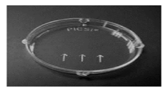
Before randomization, for all men enrolled to the study, semen analysis and sperm preparation were made. Semen samples were collected by masturbation after a 3to7 day period of sexual abstinence. Physical examination including volume, color, odor, and liquefaction were done. Microscopic examination (Olympus, C 21, Japan) was done to evaluate sperm concentration, motility, morphology, and the presence of another cellular element. Sperms were classified into progressive motile, non-progressive and Immotile. After randomization, sperm samples for ICSI were processed after microscopic examination, 1 ml of sperm gradient medium (Pure sperm, Nidacon, Sweden ) were added on fresh sample and centrifuged at 1800 rpm for 10 minutes (Heraeus 300, Osterode, Germany), supernatant was removed, and then we add 2 ml of sperm washing medium (Sage, Denmark) to sperms in the resulting pellet and centrifuged at 1800 rpm for 10 minutes to reach to a number of motile and morphologically normal sperm cells needed for assisted reproduction. Posterior morphology ICSI (PICSI) was made for the HA group only.
PICSI dishes (ORIGIO, Cooper Surgical Company, Denmark) are conventional plastic culture dishes pre-prepared with 3 microdots of powder. The powdered HA is re-hydrated by adding 5 μL droplets of fresh culture medium to each of the three microdots. A 2 μL droplet with suspension of treated spermatozoa is then connected with a pipette tip to these culture medium droplets. The PICSI dish is incubated under oil; within 5 minutes the bound spermatozoa are attached by their head to the surface of the HA-microdots and are spinning around their head.
An ICSI injecting pipette (Vitrolife, Sweden) is used to pick the best motile HA-bound sperm up and inject them one by one into an oocyte. The ICSI injecting pipette can be previously loaded with viscous medium PVP or Sperm Slow (Irvine, USA) to facilitate sperm micromanipulation. Controlled ovarian hyper stimulation (COS) was performed using long GnRH agonist protocol. In this protocol, the patients started administration of GnRH agonist, triptorelin 0.1 mg (Decapeptyl 0.1, Ferring, Egypt) on the 21 days of the cycle IM once. After 14 days of GnRH-agonist treatment, ovarian down-regulation was confirmed by measurement of serum E2 level. If the level of E2 50 pg/mL or less, gonadotrophins was started using recombinant FSH (Gonal-F, EMD Serono Inc., USA) or urinary HMG (Merional 75, IBSA Inc., Switzerland), commonly with a starting dose of 150-225 IU injected SC daily. FSH dose was adjusted according to the ovarian response, and serial E2 and transvaginal US (SONOACE X4 –MEDISON Korea.) were performed every 2-3 days from day 6 of FSH stimulation onward. When three or more follicles had reached a size 18 mm, final oocyte maturation was induced using intramuscular injection of 10,000 IU of HCG (Choriomon, IBSA Inc., Switzerland).
For collection of human cumulus cells, oocytes were aspirated under general anesthesia using transvaginal US, approximately 34-36 hours after the HCG administration (Labotect aspiration pump, Germany). Follicles were aspirated with a negative pressure of 115-120 mm Hg using a single lumen 17-gauge oocyte pick-up needle (Reproline Medical, Germany). Follicular fluid was aspirated into sterile tubes 14 ml (Falcon, Boen Healthcare Co., China). The Oocyte –cumulus cells complexes were isolated under a dissecting microscope (Zeiss Stemi 2000-C Stereo Microscope,) and washed in Gamete Buffer media (Cook, Limerick, Ireland) then washed and placed into four dishes containing fertilization medium (Cook, Limerick, Ireland) and incubated at 37 ºC in 6% CO2 for approximately 1/2 hour (C60, Labotect, Germany). For denudation, the oocyte was placed in a 100 μl drop of buffered containing hyaluronidase enzyme 80 IU/ml (Life Global, Europe) 30-45 seconds, and then the oocyte was removed and placed in 100 μl drop of Gamete Buffer media, then the corona cells were removed by gentle aspiration of the oocyte in and out of a sterile drawn pipette. When denudation was completed, the oocyte was washed in Gamete Buffer (Cook, Limerick, Ireland) and was then placed in 10 μl micro drops of the fertilization medium (Cook, Limerick, Ireland) in injection dishes, covered with 3ml of sterile equilibrated mineral oil. For the oocyte grading, the oocyte was assessed quickly for maturity & quality according to grading system using an inverted microscope with Hoffman optics (Olympus 1x71, Japan), hot stage and automatic manipulators (Narishige, japan). Maturation stages were be recorded as prophase I, metaphase I (MI), metaphase II (MII) and post mature. The naked oocytes were incubated in a culture medium at 37 ºC in 6% Co2 until the ICSI procedure. [14]
Icsi procedure:
Following sperm preparation as described before, samples were incubated until time of injection. Each oocyte was injected with a single morphologically abnormal and immobilized in polyvinyl pyrolidone (PVP) (Irvine, USA) spermatozoon. Individual sperm subjected to ICSI was examined and evaluated. The injection procedure was carried out in a sterilized dish using holding pipette and injection needle. Intra cytoplasmic sperm injection was performed according to the protocol of Van Steirteghem. [15].
Assessment of fertilization and embryo's quality was made 17± h after
microinjection. The injected oocytes were observed for any sign of damage and for the presence of pronuclei. Oocytes were named fertilized if two pronuclei (2PN) were present and the second polar body had been extruded. [16] For embryo grading and transfer, approximately 72 h after injection, the cell number and morphology of each embryo were scored according to equality of blastomeric size and proportion of nucleate fragments. Then, best day-3 embryos (Grade1) were transferred to the uterus in 30μl of Global medium (Life Global, Europe) containing 10% HSA using ET catheter (Labotect, Germany) 48–72 h after oocyte retrieval. [17]. Beta hCG titter was done for the detection of pregnancy and then was confirmed by transvaginal US examination 10-15 days after.
Statistical Analysis
The primary outcome measured was clinical pregnancy, defined as the presence of at least one intrauterine fetus with heartbeat. The secondary outcomes were, the number of collected total and MII oocytes; fertilization rate, number of G1 embryos and the number of embryos transferred.
Sample size was calculated setting the power at 80% and the two-sided confidence level at 95%. A minimal sample size of 45 couples in each group was needed. We considered a total sample size of 300 to allow for subgroup analysis according to semen analysis and to compensate for any dropout. [18]
The statistical analysis was made on the intent-to-treat (ITT) population. All statistical tests were made using a significance level of 95%. A p-value < 0>
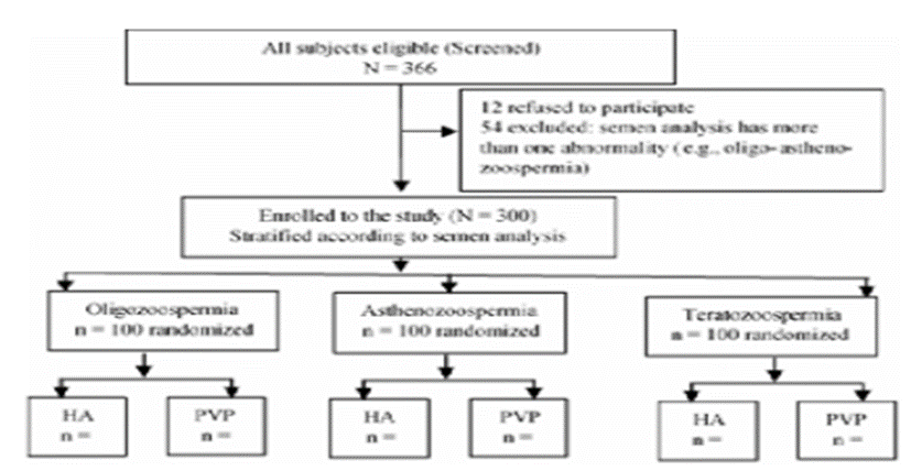
A total of 366 couples were asked to participate in the study. Twelve refused to participate, and 54 were excluded before randomization. Three hundred participants (100 with oligozoospermia, 100 with asthenozoospermia & 100 with teratozoospermia) were enrolled. Randomization was done within each semen category with 50 assigned to the HA group and 50 to the PVP group (Figure 2 ,CONSORT diagram).
Both groups were comparable with regard their baseline characteristics either overall dataset or in oligozoospermia, asthenozoospermia and teratozoospermia subsets. There was no statistically significant difference (p > 0.05) between the two groups regarding the female age, female BMI, and the duration of infertility (Table 1).
| HA group(n.=150) | PVP group (n.=150) | |||
Parameter | Mean (SD) | Mean (SD) | p | |
The entire dataset (N = 300) | Female age, years | 27.02 (5.71) | 28.17 (5.5) | 0.078 |
| Female BMI, Kg/m2 | 26.86 (4.66) | 27.1 (4.51) | 0.652 | |
| Duration of infertility, years | 7.32 (2.03) | 7.54 (2.05) | 0.335 | |
| Subgroups | Number =50 | Number = 50 | ||
| Oligozoospermia (n.=100) | Female age, years | 26.36 (6.07) | 26.94 (5.29) | 0.61 |
| Female BMI, Kg/m2 | 27.03 (4.42) | 28.23 (4.28) | 0.171 | |
| Duration of infertility, years | 7 (2.02) | 7.73 (2.13) | 0.081 | |
| Asthenozoospermia (n.=100) | Female age, years | 28.22 (5.42) | 28.98 (4.94) | 0.465 |
| Female BMI, Kg/m2 | 26.71 (4.58) | 26.33 (4.23) | 0.664 | |
| Duration of infertility, years | 7.57 (2.06) | 7.57 (2.07) | 0.993 | |
| Teratozoospermia (n.=100) | Female age, years | 26.49 (5.55) | 28.59 (6.11) | 0.076 |
| Female BMI, Kg/m2 | 26.85 (5.06) | 26.75 (4.85) | 0.92 | |
| Duration of infertility, years | 7.38 (2.02) | 7.33 (1.96) | 0.91 |
Table 1: Baseline characteristics
The controlled ovarian and the ICSI outcomes were comparable (p-values > 0.05) between the HA and PVP groups either in the overall dataset or in oligozoospermia, asthenozoospermia and teratozoospermia subsets, except for the number of fertilized oocytes, the cleavage rate, and the number of G1 embryos (p-values < 0>
Overall, the number of the collected oocytes was comparable (p-value 0.612) between the HA and PVP groups; [mean± (SD), was 6.85 (1.84) & 6.97 (2.23)] for the HA group and the PVP group, respectively. Also, overall, the number of the MII injected oocytes was comparable (p-value 0.300) between the HA and PVP groups; it was 6.29 (1.87) & 6.49 (1.44) for the HA group and the PVP group, respectively.
However, overall, the number of fertilized oocytes was higher (p-value < 0>
Moreover, in each of the oligozoospermia and teratozoospermia subsets, the number of fertilized oocytes and the fertilization rate were higher (p-values < 0> 0.05), Table 2.
Moreover, in each of the oligozoospermia, asthenozoospermia and teratozoospermia subsets, the cleavage rate, and the number of G1 embryos were higher (p-values < 0>
Overall, the number of transferred embryos was comparable (p-value 0.448) between the HA and PVP groups; it was 2.02 (0.56) & 2.07 (0.58) for the HA group and the PVP group, respectively. Moreover, in each of the oligozoospermia, asthenozoospermia and teratozoospermia subsets, the number of transferred embryos were comparable (p-values > 0.05) between the HA group and the PVP group as shown in Table 2.
HA group | PVP group | |||
Parameter | Mean (SD) | Mean (SD) | p-value | |
| Number of participants | 150 | 150 |
|
The entire dataset | Collected oocytes | 6.85 (1.84) | 6.97 (2.23) | 0.612 |
| MII injected oocytes | 6.29 (1.87) | 6.49 (1.44) | 0.3 | |
| Fertilized oocytes | 5.36 (2.08) | 4.63 (1.47) | < 0> | |
| Fertilization rate, n/MII oocytes (%) | 804/943 (85.26) | 694/973 (71.33) | < 0> | |
| Cleavage rate, n/fertilized oocytes (%) | 755/804 (93.91) | 538/694 (77.52) | < 0> | |
| G1 embryos | 3.97 (0.99) | 2.95 (0.83) | < 0> | |
| Transferred embryos | 2.02 (0.56) | 2.07 (0.58) | 0.448 | |
| Subgroups |
|
| ||
| Number of participants | 50 | 50 |
|
Oligozoospermia | Collected oocytes | 6.72 (1.74) | 7.08 (2.38) | 0.39 |
| MII injected oocytes | 6.24 (2.15) | 6.58 (1.52) | 0.363 | |
| Fertilized oocytes | 5.78 (1.69) | 5.06 (1.39) | 0.022 | |
| Fertilization rate, n/MII oocytes (%) | 289/312 (92.63) | 253/329 (76.90) | < 0> | |
| Cleavage rate, n/fertilized oocytes (%) | 268/289 (92.73) | 206/253 (81.42) | < 0> | |
| G1 embryos | 4.88 (1.09) | 3.92 (1.03) | < 0> | |
| Transferred embryos | 2.02 (0.58) | 2.08 (0.55) | 0.597 | |
Asthenozoospermia | Collected oocytes | 7.02 (1.84) | 6.82 (2.26) | 0.622 |
| MII injected oocytes | 6.34 (1.68) | 6.26 (1.47) | 0.801 | |
| Fertilized oocytes | 4.48 (1.60) | 4.58 (1.61) | 0.756 | |
| Fertilization rate, n/MII oocytes (%) | 224/317 (70.66) | 229/313 (73.16) | 0.486 | |
| Cleavage rate, n/fertilized oocytes (%) | 219/224 (97.77) | 185/229 (80.79) | < 0> | |
| G1 embryos | 4.06 (0.85) | 2.60 (0.75) | < 0> | |
| Transferred embryos | 2.06 (0.56) | 2 (0.63) | 0.616 | |
Teratozoospermia | Collected oocytes | 6.8 (1.96) | 7 (2.09) | 0.616 |
| MII injected oocytes | 6.28 (1.81) | 6.62 (1.33) | 0.279 | |
| Fertilized oocytes | 5.82 (2.04) | 4.24 (1.3) | < 0> | |
| Fertilization rate, n/MII oocytes (%) | 291/314 (92.68) | 212/331 (64.05) | < 0> | |
| Cleavage rate, n/fertilized oocytes (%) | 268/291 (92.10) | 147/212 (69.34) | < 0> | |
| G1 embryos | 2.96 (0.74) | 2.32 (0.53) | < 0> | |
| Transferred embryos | 1.98 (0.54) | 2.12 (0.58) | 0.215 |
Table 2: Controlled ovarian stimulation and ICSI outcomes
Overall, the clinical pregnancy rate, was significantly higher (p-value < 0>
Moreover, in each of the oligozoospermia, asthenozoospermia and teratozoospermia subsets, the clinical pregnancy rate, was significantly (p-values < 0>
| HA group | PVP group | p | ||
| The entire dataset | Clinical pregnancy, number (%) | 99 (66%) | 61 (40.67%) | < 0> |
| OR, estimate (95% CI) | 2.83 (1.77-4.53) | |||
| ARR, estimate (95% CI) | 25% (14-36%) | |||
| NNT, estimate (95% CI) | 4 (3-7) | |||
| Subgroups | ||||
| Oligozoospermia | Clinical pregnancy, number (%) | 35 (70%) | 23 (46%) | 0.011 |
| OR, estimate (95% CI) | 2.74 (1.2-6.23) | |||
| ARR, estimate (95% CI) | 24% (5-43%) | |||
| NNT, estimate (95% CI) | 4 (2-19) | |||
| Asthenozoospermia | Clinical pregnancy, number (%) | 33 (66) | 20 (40) | 0.006 |
| OR, estimate (95% CI) | 2.91 (1.29-6.57) | |||
| ARR, estimate (95% CI) | 26% (7-45%) | |||
| NNT, estimate (95% CI) | 4 (2-14) | |||
| Teratozoospermia | Clinical pregnancy, number (%) | 31 (62) | 18 (36) | 0.006 |
| OR, estimate (95% CI) | 2.9 (1.29-6.53) | |||
| ARR, estimate (95% CI) | 26% (7-45%) | |||
| NNT, estimate (95% CI) | 4 (2-14) | |||
Table 3: Clinical pregnancy among group
The current randomized controlled study showed that the number of fertilized oocytes was higher in the HA group than in the PVP group. Also, G1 embryos was higher in the HA group than in the PVP group. In addition, in each of the oligozoospermia and teratozoospermia subsets, the number of fertilized oocytes and the fertilization rate were significantly higher in the HA group than in the PVP group. However, in the asthenozoospermia subset, the number of fertilized oocytes and the fertilization rate were comparable between both groups. Moreover, in each of the oligozoospermia, asthenozoospermia and teratozoospermia subsets, the cleavage rate, and the number of G1 embryos were higher in the HA group than in the PVP group.
Overall, the number of transferred embryos was comparable between the HA and PVP groups. Furthermore, in each of the oligozoospermia, asthenozoospermia and teratozoospermia subsets, the number of transferred embryos were comparable between the HA group and the PVP group.
Also generally, the clinical pregnancy rate, was significantly higher in the HA group than in the PVP group. Additionally, in each of the oligozoospermia, asthenozoospermia and teratozoospermia subsets, the clinical pregnancy rate, was significantly higher in the HA group than in the PVP group.
The current study agreed with the study done by Erberelli et al., (2017), who studied 56 cases (19 ICSI and 37 PICSI) and reached similar results. Like our study, it had the advantage of classifying the different types of abnormal semen parameters (oligo, terato and asthenozoospermia). However, our study recruited a larger number than that of Erberelli et al.,
In discordance with the results of the current study, the study of Erbelli et al., (2017) showed that the number of aspirated oocytes in the PICSI group was higher in comparison to the PVP group. That is because they did not follow a specific COS protocol. However, in the present study one controlled ovarian stimulation (COS) protocol was used for all participants. [18]
Moreover, Erbelli et al., (2017) observed that the participants with teratozoospermia could benefit from the PICSI technique more than other factors sperm changes. However, in the current study, we observed the benefits in each type of sperm changes. [18]
Seemingly, the benefits of the HA binding system showed significant encouraging results only for male factor infertility as shown in the study by Majumdar (2013). [19]
Worrilow et al. (2013), did a double-blinded randomized controlled study to test the effect of PICSI on implantation, clinical pregnancy and miscarriage rates initially recruiting 802 cases and classified the cases into two main groups according to the percent of sperm bound hyaluronan in semen (>65% or 65% and less) and found a significant decrease in miscarriage rates in cases using PICSI, while clinical pregnancy rate was not significant, the later finding is different from the results of the current study; however, a possible explanation of this difference is that the current study clearly demarcates the types of semen abnormalities. [20]
Breznik et al. (2013), studied analysis of sperm DNA fragmentation, hyper activation and hyaluronan-binding ability on washed semen samples used for the insemination of oocytes in 133 cases undergoing IVF to determine the effect on both fertilization rate and embryo quality, and they had similar results to the current study in regarding both oligo and teratospermia groups. [21]
In a systematic review by Georgina et al 2018 (22), to determine the efficacy of the physiological ICSI technique (PICSI) vs. conventional ICSI in the prognosis of couples with male factor, they concluded that there is no statistically significant difference between PICSI vs. ICSI, for any of the outcomes analyzed in this study.
When comparing PICSI vs. magnetic-activated cell sorting (MACS) for abnormal sperm DNA fragmentation ICSI cases Hasanen et al 2018 (23), in their clincal trial found that PICSI and MACS are efficient techniques for sperm selection in cases with abnormal sperm DNA fragmentation. However, MACS is preferred when the females are younger than 30 years, while PICSI is preferred in older females.
However, enough information is still not available to prove the efficacy of the PICSI technique over ICSI in couples with male factor.
One advantage of this study is being randomized-controlled study with enough sample size. Furthermore, the current study categorizes three different types of male semen abnormalities separately. However, the study has some limitations as it did not report the miscarriage rate and the live-birth rate as the participant are still in the pregnancy phase.
PICSI technique provides better top-quality embryos, fertilization, and clinical pregnancy rates in cases with male factor of infertility. Further studies are needed to define specific criteria for performing PICSI taking into consideration both the expenses and time of the technique
This research did not receive any specific grant from any funding agency in the public, commercial or not-for-profit sector.
Many thanks to all staff at embryology unit, Al-Azhar university for their support during finishing of this study.
Clearly Auctoresonline and particularly Psychology and Mental Health Care Journal is dedicated to improving health care services for individuals and populations. The editorial boards' ability to efficiently recognize and share the global importance of health literacy with a variety of stakeholders. Auctoresonline publishing platform can be used to facilitate of optimal client-based services and should be added to health care professionals' repertoire of evidence-based health care resources.

Journal of Clinical Cardiology and Cardiovascular Intervention The submission and review process was adequate. However I think that the publication total value should have been enlightened in early fases. Thank you for all.

Journal of Women Health Care and Issues By the present mail, I want to say thank to you and tour colleagues for facilitating my published article. Specially thank you for the peer review process, support from the editorial office. I appreciate positively the quality of your journal.

Journal of Clinical Research and Reports I would be very delighted to submit my testimonial regarding the reviewer board and the editorial office. The reviewer board were accurate and helpful regarding any modifications for my manuscript. And the editorial office were very helpful and supportive in contacting and monitoring with any update and offering help. It was my pleasure to contribute with your promising Journal and I am looking forward for more collaboration.

We would like to thank the Journal of Thoracic Disease and Cardiothoracic Surgery because of the services they provided us for our articles. The peer-review process was done in a very excellent time manner, and the opinions of the reviewers helped us to improve our manuscript further. The editorial office had an outstanding correspondence with us and guided us in many ways. During a hard time of the pandemic that is affecting every one of us tremendously, the editorial office helped us make everything easier for publishing scientific work. Hope for a more scientific relationship with your Journal.

The peer-review process which consisted high quality queries on the paper. I did answer six reviewers’ questions and comments before the paper was accepted. The support from the editorial office is excellent.

Journal of Neuroscience and Neurological Surgery. I had the experience of publishing a research article recently. The whole process was simple from submission to publication. The reviewers made specific and valuable recommendations and corrections that improved the quality of my publication. I strongly recommend this Journal.

Dr. Katarzyna Byczkowska My testimonial covering: "The peer review process is quick and effective. The support from the editorial office is very professional and friendly. Quality of the Clinical Cardiology and Cardiovascular Interventions is scientific and publishes ground-breaking research on cardiology that is useful for other professionals in the field.

Thank you most sincerely, with regard to the support you have given in relation to the reviewing process and the processing of my article entitled "Large Cell Neuroendocrine Carcinoma of The Prostate Gland: A Review and Update" for publication in your esteemed Journal, Journal of Cancer Research and Cellular Therapeutics". The editorial team has been very supportive.

Testimony of Journal of Clinical Otorhinolaryngology: work with your Reviews has been a educational and constructive experience. The editorial office were very helpful and supportive. It was a pleasure to contribute to your Journal.

Dr. Bernard Terkimbi Utoo, I am happy to publish my scientific work in Journal of Women Health Care and Issues (JWHCI). The manuscript submission was seamless and peer review process was top notch. I was amazed that 4 reviewers worked on the manuscript which made it a highly technical, standard and excellent quality paper. I appreciate the format and consideration for the APC as well as the speed of publication. It is my pleasure to continue with this scientific relationship with the esteem JWHCI.

This is an acknowledgment for peer reviewers, editorial board of Journal of Clinical Research and Reports. They show a lot of consideration for us as publishers for our research article “Evaluation of the different factors associated with side effects of COVID-19 vaccination on medical students, Mutah university, Al-Karak, Jordan”, in a very professional and easy way. This journal is one of outstanding medical journal.

Dear Hao Jiang, to Journal of Nutrition and Food Processing We greatly appreciate the efficient, professional and rapid processing of our paper by your team. If there is anything else we should do, please do not hesitate to let us know. On behalf of my co-authors, we would like to express our great appreciation to editor and reviewers.

As an author who has recently published in the journal "Brain and Neurological Disorders". I am delighted to provide a testimonial on the peer review process, editorial office support, and the overall quality of the journal. The peer review process at Brain and Neurological Disorders is rigorous and meticulous, ensuring that only high-quality, evidence-based research is published. The reviewers are experts in their fields, and their comments and suggestions were constructive and helped improve the quality of my manuscript. The review process was timely and efficient, with clear communication from the editorial office at each stage. The support from the editorial office was exceptional throughout the entire process. The editorial staff was responsive, professional, and always willing to help. They provided valuable guidance on formatting, structure, and ethical considerations, making the submission process seamless. Moreover, they kept me informed about the status of my manuscript and provided timely updates, which made the process less stressful. The journal Brain and Neurological Disorders is of the highest quality, with a strong focus on publishing cutting-edge research in the field of neurology. The articles published in this journal are well-researched, rigorously peer-reviewed, and written by experts in the field. The journal maintains high standards, ensuring that readers are provided with the most up-to-date and reliable information on brain and neurological disorders. In conclusion, I had a wonderful experience publishing in Brain and Neurological Disorders. The peer review process was thorough, the editorial office provided exceptional support, and the journal's quality is second to none. I would highly recommend this journal to any researcher working in the field of neurology and brain disorders.
Dear Agrippa Hilda, Journal of Neuroscience and Neurological Surgery, Editorial Coordinator, I trust this message finds you well. I want to extend my appreciation for considering my article for publication in your esteemed journal. I am pleased to provide a testimonial regarding the peer review process and the support received from your editorial office. The peer review process for my paper was carried out in a highly professional and thorough manner. The feedback and comments provided by the authors were constructive and very useful in improving the quality of the manuscript. This rigorous assessment process undoubtedly contributes to the high standards maintained by your journal.

International Journal of Clinical Case Reports and Reviews. I strongly recommend to consider submitting your work to this high-quality journal. The support and availability of the Editorial staff is outstanding and the review process was both efficient and rigorous.

Thank you very much for publishing my Research Article titled “Comparing Treatment Outcome Of Allergic Rhinitis Patients After Using Fluticasone Nasal Spray And Nasal Douching" in the Journal of Clinical Otorhinolaryngology. As Medical Professionals we are immensely benefited from study of various informative Articles and Papers published in this high quality Journal. I look forward to enriching my knowledge by regular study of the Journal and contribute my future work in the field of ENT through the Journal for use by the medical fraternity. The support from the Editorial office was excellent and very prompt. I also welcome the comments received from the readers of my Research Article.

Dear Erica Kelsey, Editorial Coordinator of Cancer Research and Cellular Therapeutics Our team is very satisfied with the processing of our paper by your journal. That was fast, efficient, rigorous, but without unnecessary complications. We appreciated the very short time between the submission of the paper and its publication on line on your site.

I am very glad to say that the peer review process is very successful and fast and support from the Editorial Office. Therefore, I would like to continue our scientific relationship for a long time. And I especially thank you for your kindly attention towards my article. Have a good day!

"We recently published an article entitled “Influence of beta-Cyclodextrins upon the Degradation of Carbofuran Derivatives under Alkaline Conditions" in the Journal of “Pesticides and Biofertilizers” to show that the cyclodextrins protect the carbamates increasing their half-life time in the presence of basic conditions This will be very helpful to understand carbofuran behaviour in the analytical, agro-environmental and food areas. We greatly appreciated the interaction with the editor and the editorial team; we were particularly well accompanied during the course of the revision process, since all various steps towards publication were short and without delay".

I would like to express my gratitude towards you process of article review and submission. I found this to be very fair and expedient. Your follow up has been excellent. I have many publications in national and international journal and your process has been one of the best so far. Keep up the great work.

We are grateful for this opportunity to provide a glowing recommendation to the Journal of Psychiatry and Psychotherapy. We found that the editorial team were very supportive, helpful, kept us abreast of timelines and over all very professional in nature. The peer review process was rigorous, efficient and constructive that really enhanced our article submission. The experience with this journal remains one of our best ever and we look forward to providing future submissions in the near future.

I am very pleased to serve as EBM of the journal, I hope many years of my experience in stem cells can help the journal from one way or another. As we know, stem cells hold great potential for regenerative medicine, which are mostly used to promote the repair response of diseased, dysfunctional or injured tissue using stem cells or their derivatives. I think Stem Cell Research and Therapeutics International is a great platform to publish and share the understanding towards the biology and translational or clinical application of stem cells.

I would like to give my testimony in the support I have got by the peer review process and to support the editorial office where they were of asset to support young author like me to be encouraged to publish their work in your respected journal and globalize and share knowledge across the globe. I really give my great gratitude to your journal and the peer review including the editorial office.

I am delighted to publish our manuscript entitled "A Perspective on Cocaine Induced Stroke - Its Mechanisms and Management" in the Journal of Neuroscience and Neurological Surgery. The peer review process, support from the editorial office, and quality of the journal are excellent. The manuscripts published are of high quality and of excellent scientific value. I recommend this journal very much to colleagues.

Dr.Tania Muñoz, My experience as researcher and author of a review article in The Journal Clinical Cardiology and Interventions has been very enriching and stimulating. The editorial team is excellent, performs its work with absolute responsibility and delivery. They are proactive, dynamic and receptive to all proposals. Supporting at all times the vast universe of authors who choose them as an option for publication. The team of review specialists, members of the editorial board, are brilliant professionals, with remarkable performance in medical research and scientific methodology. Together they form a frontline team that consolidates the JCCI as a magnificent option for the publication and review of high-level medical articles and broad collective interest. I am honored to be able to share my review article and open to receive all your comments.

“The peer review process of JPMHC is quick and effective. Authors are benefited by good and professional reviewers with huge experience in the field of psychology and mental health. The support from the editorial office is very professional. People to contact to are friendly and happy to help and assist any query authors might have. Quality of the Journal is scientific and publishes ground-breaking research on mental health that is useful for other professionals in the field”.

Dear editorial department: On behalf of our team, I hereby certify the reliability and superiority of the International Journal of Clinical Case Reports and Reviews in the peer review process, editorial support, and journal quality. Firstly, the peer review process of the International Journal of Clinical Case Reports and Reviews is rigorous, fair, transparent, fast, and of high quality. The editorial department invites experts from relevant fields as anonymous reviewers to review all submitted manuscripts. These experts have rich academic backgrounds and experience, and can accurately evaluate the academic quality, originality, and suitability of manuscripts. The editorial department is committed to ensuring the rigor of the peer review process, while also making every effort to ensure a fast review cycle to meet the needs of authors and the academic community. Secondly, the editorial team of the International Journal of Clinical Case Reports and Reviews is composed of a group of senior scholars and professionals with rich experience and professional knowledge in related fields. The editorial department is committed to assisting authors in improving their manuscripts, ensuring their academic accuracy, clarity, and completeness. Editors actively collaborate with authors, providing useful suggestions and feedback to promote the improvement and development of the manuscript. We believe that the support of the editorial department is one of the key factors in ensuring the quality of the journal. Finally, the International Journal of Clinical Case Reports and Reviews is renowned for its high- quality articles and strict academic standards. The editorial department is committed to publishing innovative and academically valuable research results to promote the development and progress of related fields. The International Journal of Clinical Case Reports and Reviews is reasonably priced and ensures excellent service and quality ratio, allowing authors to obtain high-level academic publishing opportunities in an affordable manner. I hereby solemnly declare that the International Journal of Clinical Case Reports and Reviews has a high level of credibility and superiority in terms of peer review process, editorial support, reasonable fees, and journal quality. Sincerely, Rui Tao.

Clinical Cardiology and Cardiovascular Interventions I testity the covering of the peer review process, support from the editorial office, and quality of the journal.
