AUCTORES
Globalize your Research

Research | DOI: https://doi.org/10.31579/2578-8949/099
1 Dermatology and Andrology department, Faculty of Medicine, Minia University, Egypt.
2 Pathology and Laboratory Medicine, London Health Sciences Centre, Schulich School of Medicine & Dentistry, Western University, London, Canada.
*Corresponding Author: Sherif S. Awad, Dermatology and Andrology department, Faculty of Medicine, Minia University, Egypt.
Citation: A Fabrice, A Hugues, D Juste, D Berenice, A Nadege, at el. (2022) Epidemiological profile of tumor dermatoses in the hospital setting in Cotonou (Benin) from 2009 to 2018. Dermatology and Dermatitis. 7(2); Doi:10.31579/2578-8949/099
Copyright: ©2022 Sherif S. Awad, this is an open-access article distributed under the terms of The Creative Commons. Attribution License, which permits unrestricted use, distribution, and reproduction in any medium, provided the original author and source are credited.
Received: 26 May 2022 | Accepted: 25 April 2022 | Published: 09 May 2022
Keywords: vitiligo; immunomarkers; melanoblasts;melanocyte stem cells
Melanocytes are specialized neural crest-derived cells that are responsible for skin pigmentation. In vitiligo, loss of functioning epidermal melanocytes results in loss of pigment. Based on published studies, many transcription factors including microphtalmia-associated transcription factor, SRY (Sex Determining Region Y)-Box 10 are responsible for differentiation of melanocytes from neural crest cells as well as development of melanoblasts). However, the pathogenesis of loss of pigmentation in skin still unclear.
We studied the expression of several melanocyte-related proteins and markers in vitiligo samples and comparing them to normally pigmented skin samples. This may provide a more insight into the pathogenesis of vitiligo which is a still controversy.
We assessed the expression of six antibodies including S100, Melan-A (to detect fully formed, functioning melanocytes), CD117 (a specific relevant tyrosinase inducer), MITF and SOX-10 (important related transcription factors), and BCL-2 (stem cell markers) in 32 skin samples.
We demonstrated significant decline in the expression of all examined immunomarkers in the skin of vitiligo when compared to normal pigmented skin.
Our data may speculate that vitiligo pathogenesis does not involve only destruction of functioning active melanocytes, but also, other steps of cellular stimulation of induction of melanocytes and melanogensis are significantly affected as well.
microphtalmia-associated transcription factor (MITF), SRY (Sex Determining Region Y)-Box 10 (SOX10), tyrosine kinase receptor KIT (c-kit), melanocyte stem cells (MelSCs), Dopachrome tautomerase gene (Dct),
Melanocytes are specialized neural crest-derived cells that are responsible for skin pigmentation. The origin of melanocytes can be directly from neural crest cells migrating at the level of the skin through a dorsolateral migratory pathway, or from Schwann cell progenitors present in the peripheral nerves located at the level of the skin. Melanocytes reside in the basal layer of epidermis where they form the epidermal melanin units. The ratio of melanocytes to keratinocytes is 1: 10 in the epidermal basal layer (Haass et al. 2005). Melanocytes produce melanin in melanosomes and transfer it to neighboring keratinocytes by using the dendrites, giving the skin color, and protecting the genetic material of keratinocytes from damage caused by UV radiation (Pisarchik A, Wortsman J 2004).
Melanoblasts are the melanocyte precursor cells and arise during gastrulation of embryogenesis at the dorsal edge of the neural crest (Thomas AJ 2009). The differentiation of melanocytes from neural crest cells as well as development of melanoblasts is controlled through complex molecular mechanisms mediated by a network of transcription factors, including microphtalmia-associated transcription factor (Mitf), SRY (Sex Determining Region Y)-Box 10 (SOX10), Paired Box 3 and Wnt proteins (Hou, Ling & Pavan 2008).
A key factor during melanocyte differentiation is microphthalmia associated transcription factor (Mitf) (Widlund HR 2003), (Hemesath, Timothy & Steingrímsson, Eiríkur & Mcgill, Gaël & J Hansen, M & Vaught, J & Hodgkinson 1994). MITF controls the expression of key pigment synthetic genes including TRP-1, dopachrome tautomerase and tyrosinase (Lin, Connie et al 2002). Various extracellular signaling pathways converge on MITF to control both migration and survival of melanoblasts (Tabone-Eglinger S., et al 2012).
Dependency on tyrosine kinase receptor KIT (c-kit) receptor and its ligand stem cell factor (SCF) is well known (Murphy, M., Reid, K. et al 1992) ,(Cable, J., et al 1995) ,(Sarin, Kavita & E Artandi 2007). In the bulge region of the hair follicle, melanoblasts can differentiate into melanocytes or can remain as melanocyte stem cells (MelSCs) (Kubic D, Jennifer, et al 2008).
Sox10 is also a neural crest transcription factor crucial for specification, maturation, and maintenance of Schwann cells and melanocytes. One of the studies suggested that SOX10 binding sites contribute significantly to the cAMP responsiveness of the Mitf promoter (Huber, W.E., et al 2003). Some of in vitro and in vivo tests showed that some melanogenic genes are stimulated not only by Mitf but also directly by SOX10, a regulator of MITF, suggesting the existence of feed-forward loops between SOX10 and Mitf target genes. Without functional MITF, however, melanocytes can never mature and become pigmented, but when they remain unpigmented because they have mutations in the melanin biosynthetic genes, they stay alive and thrive (Bauer GL, et al 2009).
Melanocytes molecularly are recognizable by identification of melanocyte-specific proteins as TYR, TYRP1, TYRP2, melanosomal matrix proteins as Pmel17, MART-1, and transcription factors such as microphthalmia transcription factor (MITF) (Plonka PM, et al 2009).
In vitiligo, loss of functioning epidermal melanocytes results in loss of pigment and induction of leukoderma characterized by disfiguring macules and patches of depigmented skin.(Arican O and Kurutas EB 2008).
The aim of this work is to monitor the expression of several melanocyte related proteins in normal skin and to compare that to the expression in vitiligo skin. This may provide a more insight into the pathogenesis of vitiligo which is a still controversy.
Clinical data
The study included 32 skin samples obtained using 4mm punch probes. Half of them were obtained from center of depigmented vitiligo skin of non-segmental vitiligo cases and the other half from normally pigmented skin away from the lesions. Biopsies were immediately fixed in 10% formalin, routinely processed and embedded in paraffin blocks.
Immunohistochemical staining and scoring:
Immunohistochemical staining was performed on the paraffin-embedded specimens using a semi-automated Discovery autostainer (Dako EnVision FLEX+ detection system) and EnVision FLEX+ DAB+ Chromogen.
Tissues were exposed to 7 different primary antibodies including: 1-S100: polyclonal rabbit anti-S100 (code no.: IR504, Dako, North America) is used. Melanocytic cells were scored positive if they showed cytoplasmic or nuclear staining. 2- Melan-A: FLEX monoclonal Mouse Anti-Human Melan-A (code IR633, Dako, North America) is used. Cells were scored positive if they showed cytoplasmic staining. 3- CD117: polyclonal Rabbit Anti-Human CD117 (c-kit) antibody (code no.: A4502, Dako, North America) at a dilution 1:100 is used. Cells were scored positive if they showed cytoplasmic staining. 4- MITF (microphthalmia transcription factor): monoclonal Mouse Anti-Human MITF antibody (code no.: M3621, Dako, North America) at a dilution of 1:100 is used. Cells were scored positive if they showed nuclear staining. 5- SOX10 monoclonal Mouse Antibody SOX10 (code: SC-365692, Santa Cruz Biotechnology, Europe) at a dilution of 1:300 is used. Cells were scored positive if they showed cytoplasmic or nuclear staining. 6- BCL-2: monoclonal Mouse Anti-Human BCL-2 Oncoprotein (code no.: IR614, Dako, North America) is used. Cells were scored positive if they showed cytoplasmic or nuclear staining.
Slides scanning:
The slides were examined and scanned by using Aperio glass slide scanner at Department of Pathology and laboratory Medicine at Western University. The ScanScope scans microscope slides at 20X, 40X to a digital image that can be viewed with software that simulates the use of a light microscope. The svs files can be viewed and manipulated using a free software program called ImageScope. Representative photographs were captured using Aperio ScanScope™ technology at equivalent exposure times.
Immunohistochemical scoring:
Evaluation of the epidermal expression was carried by counting the number of positively stained cells within the epidermis in 1 mm length of stained tissue in the obtained digital slide-photos and multiplying the number by the intensity of positive staining; 1 for mild staining, 2 for moderate intensity and 3 for strong. Three independent doctors carried out the evaluation and median value for each case was recorded for further statistical evaluation
Statistical analysis
Clinical data were compared across the two groups using analysis of variance for differences in clinical parameters including age, sex, skin type, duration and activity of the disease and family history. A two-way repeated measures ANOVA with an interaction test and pairwise differences were used for comparing parametric quantitative data of two groups regarding the differences in the expression of six immunomarkers. All tests were two-sided, with a P-value of <0>
Clinical data
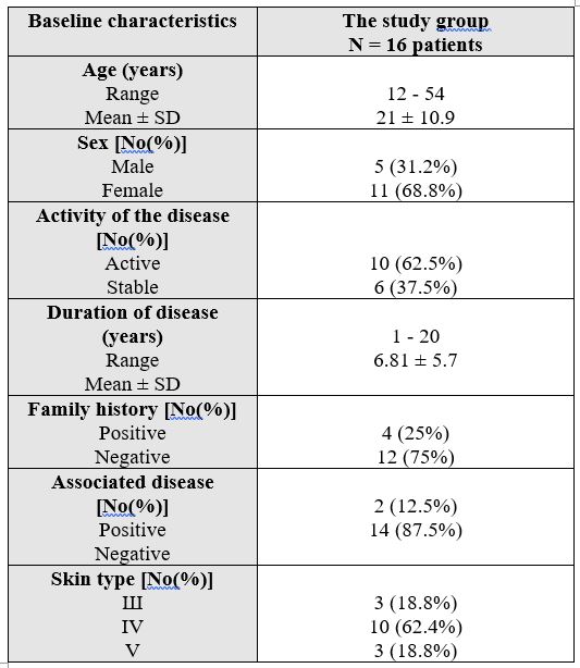
The present study included 16 patients with vitiligo vulgaris. Eleven female patients (68.8%) and 5 males (31.2%) were included in this study. The age of these patients ranged from 12 to 54 years old (mean 21 ± 10.9). The disease duration ranged from 1 to 20 years (6.81 ± 5.7). A positive family history of vitiligo presented in 4 patients (25%). Associated autoimmune diseases presented in 2 patients (12.5%) (one patient had alopecia areata, the other patient had idiopathic thrombocytopenic purpura). As regard skin type, 3 patients (18.8%) were skin type III, 10 patients (62.4%) were skin type IV and 3 patients (18.8%) were skin type V (Table 1).
3.2 Results of immunohistochemistry staining:
S100 expression:
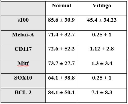
Examining the epidermis of the stained skin samples revealed that S100 positively stained cells were seen within the epidermis and at the basal layer. Scattered Cells were more abundant in normal skin compared to vitiligo skin which also contained several positively stained cells within the epidermis yet away from basal layer. S100 values in vitiliginous lesion ranged from 12 to 120 (mean 45.4 ± 34.23). In the non-lesional normally pigmented skin, it ranged from 45 to 144 (mean 85.6 ± 30.9) with statistically significant difference (p = 0.001), compared to vitiliginous lesion (Figure 1).

Melan-A expression:
Melan-A positively stained cells were demonstrated only within basal cells. Melan-A stained cells were minimal in vitiliginous lesion with values ranged from 0 to 4 (0.25 ± 1). In the non-lesional normally pigmented skin, it ranged from 15 to 135 (71.4 ± 32.7) with statistically significant difference (p = 0.001) (Figure 2).

CD-117 expression:
CD 117 (C-Kit) stained cells were demonstrated in the basal layer only. CD 117 stained cells in vitiliginous lesion were scanty and ranged from 0 to 10 (mean 1.12 ± 2.8). In the non-lesional normally pigmented skin, it ranged from 14 to 174 (mean 72.6 ± 52.3) with statistically significant difference (p = 0.001) (Figure 3).

MITF expression:
Mitf revealed positively stained cells within the basal layer. MITF stained cells showed values in vitiliginous lesion ranging from 0 to 12 (1.3 ± 3.4). In the non-lesional normally pigmented skin, it ranged from 7 to 108 (73.7 ± 27.7) with statistically significant difference (p = 0.001) (Figure 4)

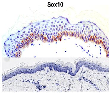
SOX-10 expression:
SOX10 revealed positively stained cells within the basal layer. SOX10 stained cells in vitiliginous lesion were minimal with values ranged from 0 to 4 (0.25 ± 1). In the non-lesional normally pigmented skin, it ranged from 14 to 180 (64.1 ± 38.8) with statistically significant difference (p = 0.001) (Figure 5).
BCL-2 expression:
BCL-2 revealed positively stained cells within the basal layer. BCL-2 values in vitiliginous lesion ranged from 0 to 22 (7.1 ± 8.3). In the non-lesional normally pigmented skin, it ranged from 10 to 180 (84.1 ± 50.1) with statistically significant difference (p = 0.001) (Figure 6).

The overall expression values in vitiligo and normal skin are summarized in (Table 2 and Figure 7).

Correlation of expression of all markers in epidermis of diseased skin specimens:
By comparing the expression of all markers in the epidermis of vitiliginous specimens, significant decline was demonstrated. However, there is a significant difference between S100 & BC12 in expression in vitiligo specimens, but none of the others.
Melanocyte is a specialized cell which evolves from primitive skin stem cells and goes through a long and complicated process of differentiation, proliferation, migration, maturation and melanin synthesis. This study targeted the demonstration of expression of several melanocytes related proteins and markers in vitiligo samples and comparing them to normally pigmented skin.
S-100 and Melan A were used to detect fully formed and functioning melanocytes, CD117 was utilized as a specific relevant tyrosinase inducer, SOX10 and MITF were examined as important related transcription factors, and BCL-2 as stem cell markers.
It is known that, S100 protein can be expressed in different cells including fibroblasts, myoepithelial cells, histocytes and Langerhans cells in normal skin biopsies.(Xia J, et al 2016). We demonstrated that loss of S100 staining in vitiligo was mainly at basal layers as other non-melanocytic cells are still present intra-epidermally like Langerhans cells. Yet, significant drop in total values of expression in the epidermis was demonstrated due to the loss of functioning melanocytes located in the basal layer.
On the other hand, Melan-A expression is known to be restricted to melanin-producing cells, including normal and transformed skin melanocytes (Kawakami Y, et al 1994). Melan-A may be a melanosomal membrane protein, although its sequence does not bear any apparent homology to other known melanosomal proteins and does not possess melanogenic enzymatic activities(Kawakami Y, et al 1997). Our study was clearly demonstrating that Melan-A expression significantly decreased in vitiligo skin compared to normal skin. This data is comparable to previously reported studies and minimal persistence of some positive cells is also recorded before (Mandelcorn-Monson RL, et al 2003) (Park OJ, et al 2016).
CD117 was also examined as it is a known important and specific inducer of melanocytes and melanogenesis. Significant drop in expression of this was clearly demonstrated in vitiligo. The intracellular signaling from the c-Kit plays a critical role in the development of melanocytes. Downstream signal transduction molecules can be activated through the formation of the c-kit/SCF complex, and then regulate gene expression and cell growth, proliferation and differentiation (Nishikawa S, et al 1991), (Grabbe J, et al 1994), (Galli SJ, et al 1995). There was a significant decline in C-kit expression in vitiligo compared to normal skin and this decline is similar to previous reports although was non-significant in many cases (Mandelcorn-Monson RL, et al 2003),(Park O, et al 2016).
Transcription factors and proteins are required during the process of melanocytes induction and formation from precursors. The microphthalmia-associated transcription factor (Mitf) protein is a key regulator of melanocyte development. The function of Mitf has been particularly well-studied in melanocytes , and it has been shown that Mitf is involved in the maintenance and self-renewal of melanocyte stem cells (Lang D1, et al 2005) (Loercher AE, et al 2005). Mitf can efficiently transactivate the melanogenesis enzyme genes, such as tyrosinase and tyrosinase-related protein 1 (TRP-1) in cultured cells (Yasumoto K, et al 1994), (Yasumoto K, et al 1997) ,(Amae S, et al 1998) , (Shibahara S, et al 1999). A significant drop in expression of this factor was demonstrated in vitiligo samples compared to normal pigmented skin. Bhardwaj et al has been described similar findings in a recent study as well (Bhardwaj S, et al 2017).
SOX10 transcription factor belongs to the Sox-family of transcription factors important in the development and maintenance of melanocytes. It was significantly declined in vitiligo compared to expression in normal skin. In the melanocyte lineage, SOX10 directly regulates the expression of the Mitf gene (Bondurand N1, et al 2000), (Potterf SB, et al 2000), (Lee M, Goodall J, et al 2000) . and SOX10 also regulates the expression of the Dopachrome tautomerase gene (Dct), which functions in melanin biosynthesis (Britsch S, et al 2001) ,(Potterf SB, et al 2001) . It was demonstrated the elevation of expression of sox 10 in melanocytic tumors (Shin J, et al 2012) yet studying its expression in vitiligo is lacking and this could be first study to show its decline in vitiligo skin.
Stem cells are required to provide generations of melanocytes needed for the pigmentation process. No single marker is currently universally accepted as an identifier of all melanocyte stem cells (Bonchak JG, et al 2014) and here we examined for BCL-2. The proteins of the Bcl-2 family are important regulators of programmed cell death (Oltvai ZN, Milliman CL 1993) . BCL-2 interacts with Mitf to maintain survival of melanocyte (McGill GG, et al 2002). There is an indispensible role of BCL-2 in the development and maintenance of melanocyte stem cells (Hallsson JH, et al 2007) and so; mice null bcls lose pigment and melanoblasts decline on lose of BCL-2 (Mak SS, et al 2005). In our study BCL-2 was significantly decreased in the vitiliginous areas. Although some others, previously, denied any dysregulation of apoptosis molecules including BCL-2 in vitiligo (van den Wijngaar, et al 2000).
This study demonstrated the significant decline of several proteins and markers from the skin of vitiligo when compared to normal pigmented skin. It is comparable to many previous reports regarding MART-1, S100, and for a few reports regarding C-Kit expression or other transcription factors like Mitf yet it is the first report to signify the decline of SOX10, and BCL-2 in vitiligo skin.
In conclusion, Vitiligo skin lacks melanocytes and subsequently loses its melanocyte specific markers and proteins like Melan-A significantly. S100, although non-specific and can stain different types of cells within the epidermis, yet its expression in vitiligo also showed a significant drop compared to non lesional skin. The vital melanogenic factor C-Kit protein again demonstrated significant decline. Transcription factors and proteins required for induction of melanocyte and melanogenesis, Mitf and SOX10 were also significantly decreased. Stem cell related proteins and markers, BCL-2, significantly drop in vitiligo skin compared to non lesional skin and this may explain the persistent lack of the repigmentation in many cases and the characteristic recalcitrance of the disease.
Such findings may demonstrate that vitiligo pathogenesis does not involve only destruction of functioning active melanocytes, yet, other steps of early cellular stimulation of induction and melanocyte precursors are targeted and significantly affected as well.
This is a possible new view demonstrating that vitiligo could be a problem of homeostasis. In normal skin, not only functioning melanocyte proteins like Melan-A and CD117 exist but precursor and MelSCs related proteins like SOX-10, Mitf, BCL-2 also exist, so as to continually provide new generations of pigment producing cells to keep this pigmentary homeostasis. But in vitiligo, loss of those melanogenic machinery components leads to stoppage of production process from a very early step. Vitiligo skin cannot maintain the usual existence of melanocytes in the skin through continual stimulation of melanocyte precursor’s lineage due to lack of required precursors and their relevant proteins.
None
Clearly Auctoresonline and particularly Psychology and Mental Health Care Journal is dedicated to improving health care services for individuals and populations. The editorial boards' ability to efficiently recognize and share the global importance of health literacy with a variety of stakeholders. Auctoresonline publishing platform can be used to facilitate of optimal client-based services and should be added to health care professionals' repertoire of evidence-based health care resources.

Journal of Clinical Cardiology and Cardiovascular Intervention The submission and review process was adequate. However I think that the publication total value should have been enlightened in early fases. Thank you for all.

Journal of Women Health Care and Issues By the present mail, I want to say thank to you and tour colleagues for facilitating my published article. Specially thank you for the peer review process, support from the editorial office. I appreciate positively the quality of your journal.

Journal of Clinical Research and Reports I would be very delighted to submit my testimonial regarding the reviewer board and the editorial office. The reviewer board were accurate and helpful regarding any modifications for my manuscript. And the editorial office were very helpful and supportive in contacting and monitoring with any update and offering help. It was my pleasure to contribute with your promising Journal and I am looking forward for more collaboration.

We would like to thank the Journal of Thoracic Disease and Cardiothoracic Surgery because of the services they provided us for our articles. The peer-review process was done in a very excellent time manner, and the opinions of the reviewers helped us to improve our manuscript further. The editorial office had an outstanding correspondence with us and guided us in many ways. During a hard time of the pandemic that is affecting every one of us tremendously, the editorial office helped us make everything easier for publishing scientific work. Hope for a more scientific relationship with your Journal.

The peer-review process which consisted high quality queries on the paper. I did answer six reviewers’ questions and comments before the paper was accepted. The support from the editorial office is excellent.

Journal of Neuroscience and Neurological Surgery. I had the experience of publishing a research article recently. The whole process was simple from submission to publication. The reviewers made specific and valuable recommendations and corrections that improved the quality of my publication. I strongly recommend this Journal.

Dr. Katarzyna Byczkowska My testimonial covering: "The peer review process is quick and effective. The support from the editorial office is very professional and friendly. Quality of the Clinical Cardiology and Cardiovascular Interventions is scientific and publishes ground-breaking research on cardiology that is useful for other professionals in the field.

Thank you most sincerely, with regard to the support you have given in relation to the reviewing process and the processing of my article entitled "Large Cell Neuroendocrine Carcinoma of The Prostate Gland: A Review and Update" for publication in your esteemed Journal, Journal of Cancer Research and Cellular Therapeutics". The editorial team has been very supportive.

Testimony of Journal of Clinical Otorhinolaryngology: work with your Reviews has been a educational and constructive experience. The editorial office were very helpful and supportive. It was a pleasure to contribute to your Journal.

Dr. Bernard Terkimbi Utoo, I am happy to publish my scientific work in Journal of Women Health Care and Issues (JWHCI). The manuscript submission was seamless and peer review process was top notch. I was amazed that 4 reviewers worked on the manuscript which made it a highly technical, standard and excellent quality paper. I appreciate the format and consideration for the APC as well as the speed of publication. It is my pleasure to continue with this scientific relationship with the esteem JWHCI.

This is an acknowledgment for peer reviewers, editorial board of Journal of Clinical Research and Reports. They show a lot of consideration for us as publishers for our research article “Evaluation of the different factors associated with side effects of COVID-19 vaccination on medical students, Mutah university, Al-Karak, Jordan”, in a very professional and easy way. This journal is one of outstanding medical journal.

Dear Hao Jiang, to Journal of Nutrition and Food Processing We greatly appreciate the efficient, professional and rapid processing of our paper by your team. If there is anything else we should do, please do not hesitate to let us know. On behalf of my co-authors, we would like to express our great appreciation to editor and reviewers.

As an author who has recently published in the journal "Brain and Neurological Disorders". I am delighted to provide a testimonial on the peer review process, editorial office support, and the overall quality of the journal. The peer review process at Brain and Neurological Disorders is rigorous and meticulous, ensuring that only high-quality, evidence-based research is published. The reviewers are experts in their fields, and their comments and suggestions were constructive and helped improve the quality of my manuscript. The review process was timely and efficient, with clear communication from the editorial office at each stage. The support from the editorial office was exceptional throughout the entire process. The editorial staff was responsive, professional, and always willing to help. They provided valuable guidance on formatting, structure, and ethical considerations, making the submission process seamless. Moreover, they kept me informed about the status of my manuscript and provided timely updates, which made the process less stressful. The journal Brain and Neurological Disorders is of the highest quality, with a strong focus on publishing cutting-edge research in the field of neurology. The articles published in this journal are well-researched, rigorously peer-reviewed, and written by experts in the field. The journal maintains high standards, ensuring that readers are provided with the most up-to-date and reliable information on brain and neurological disorders. In conclusion, I had a wonderful experience publishing in Brain and Neurological Disorders. The peer review process was thorough, the editorial office provided exceptional support, and the journal's quality is second to none. I would highly recommend this journal to any researcher working in the field of neurology and brain disorders.
Dear Agrippa Hilda, Journal of Neuroscience and Neurological Surgery, Editorial Coordinator, I trust this message finds you well. I want to extend my appreciation for considering my article for publication in your esteemed journal. I am pleased to provide a testimonial regarding the peer review process and the support received from your editorial office. The peer review process for my paper was carried out in a highly professional and thorough manner. The feedback and comments provided by the authors were constructive and very useful in improving the quality of the manuscript. This rigorous assessment process undoubtedly contributes to the high standards maintained by your journal.

International Journal of Clinical Case Reports and Reviews. I strongly recommend to consider submitting your work to this high-quality journal. The support and availability of the Editorial staff is outstanding and the review process was both efficient and rigorous.

Thank you very much for publishing my Research Article titled “Comparing Treatment Outcome Of Allergic Rhinitis Patients After Using Fluticasone Nasal Spray And Nasal Douching" in the Journal of Clinical Otorhinolaryngology. As Medical Professionals we are immensely benefited from study of various informative Articles and Papers published in this high quality Journal. I look forward to enriching my knowledge by regular study of the Journal and contribute my future work in the field of ENT through the Journal for use by the medical fraternity. The support from the Editorial office was excellent and very prompt. I also welcome the comments received from the readers of my Research Article.

Dear Erica Kelsey, Editorial Coordinator of Cancer Research and Cellular Therapeutics Our team is very satisfied with the processing of our paper by your journal. That was fast, efficient, rigorous, but without unnecessary complications. We appreciated the very short time between the submission of the paper and its publication on line on your site.

I am very glad to say that the peer review process is very successful and fast and support from the Editorial Office. Therefore, I would like to continue our scientific relationship for a long time. And I especially thank you for your kindly attention towards my article. Have a good day!

"We recently published an article entitled “Influence of beta-Cyclodextrins upon the Degradation of Carbofuran Derivatives under Alkaline Conditions" in the Journal of “Pesticides and Biofertilizers” to show that the cyclodextrins protect the carbamates increasing their half-life time in the presence of basic conditions This will be very helpful to understand carbofuran behaviour in the analytical, agro-environmental and food areas. We greatly appreciated the interaction with the editor and the editorial team; we were particularly well accompanied during the course of the revision process, since all various steps towards publication were short and without delay".

I would like to express my gratitude towards you process of article review and submission. I found this to be very fair and expedient. Your follow up has been excellent. I have many publications in national and international journal and your process has been one of the best so far. Keep up the great work.

We are grateful for this opportunity to provide a glowing recommendation to the Journal of Psychiatry and Psychotherapy. We found that the editorial team were very supportive, helpful, kept us abreast of timelines and over all very professional in nature. The peer review process was rigorous, efficient and constructive that really enhanced our article submission. The experience with this journal remains one of our best ever and we look forward to providing future submissions in the near future.

I am very pleased to serve as EBM of the journal, I hope many years of my experience in stem cells can help the journal from one way or another. As we know, stem cells hold great potential for regenerative medicine, which are mostly used to promote the repair response of diseased, dysfunctional or injured tissue using stem cells or their derivatives. I think Stem Cell Research and Therapeutics International is a great platform to publish and share the understanding towards the biology and translational or clinical application of stem cells.

I would like to give my testimony in the support I have got by the peer review process and to support the editorial office where they were of asset to support young author like me to be encouraged to publish their work in your respected journal and globalize and share knowledge across the globe. I really give my great gratitude to your journal and the peer review including the editorial office.

I am delighted to publish our manuscript entitled "A Perspective on Cocaine Induced Stroke - Its Mechanisms and Management" in the Journal of Neuroscience and Neurological Surgery. The peer review process, support from the editorial office, and quality of the journal are excellent. The manuscripts published are of high quality and of excellent scientific value. I recommend this journal very much to colleagues.

Dr.Tania Muñoz, My experience as researcher and author of a review article in The Journal Clinical Cardiology and Interventions has been very enriching and stimulating. The editorial team is excellent, performs its work with absolute responsibility and delivery. They are proactive, dynamic and receptive to all proposals. Supporting at all times the vast universe of authors who choose them as an option for publication. The team of review specialists, members of the editorial board, are brilliant professionals, with remarkable performance in medical research and scientific methodology. Together they form a frontline team that consolidates the JCCI as a magnificent option for the publication and review of high-level medical articles and broad collective interest. I am honored to be able to share my review article and open to receive all your comments.

“The peer review process of JPMHC is quick and effective. Authors are benefited by good and professional reviewers with huge experience in the field of psychology and mental health. The support from the editorial office is very professional. People to contact to are friendly and happy to help and assist any query authors might have. Quality of the Journal is scientific and publishes ground-breaking research on mental health that is useful for other professionals in the field”.

Dear editorial department: On behalf of our team, I hereby certify the reliability and superiority of the International Journal of Clinical Case Reports and Reviews in the peer review process, editorial support, and journal quality. Firstly, the peer review process of the International Journal of Clinical Case Reports and Reviews is rigorous, fair, transparent, fast, and of high quality. The editorial department invites experts from relevant fields as anonymous reviewers to review all submitted manuscripts. These experts have rich academic backgrounds and experience, and can accurately evaluate the academic quality, originality, and suitability of manuscripts. The editorial department is committed to ensuring the rigor of the peer review process, while also making every effort to ensure a fast review cycle to meet the needs of authors and the academic community. Secondly, the editorial team of the International Journal of Clinical Case Reports and Reviews is composed of a group of senior scholars and professionals with rich experience and professional knowledge in related fields. The editorial department is committed to assisting authors in improving their manuscripts, ensuring their academic accuracy, clarity, and completeness. Editors actively collaborate with authors, providing useful suggestions and feedback to promote the improvement and development of the manuscript. We believe that the support of the editorial department is one of the key factors in ensuring the quality of the journal. Finally, the International Journal of Clinical Case Reports and Reviews is renowned for its high- quality articles and strict academic standards. The editorial department is committed to publishing innovative and academically valuable research results to promote the development and progress of related fields. The International Journal of Clinical Case Reports and Reviews is reasonably priced and ensures excellent service and quality ratio, allowing authors to obtain high-level academic publishing opportunities in an affordable manner. I hereby solemnly declare that the International Journal of Clinical Case Reports and Reviews has a high level of credibility and superiority in terms of peer review process, editorial support, reasonable fees, and journal quality. Sincerely, Rui Tao.

Clinical Cardiology and Cardiovascular Interventions I testity the covering of the peer review process, support from the editorial office, and quality of the journal.

Clinical Cardiology and Cardiovascular Interventions, we deeply appreciate the interest shown in our work and its publication. It has been a true pleasure to collaborate with you. The peer review process, as well as the support provided by the editorial office, have been exceptional, and the quality of the journal is very high, which was a determining factor in our decision to publish with you.

The peer reviewers process is quick and effective, the supports from editorial office is excellent, the quality of journal is high. I would like to collabroate with Internatioanl journal of Clinical Case Reports and Reviews journal clinically in the future time.

Clinical Cardiology and Cardiovascular Interventions, I would like to express my sincerest gratitude for the trust placed in our team for the publication in your journal. It has been a true pleasure to collaborate with you on this project. I am pleased to inform you that both the peer review process and the attention from the editorial coordination have been excellent. Your team has worked with dedication and professionalism to ensure that your publication meets the highest standards of quality. We are confident that this collaboration will result in mutual success, and we are eager to see the fruits of this shared effort.

Dear Dr. Jessica Magne, Editorial Coordinator 0f Clinical Cardiology and Cardiovascular Interventions, I hope this message finds you well. I want to express my utmost gratitude for your excellent work and for the dedication and speed in the publication process of my article titled "Navigating Innovation: Qualitative Insights on Using Technology for Health Education in Acute Coronary Syndrome Patients." I am very satisfied with the peer review process, the support from the editorial office, and the quality of the journal. I hope we can maintain our scientific relationship in the long term.

Dear Monica Gissare, - Editorial Coordinator of Nutrition and Food Processing. ¨My testimony with you is truly professional, with a positive response regarding the follow-up of the article and its review, you took into account my qualities and the importance of the topic¨.

Dear Dr. Jessica Magne, Editorial Coordinator 0f Clinical Cardiology and Cardiovascular Interventions, The review process for the article “The Handling of Anti-aggregants and Anticoagulants in the Oncologic Heart Patient Submitted to Surgery” was extremely rigorous and detailed. From the initial submission to the final acceptance, the editorial team at the “Journal of Clinical Cardiology and Cardiovascular Interventions” demonstrated a high level of professionalism and dedication. The reviewers provided constructive and detailed feedback, which was essential for improving the quality of our work. Communication was always clear and efficient, ensuring that all our questions were promptly addressed. The quality of the “Journal of Clinical Cardiology and Cardiovascular Interventions” is undeniable. It is a peer-reviewed, open-access publication dedicated exclusively to disseminating high-quality research in the field of clinical cardiology and cardiovascular interventions. The journal's impact factor is currently under evaluation, and it is indexed in reputable databases, which further reinforces its credibility and relevance in the scientific field. I highly recommend this journal to researchers looking for a reputable platform to publish their studies.

Dear Editorial Coordinator of the Journal of Nutrition and Food Processing! "I would like to thank the Journal of Nutrition and Food Processing for including and publishing my article. The peer review process was very quick, movement and precise. The Editorial Board has done an extremely conscientious job with much help, valuable comments and advices. I find the journal very valuable from a professional point of view, thank you very much for allowing me to be part of it and I would like to participate in the future!”

Dealing with The Journal of Neurology and Neurological Surgery was very smooth and comprehensive. The office staff took time to address my needs and the response from editors and the office was prompt and fair. I certainly hope to publish with this journal again.Their professionalism is apparent and more than satisfactory. Susan Weiner

My Testimonial Covering as fellowing: Lin-Show Chin. The peer reviewers process is quick and effective, the supports from editorial office is excellent, the quality of journal is high. I would like to collabroate with Internatioanl journal of Clinical Case Reports and Reviews.

My experience publishing in Psychology and Mental Health Care was exceptional. The peer review process was rigorous and constructive, with reviewers providing valuable insights that helped enhance the quality of our work. The editorial team was highly supportive and responsive, making the submission process smooth and efficient. The journal's commitment to high standards and academic rigor makes it a respected platform for quality research. I am grateful for the opportunity to publish in such a reputable journal.

My experience publishing in International Journal of Clinical Case Reports and Reviews was exceptional. I Come forth to Provide a Testimonial Covering the Peer Review Process and the editorial office for the Professional and Impartial Evaluation of the Manuscript.

I would like to offer my testimony in the support. I have received through the peer review process and support the editorial office where they are to support young authors like me, encourage them to publish their work in your esteemed journals, and globalize and share knowledge globally. I really appreciate your journal, peer review, and editorial office.

Dear Agrippa Hilda- Editorial Coordinator of Journal of Neuroscience and Neurological Surgery, "The peer review process was very quick and of high quality, which can also be seen in the articles in the journal. The collaboration with the editorial office was very good."

I would like to express my sincere gratitude for the support and efficiency provided by the editorial office throughout the publication process of my article, “Delayed Vulvar Metastases from Rectal Carcinoma: A Case Report.” I greatly appreciate the assistance and guidance I received from your team, which made the entire process smooth and efficient. The peer review process was thorough and constructive, contributing to the overall quality of the final article. I am very grateful for the high level of professionalism and commitment shown by the editorial staff, and I look forward to maintaining a long-term collaboration with the International Journal of Clinical Case Reports and Reviews.

To Dear Erin Aust, I would like to express my heartfelt appreciation for the opportunity to have my work published in this esteemed journal. The entire publication process was smooth and well-organized, and I am extremely satisfied with the final result. The Editorial Team demonstrated the utmost professionalism, providing prompt and insightful feedback throughout the review process. Their clear communication and constructive suggestions were invaluable in enhancing my manuscript, and their meticulous attention to detail and dedication to quality are truly commendable. Additionally, the support from the Editorial Office was exceptional. From the initial submission to the final publication, I was guided through every step of the process with great care and professionalism. The team's responsiveness and assistance made the entire experience both easy and stress-free. I am also deeply impressed by the quality and reputation of the journal. It is an honor to have my research featured in such a respected publication, and I am confident that it will make a meaningful contribution to the field.

"I am grateful for the opportunity of contributing to [International Journal of Clinical Case Reports and Reviews] and for the rigorous review process that enhances the quality of research published in your esteemed journal. I sincerely appreciate the time and effort of your team who have dedicatedly helped me in improvising changes and modifying my manuscript. The insightful comments and constructive feedback provided have been invaluable in refining and strengthening my work".

I thank the ‘Journal of Clinical Research and Reports’ for accepting this article for publication. This is a rigorously peer reviewed journal which is on all major global scientific data bases. I note the review process was prompt, thorough and professionally critical. It gave us an insight into a number of important scientific/statistical issues. The review prompted us to review the relevant literature again and look at the limitations of the study. The peer reviewers were open, clear in the instructions and the editorial team was very prompt in their communication. This journal certainly publishes quality research articles. I would recommend the journal for any future publications.

Dear Jessica Magne, with gratitude for the joint work. Fast process of receiving and processing the submitted scientific materials in “Clinical Cardiology and Cardiovascular Interventions”. High level of competence of the editors with clear and correct recommendations and ideas for enriching the article.

We found the peer review process quick and positive in its input. The support from the editorial officer has been very agile, always with the intention of improving the article and taking into account our subsequent corrections.

My article, titled 'No Way Out of the Smartphone Epidemic Without Considering the Insights of Brain Research,' has been republished in the International Journal of Clinical Case Reports and Reviews. The review process was seamless and professional, with the editors being both friendly and supportive. I am deeply grateful for their efforts.

To Dear Erin Aust – Editorial Coordinator of Journal of General Medicine and Clinical Practice! I declare that I am absolutely satisfied with your work carried out with great competence in following the manuscript during the various stages from its receipt, during the revision process to the final acceptance for publication. Thank Prof. Elvira Farina

Dear Jessica, and the super professional team of the ‘Clinical Cardiology and Cardiovascular Interventions’ I am sincerely grateful to the coordinated work of the journal team for the no problem with the submission of my manuscript: “Cardiometabolic Disorders in A Pregnant Woman with Severe Preeclampsia on the Background of Morbid Obesity (Case Report).” The review process by 5 experts was fast, and the comments were professional, which made it more specific and academic, and the process of publication and presentation of the article was excellent. I recommend that my colleagues publish articles in this journal, and I am interested in further scientific cooperation. Sincerely and best wishes, Dr. Oleg Golyanovskiy.

Dear Ashley Rosa, Editorial Coordinator of the journal - Psychology and Mental Health Care. " The process of obtaining publication of my article in the Psychology and Mental Health Journal was positive in all areas. The peer review process resulted in a number of valuable comments, the editorial process was collaborative and timely, and the quality of this journal has been quickly noticed, resulting in alternative journals contacting me to publish with them." Warm regards, Susan Anne Smith, PhD. Australian Breastfeeding Association.

Dear Jessica Magne, Editorial Coordinator, Clinical Cardiology and Cardiovascular Interventions, Auctores Publishing LLC. I appreciate the journal (JCCI) editorial office support, the entire team leads were always ready to help, not only on technical front but also on thorough process. Also, I should thank dear reviewers’ attention to detail and creative approach to teach me and bring new insights by their comments. Surely, more discussions and introduction of other hemodynamic devices would provide better prevention and management of shock states. Your efforts and dedication in presenting educational materials in this journal are commendable. Best wishes from, Farahnaz Fallahian.

Dear Maria Emerson, Editorial Coordinator, International Journal of Clinical Case Reports and Reviews, Auctores Publishing LLC. I am delighted to have published our manuscript, "Acute Colonic Pseudo-Obstruction (ACPO): A rare but serious complication following caesarean section." I want to thank the editorial team, especially Maria Emerson, for their prompt review of the manuscript, quick responses to queries, and overall support. Yours sincerely Dr. Victor Olagundoye.

Dear Ashley Rosa, Editorial Coordinator, International Journal of Clinical Case Reports and Reviews. Many thanks for publishing this manuscript after I lost confidence the editors were most helpful, more than other journals Best wishes from, Susan Anne Smith, PhD. Australian Breastfeeding Association.

Dear Agrippa Hilda, Editorial Coordinator, Journal of Neuroscience and Neurological Surgery. The entire process including article submission, review, revision, and publication was extremely easy. The journal editor was prompt and helpful, and the reviewers contributed to the quality of the paper. Thank you so much! Eric Nussbaum, MD

Dr Hala Al Shaikh This is to acknowledge that the peer review process for the article ’ A Novel Gnrh1 Gene Mutation in Four Omani Male Siblings, Presentation and Management ’ sent to the International Journal of Clinical Case Reports and Reviews was quick and smooth. The editorial office was prompt with easy communication.

Dear Erin Aust, Editorial Coordinator, Journal of General Medicine and Clinical Practice. We are pleased to share our experience with the “Journal of General Medicine and Clinical Practice”, following the successful publication of our article. The peer review process was thorough and constructive, helping to improve the clarity and quality of the manuscript. We are especially thankful to Ms. Erin Aust, the Editorial Coordinator, for her prompt communication and continuous support throughout the process. Her professionalism ensured a smooth and efficient publication experience. The journal upholds high editorial standards, and we highly recommend it to fellow researchers seeking a credible platform for their work. Best wishes By, Dr. Rakhi Mishra.

Dear Jessica Magne, Editorial Coordinator, Clinical Cardiology and Cardiovascular Interventions, Auctores Publishing LLC. The peer review process of the journal of Clinical Cardiology and Cardiovascular Interventions was excellent and fast, as was the support of the editorial office and the quality of the journal. Kind regards Walter F. Riesen Prof. Dr. Dr. h.c. Walter F. Riesen.

Dear Ashley Rosa, Editorial Coordinator, International Journal of Clinical Case Reports and Reviews, Auctores Publishing LLC. Thank you for publishing our article, Exploring Clozapine's Efficacy in Managing Aggression: A Multiple Single-Case Study in Forensic Psychiatry in the international journal of clinical case reports and reviews. We found the peer review process very professional and efficient. The comments were constructive, and the whole process was efficient. On behalf of the co-authors, I would like to thank you for publishing this article. With regards, Dr. Jelle R. Lettinga.

Dear Clarissa Eric, Editorial Coordinator, Journal of Clinical Case Reports and Studies, I would like to express my deep admiration for the exceptional professionalism demonstrated by your journal. I am thoroughly impressed by the speed of the editorial process, the substantive and insightful reviews, and the meticulous preparation of the manuscript for publication. Additionally, I greatly appreciate the courteous and immediate responses from your editorial office to all my inquiries. Best Regards, Dariusz Ziora

Dear Chrystine Mejia, Editorial Coordinator, Journal of Neurodegeneration and Neurorehabilitation, Auctores Publishing LLC, We would like to thank the editorial team for the smooth and high-quality communication leading up to the publication of our article in the Journal of Neurodegeneration and Neurorehabilitation. The reviewers have extensive knowledge in the field, and their relevant questions helped to add value to our publication. Kind regards, Dr. Ravi Shrivastava.

Dear Clarissa Eric, Editorial Coordinator, Journal of Clinical Case Reports and Studies, Auctores Publishing LLC, USA Office: +1-(302)-520-2644. I would like to express my sincere appreciation for the efficient and professional handling of my case report by the ‘Journal of Clinical Case Reports and Studies’. The peer review process was not only fast but also highly constructive—the reviewers’ comments were clear, relevant, and greatly helped me improve the quality and clarity of my manuscript. I also received excellent support from the editorial office throughout the process. Communication was smooth and timely, and I felt well guided at every stage, from submission to publication. The overall quality and rigor of the journal are truly commendable. I am pleased to have published my work with Journal of Clinical Case Reports and Studies, and I look forward to future opportunities for collaboration. Sincerely, Aline Tollet, UCLouvain.

Dear Ms. Mayra Duenas, Editorial Coordinator, International Journal of Clinical Case Reports and Reviews. “The International Journal of Clinical Case Reports and Reviews represented the “ideal house” to share with the research community a first experience with the use of the Simeox device for speech rehabilitation. High scientific reputation and attractive website communication were first determinants for the selection of this Journal, and the following submission process exceeded expectations: fast but highly professional peer review, great support by the editorial office, elegant graphic layout. Exactly what a dynamic research team - also composed by allied professionals - needs!" From, Chiara Beccaluva, PT - Italy.

Dear Maria Emerson, Editorial Coordinator, we have deeply appreciated the professionalism demonstrated by the International Journal of Clinical Case Reports and Reviews. The reviewers have extensive knowledge of our field and have been very efficient and fast in supporting the process. I am really looking forward to further collaboration. Thanks. Best regards, Dr. Claudio Ligresti

Dear Chrystine Mejia, Editorial Coordinator, Journal of Neurodegeneration and Neurorehabilitation. “The peer review process was efficient and constructive, and the editorial office provided excellent communication and support throughout. The journal ensures scientific rigor and high editorial standards, while also offering a smooth and timely publication process. We sincerely appreciate the work of the editorial team in facilitating the dissemination of innovative approaches such as the Bonori Method.” Best regards, Dr. Matteo Bonori.

I recommend without hesitation submitting relevant papers on medical decision making to the International Journal of Clinical Case Reports and Reviews. I am very grateful to the editorial staff. Maria Emerson was a pleasure to communicate with. The time from submission to publication was an extremely short 3 weeks. The editorial staff submitted the paper to three reviewers. Two of the reviewers commented positively on the value of publishing the paper. The editorial staff quickly recognized the third reviewer’s comments as an unjust attempt to reject the paper. I revised the paper as recommended by the first two reviewers.

Dear Maria Emerson, Editorial Coordinator, Journal of Clinical Research and Reports. Thank you for publishing our case report: "Clinical Case of Effective Fetal Stem Cells Treatment in a Patient with Autism Spectrum Disorder" within the "Journal of Clinical Research and Reports" being submitted by the team of EmCell doctors from Kyiv, Ukraine. We much appreciate a professional and transparent peer-review process from Auctores. All research Doctors are so grateful to your Editorial Office and Auctores Publishing support! I amiably wish our article publication maintained a top quality of your International Scientific Journal. My best wishes for a prosperity of the Journal of Clinical Research and Reports. Hope our scientific relationship and cooperation will remain long lasting. Thank you very much indeed. Kind regards, Dr. Andriy Sinelnyk Cell Therapy Center EmCell