AUCTORES
Globalize your Research

Research Article | DOI: https://doi.org/10.31579/2578-8965/102
1Omam Hospital, Department of Gynecology and Obstetrics, 2 Elbatal Ahmed Abdelaziz St., 12611 Giza, Egypt
2Department of Primary Care and Population Health, University of Nicosia Medical School, 2408, Nicosia, Cyprus
3Ain Shams University, Faculty of Medicine, Department of Pathology, 38, Abbasia, Al Wailli, 11566 Cairo, Egypt
4President Scientific Council Egyptian Board of Pathology
5St George’s, University of London MBBS Programme at the University of Nicosia Medical School, 2408, Nicosia, Cyprus
6University of Nicosia - Medical School, and Aretaeio Hospital, 2024, Nicosia, Cyprus
*Corresponding Author: Vasilios Tanos, University of Nicosia - Medical School, and Aretaeio Hospital, 2024, Nicosia, Cyprus
Citation: Sayed El-Akhras, Mohamed A. elenen, Christiana Demetriou C., Nafissa M.A El Badawy, Balami S., Tanos V. (2022) Correlation of Bladder Wall Endometriosis Histological Location, To Infertility Patients’ Clinical Characteristics and Severity of Peritoneal Endometriosis. J. Obstetrics Gynecology and Reproductive Sciences; 6(2) DOI:10.31579/2578-8965/102
Copyright: © 2022, Vasilios Tanos, This is an open access article distributed under the Creative Commons Attribution License, which permits unrestricted use, distribution, and reproduction in any medium, provided the original work is properly cited.
Received: 25 October 2021 | Accepted: 30 November 2021 | Published: 05 January 2022
Keywords: bladder endometriosis; bladder wall endometriosis; peritoneal endometriosis; bladder endometriosis and patients’ clinical characteristics, histopathology and bladder endometriosis
Study question: What is the correlation of bladder wall endometriosis histological location, to the severity of peritoneal endometriosis in infertility patients?
Summary answer: Secondary infertility, back pain, micturition problems, history of ectopic pregnancy and number of abortions can probably be considered as high-risk factors for bladder wall endometriosis for infertility patients.
What is known already: Bladder and/or ureter endometriosis occur in 70–85% among patients with deep infiltrating endometriosis. The knowledge regarding the bladder wall involvement with endometriosis in association to peritoneal endometriosis and infertility patients’ clinical characteristics is limited.
Study design, size, duration: Retrospective, longitudinal cohort, Sixty-six, primary and secondary infertility patients, collection of surgical and clinical data between 2010 to 2018.
Participants/materials, setting, and methods: An experienced histopathologist on endometriosis was asked to review all the patients’ histopathological results. The histopathological reported findings were reviewed prior to the study to reassure the bladder wall depth of endometriosis involvement.
The operation and tissue macroscopic description reports before processing were also reviewed. Attention was paid for possible discrepancies or missed important data that could influence the histopathological results. In cases where results were equivocal, the paraffin blocks were available for additional sections for reassuring the diagnosis. An extra effort was made to meticulously observe and identify the involvement of the bladder serosa, muscularis and mucosa with endometriotic cells and glands.
Main results and the role of chance: Primary infertility was the indication for the current laparoscopic surgeries in 32 out of 66 (48.5%) patients and secondary infertility for the rest of the group. The highest incidence of bladder endometriosis (BE) was detected on the serosa of 12 patients and in the detrusor muscle (DM) of 11 cases. Bladder serosa endometriosis (BSE) was significantly more prominent among patients with history of ectopic pregnancy (p=0.004) and among patients with secondary infertility (p=0.029). Destrusor muscle endometriosis (DME) was significantly more frequent (p=0.012) in patients with increasing number of abortions. DME highest rates of 37.7% were observed among the severe spread of abdominal endometriosis as compared to 19% of the cases with bladder serosa endometriosis. No statistically significant difference found between serosa and detrusor muscle endometriosis involvement, when compared to severity and spread of endometriosis within the abdominal cavity. Back pain was most prominent with statistical significant difference (p=0.007) in 8 patients with BSE + DME as compared with other groups of patients (4 BSE, 3 DME and 3 BME+DME patients). Among 30 cases with an ovarian endometrioma detected by TVU, DME was diagnosed in 13 patients, in serosa of 10, and in serosa and DM of 6 patients. Statistical analysis was performed using Pearson chi-square, Fisher’s exact tests and the Kruskal-Wallis test by STATA version 15 SE (StataCorp. 2017).
Limitations, reasons for caution: This is a cohort retrospective study. There is a possibility that other areas with endometriosis were also involved in the BW other than those diagnosed and treated. The mixture of patients with primary and secondary infertility could also affect the results, although statistical analysis did not show any significance in BWE, clinical symptoms and surgical findings. BE is rarely an isolated condition, and other forms of endometriosis are frequently concomitant
Wider implications of the findings: Detrusor muscle endometriosis involvement was in 68% and bladder serosa in 32% of all cases with bladder endometriosis and infertility investigated. The severity of the peritoneal endometriosis can probably direct to meticulous intraoperative investigation for bladder endometriosis.
Study funding/competing interest(s): This study has been performed without any funding
Bladder Endometriosis in infertility patients
Urinary tract endometriosis is evident in approximately 1% of women with endometriosis (Leone Roberti Maggiore et al., 2017). Its prevalence increases to 19–53% among patients with deep infiltrating endometriosis (DIE) (Gabriel et al., 2011; Knabben et al., 2015). DIE is a particular form of endometriosis that penetrates > 5 mm under the peritoneal surface (Koninckx and Martin, 1994). Bladder and/or ureter endometriosis occur in 70–85% of cases with severe endometriosis (Knabben et al., 2015).
The anatomical proximity of the bladder to the anterior cul-de-sac and uterus as well as the standing posture and the effects of gravity, have been suspected to contribute in the development of endometriosis in the vesicovaginal septum (Vercellini et al., 2002). The distance between the bladder and anterior uterine wall seems to be a crucial factor since no BE was detected in cases with retroverted uterus (Vercellini et al., 2002). Spontaneous growth of bladder peritoneum and interstitial endometriotic nodules is possible, induced by oestrogens during artificial reproductive techniques and may cause obstetrical complications especially during caesarean section (Somigliana et al., 2015a; Leone Roberti Maggiore et al., 2016). Trans-tubal regurgitation of menstrual endometrium facilitates implantation on peritoneal surfaces.
BE is defined as the presence of endometrial glands and stroma in the detrusor muscle. The dome and the base are the most frequently affected sites. Primary BE is extremely rare. It presents as a spontaneously occurring manifestation among a generalized pelvic endometriosis. So far, the development of adenomyotic nodules found in the verumontanum, trigone, ureterovesical junction and lateral wall of the bladder has been attributed to Mullerian duct remnant metaplasia (Vigano et al,. 2009). Secondary BE is considered a dissemination and progression of cells due to iatrogenic causes, occurring after pelvic surgery, such as caesarean delivery or hysterectomy or secondary to other forms of pelvic endometriosis (Somigliana et al., 2007). BE is not an independent form of the disease and at least one other site of the abdomen is involved Abrao et al., 2009). According to ASRM staging system, the presence of BE is classified as stage IV, as DIE.
When endometrial glands and stroma are present within the detrusor muscle, it is defined as bladder adenomyosis. Ninety percent of patients with BE, usually complain of urinary frequency, dysuria and less frequently of bladder pain, urgency and haematuria Abrao et al., 2009). Hence, dysuria, pelvic pain and micturition problems in absence of urinary tract infection should raise suspicion and direct the mode of management in these patients.
BE as an isolated pathology does not seem to cause infertility. However, it is reported that surgical interventions in cases with deep infiltrating endometriosis and BE lesions have been increasing the IVF pregnancy rates to 42–44% (Meuleman et al., 2009; . Soriano et al., 2011). In case of fertility treatment, it has been calculated that only 20–25% of women may really benefit from an isolated bladder nodule resection (Meuleman et al., 2009). IVF appears to be more effective and less risky compared to bladder surgery in patients with moderate pelvic endometriosis and BE (Somigliana & Garcia-Velasco, 2015b).
In this cohort study, the impact of abdominal endometriosis on the bladder wall depth of endometriosis involvement has been investigated among infertility patients. All patients were diagnosed with BE. The extent of the peritoneal endometriotic lesions diagnosed by laparoscopy was corelated to BE according to histopathological findings, including the bladder serosa, detrusor muscle and mucosa. Patients’ age, past health problems and operations, as well as present laparoscopic surgery results were investigated in relation to BE involvement. A potential association between patients’ parity, gravidity, infertility, pain status, and menstruation characteristics with the bladder wall endometriosis involvement was also investigated.
Sixty-six patients operated in Omam private Hospital in Cairo, diagnosed with bladder endometriosis (BE) between 2010 and 2018 were included in this cohort study. Patients records, including age, gravidity, parity, obstetrical outcome, history of health problems and operations, clinical symptoms and examination results, current minimally invasive surgery indication and results, and histopathological reports were extracted, and compiled into a database. Tables I, II and III demonstrate in detail patients’ clinical records and histopathological examination results in relation to BE involvement.
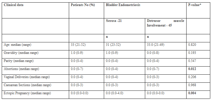
* P-value compares each variable between bladder endometriosis categories.
As the numbers are no low, table 1 would benefit from showing n numbers and mean in addition to median and range. Also ensuring that the abbreviation and terminology are consistent between the tables and the
text in the paper.
Why isn't the TVU data presented in the tables (polyps, fibroid)?

Endoma = endometrioma
* P-value compares each variable between bladder endometriosis categories
* P-value from a Pearson chi-square test
For categorical variables, the Pearson’s Chi-square test was used. For continuous non-normally distributed variables, the Kruskal-Wallis test was used.
∞ Only among non-virgin participants (ntotal=60, nserosa=20, nDetrusor muscle Involvement within=37)

BSE = Bladder Serosa Endometriosis
DME = Detrusor Muscle Endometriosis
BME = Bladder Mucosa Endometriosis
Surgical Management
All patients underwent laparoscopy and were diagnosed with an endometriotic lesion on the bladder serosa mainly on the anterior and fundal walls. Since the nodule develops from the outer layer of the bladder wall towards the detrusor muscle inner layer, local excision was performed until free from endometriosis margins were visually confirmed. In excisions that involved only the bladder serosa, a single layer wound closure was performed, while for interstitial excisions, the wound was closed in 2 layers, using 2.0 and / or 3.0 PDS sutures. The bladder serosa integrity was then examined by infusing into the bladder 300 ml of normal saline stained with methylene blue dye. The serosa part of the specimen was designated by placing a suture, assisting histopathologist orientation. Cystoscopy was performed in 15 cases and 11 cases underwent ureteric stenting.
Histopathology
The histopathological reported findings were reviewed prior to the study to reassure the bladder wall depth of involvement. An experienced histopathologist on endometriosis was asked to review all the patients’ histopathological results. The operation and tissue macroscopic description reports before processing were also reviewed. Attention was paid for possible discrepancies or missed important data that could influence the histopathological results. Light microscopy using x40 x100 magnification was performed on all the patients’ sections slides. In cases where results were equivocal, the paraffin blocks were available for additional sections for reassuring the diagnosis. An extra effort was made to meticulously observe and identify the involvement of the bladder serosa, muscularis and mucosa with endometriotic cells and glands.
Demographic, reproductive and medical history variables were compared between women with different bladder wall endometriosis and adenomyosis involvement using univariate tests. Bladder endometriosis was investigated according to depth of BWE involvement and according to four histopathological localizations of endometriosis
(Serosa [BSE]
Detrusor muscle [DME])
(Serosa [BSE] vs. Detrusor muscle [DME]
Mucosa [BME] & Detrusor muscle [DME]).
For categorical variables, the Pearson chi-square or the Fisher’s exact tests were used, the latter being reserved for comparisons with small sample numbers. For continuous non-normally distributed variables, the Kruskal-Wallis test was used. The statistical significance threshold was set at 0.05. Statistically significant associations with depth of BWE involvement were also tested with univariate logistic regression analysis. All statistical analyses were performed using STATA version 15 SE (StataCorp. 2017. Stata Statistical Software: Release 15. College Station, TX: StataCorp LLC).
Ethical approval: The study was examined and approved by the internal Bioethics committee of the OMAM Hospital, Cairo.
Among 66 patients diagnosed with bladder biopsy confirmed endometriosis, in 21 cases, endometriosis was isolated on the bladder serosa (BSE) and in 45 cases endometriosis was found within the detrusor muscle (DME) (Tables I&II). The frequency of bladder endometriosis (BE) involvement according to histological findings on bladder serosa and detrusor muscle, was investigated in association with age, gravidity, parity, number of abortions, ectopic pregnancies, normal deliveries and caesarean sections, as demonstrated in Table I. The frequencies of BSE and DME in association to menstruation, pain status, past medical and surgical history, management and endometriosis spread within the abdominal cavity are presented in Table II.
The average patients’ age recorded for BSE was 31 years and for DME 33 years. No significance was found between the patients’ age in both groups. No significance was found among the two groups for gravidity, parity, vaginal and caesarean section mode of delivery (Table I). BSE was significantly more prominent among patients with history of ectopic pregnancy (p=0.004) (Table I) and among patients with secondary infertility (p=0.029) (Table II). DME was significantly more frequent (p=0.012) in patients with increasing number of abortions (Table I).
No significance was found between serosa and detrusor muscle endometriosis involvement, when compared to severity and spread of endometriosis within the abdominal cavity (Table II).
Micturition problems were reported by 5 (7.6%) patients, urinary tract infection was diagnosed in 9 (13.8%) patients and vaginitis in 8 (12.1%) out of the 66 patients. Looking at the four histopathological BWE localisation categories, among 5 patients with micturition problems, endometriosis was diagnosed in 1 patient in each 4 histological locations, i.e serosa, serosa + DM, DM + mucosa and mucosa. In 8 patients with vaginitis, BE was mostly detected in the bladder serosa and non in mucosa and DM (Table III).
Seven patients reported a past-operations history of appendicectomy and 13 had caesarean sections, BE was mostly detected in BW and serosa. Ten patients had ovarian endometrioma surgery, 11 patients had laparoscopic surgery and 12 patients had laparotomies for various health issues, and BE was mostly detected in bladder serosa and DM. None of the past surgeries reported were found to have any statistically significant correlation with the BWE subgroups diagnosis (Table II).
Since all patients with BE were stage IV according to ASRM endometriosis classification. In order to be able to fulfil the scope of our study, the severity of endometriosis within the abdominal cavity was defined as mild when 20 cases presented only multiple foci of endometriosis in the pelvis, moderate when in 25 patients endometrioma was present and severe when 21 patients had deep infiltrating nodule/s. Pearson x2 test and Kruskal Wallis test used to compare each variable mild, moderate and severe endometriosis between BE histological locations did not reach any significance (Table II). Although missing of any significance, the statistical model showed a higher risk of DME when severe endometriosis was reported in the abdominal cavity.
In association with the four categories, the frequency of menstrual abnormalities and pain, including abdominal pain dysmenorrhoea, dyspareunia and dysuria were associated with DME involvement, without any significance. However, back pain was most prominent and significance (p=0.007) in 8 patients with BSE + DME as compared to BSE found in the serosa of 4 patients; in DME of 3 patients and BME + DME of 3 patients. (Table III). An association between the BWE and patients’ past history of health problems and operations, as well as current management findings and operative approach, did not show any significance among the endometriosis histological locations across the BW as defined above.
The highest incidence of BE was detected in the DM in 9 patients with primary (I) infertility and in the bladder serosa of 7 patients with secondary (II) infertility (Table III). No significance was noted among the I and II infertility patients and the histological locations of endometriosis on the serosa, DM and mucosa. Among patients with urinary tract infection (UTI), endometriosis was mostly diagnosed with significance (p = 0.011), in patients with endometriosis on the serosa and DM as compared to women with endometriosis throughout the BW (serosa, DM and mucosa). No UTI was reported among patients with DME alone (Table III).
Primary infertility was the indication for the current laparoscopic surgeries in 32 out of 66 (48.5%) patients and secondary infertility for the rest of the group. The highest incidence of BE was detected on the serosa of 12 patients and in the DM of 11 cases. Preoperative uterine imaging by TVU revealed that in 7 cases with benign pathologies like fibroids and / or polyps, there was no correlation to any BE histological locations either. Among 30 cases with an ovarian endometrioma detected by TVU, DME was diagnosed in 13 patients, in serosa of 10, and in serosa and DM of 6 patients.
A group of 8 patients converted from laparoscopy to laparotomy. In 58 patients that underwent laparoscopic surgery DME was evident in 21 cases, in BSE in 17 women, BSE and DME in 15 women and BME and DME in 3 patients. However, no significance was found among the BE histological locations and laparoscopic surgeries. Pelvic and abdominal endometriosis, stripping of endometrioma and cystoscopy were diagnosed and performed in 31 (47%) patients and BSE and DME was diagnosed in 38 – 56.5% of the cases without any significance. Among 56 patients who underwent hysteroscopy, BSE was detected in 21 cases, in DME in 23 cases, in BSE+DME in 15 and BME+DME in 4 cases, however without reaching any significance (Table III).
Univariate logistic regression analysis of endometriosis on the bladder serosa and DM associated to menstruation, pain status, past medical and surgical history, management and endometriosis spread within the abdominal cavity. No significance were found among patients’ age and obstetrical history when univariate logistic regression analysis was performed, comparing BSE and DME. BSE of 33.3% was significantly more prevalent (p=0.036), among patients with secondary infertility as compared to DME involvement 11.1%. No significance between BSE and DME were detected for the rest of the 27 parameters underwent regression analysis.
In this cohort study we tried to identify if histopathological locations of BWE were associated with patients’ age, obstetrical, medical and operation history, clinical characteristics, management and spread of endometriosis within the abdominal cavity. Among the 66 patients with BE, histological sections diagnosed endometriosis in only 4 cases on the bladder mucosa 6%, in 21 cases on the serosa 32%, and in 45 cases within the DM 64%. Many sections (19/66) 29% presented endometriosis in more than one layer of the BW. The high incidence of 68.2% of DM endometriosis involvement, agrees with earlier studies which presented similar results (Leone Roberti Maggiore et al., 2017). The fact that no significance was found between BSE and DME involvement, when compared to severity and spread of endometriosis within the abdominal cavity, complies with the current experience of symptoms variability and abdominal endometriosis severity (Table II). Although without any statistical significance, the higher risk of DME when severe endometriosis was reported in the abdominal cavity draws the attention for meticulous intraoperative BWE investigation. The statistically significant more frequent DME in patients with increasing number of abortions (p=0.012) seems to mimic the development of adenomyosis within the myometrium. The anatomical proximity of the bladder and uterus allows for the increased risk of trauma during the uterine intracavitary interventions and subsequently increased risk of developing DME, i.e. bladder wall adenomyosis. Circumstantial evidence suggests that microtraumatizations allow for the activation of the ‘tissue injury and repair’ mechanism which stimulates local estrogen production. Consequently, leading to permanent hyperperistalsis and self-perpetuation of the disease process. (Leyendecker G. et.al., 2009)
There are higher rates of pain, infertility, micturition problems, past operations and severity of endometriosis findings within the abdominal cavity in patients with DME, as compared to those with BSE. Despite the findings lacking significance, this supports previous literature findings and reassures the study’s results (Leone Roberti Maggiore et al., 2017). Indeed, back pain was most prominent (p=0.007) in 8 patients with BSE + DME as compared to endometriosis in the other histopathological locations of BWE; denoting the importance of BWE involvement within the serosa and DM and might be considered as a significant pre-operative symptom. This can be attributed to the neuroanatomy of the pelvis. De Sousa et al. demonstrated the spread of endometriosis from the uterine cavity along the autonomic nerves in the pelvis into the lumbosacral plexus [28]. Further spread of the endometriotic lesions into the spinal nerves and even the dura of the spinal cord was proposed to be a possible aetiology of DIE [1,28].
The backpain symptom may direct patients to 3D ultrasound (US) and magnetic resonance imaging (MRI) focusing on BWE diagnosis. It has been suggested that 3D US acquisition may improve endometriotic nodule localisation and evaluation of its size, volume, and infiltration of the bladder wall in comparison to two-dimensional transvaginal ultrasound (TVS) (Thonnon et al., 2015). TVS is the most accurate in defining the size of the lesions, infiltration of the DM, and continuity with extravesical lesions (Fedele et al., 1997). Also reports high accuracy, reproducibility and specificity but fair sensitivity in the diagnosis of BE (Tammaa et al., 2015). BWE in TVS appears as a filling defect of the posterior wall with iso/hypoechoic protrusions into the lumen, usually not vascularised (Leone Roberti Maggiore et al., 2017).
The higher rates of UTI (p=0.011) in patients with BSE+DME reflects the urinary bladder dysfunction due to BWE and to chronic inflammation caused by endometriosis. Dysuria has been reported in 21–69% of patients with BWE. Bladder pain and frequency and less commonly haematuria, urgency, and urinary incontinence are symptoms associated with the presence of BWE (Villa et al., 2007;,Leone Roberti Maggiore et al., 2015). Haematuria was reported in only 2 women out of the 66 patients. Patients experiencing urgency and/or urinary frequency, dysuria provoked during bladder filling, raises the suspicion of a DME.
In general, DIE is associated with lower urinary tract symptoms and the incidence ranges between 2% and 77% (Bonneau et al., 2013;,Ballester et al., 2014). Endometriosis and interstitial cystitis have been associated with recurrent cystitis and overactive bladder and symptoms of chronic bladder and pelvic pain or discomfort, and may be accompanied with persistent urge to void or frequency in the absence of any identifiable pathology or infection (van de Merwe et al., 2008;,Hanno et al., 2010). Univariate logistic regression analysis did not show any significant correlation between DME and UTI micturition problems or vaginitis. A recent study reported no difference in the rate of urgency, urinary frequency, voiding symptoms and bladder pain between patient with posterior endometriosis plus BE compared with those with posterior endometriosis only (Panel et al., 2016). Women of reproductive age complaining of lower urinary tract symptoms, particularly in combination with dysmenorrhoea, back pain and/or anterior sensitivity during vaginal examination, should be always considered as high risk for BWE. 2/3D TVS and MRI should follow although absence of endometriotic nodules in imaging cannot exclude BWE, hence, laparoscopy and eventually cystoscopy are necessary for final diagnosis and treatment.
In 15 out of 66 cases, cystoscopy was performed to rule out endometriotic lesions on the bladder mucosa. Four cases advancing from the bladder serosa towards the DM and appearing on the mucosa were diagnosed. Typical red or bluish nodules have been observed but not any ulcerations, which comes into agreement with other studies (Thonnon et al., 2015;,Fedele et al., 1997). Cystoscopy has limited value for screening or routine purposes in women with endometriosis and should be reserved for those cases at high risk for BME. Identifying the endometriotic lesion position, especially those close to the trigonum and ureteral ostia, inadvertent trauma can be avoided during surgery. Cystoscopy is mandatory in cases with suspicion of malignancy, excluding bladder carcinoma, varices, papillomas, angiomas, and detrusor mesenchymal tumours (Leone Roberti Maggiore et al., 2017).
The highest incidence of BE involving only the serosa in 21 patients and only the DM of 23 cases follows the results of other studies that BWE presents a severe form of endometriosis with bad prognosis for fertility potential [(Vercellini et al., 2014a;,Vercellini et al., 2014b)]. Logistic regression analysis as presented in the results, demonstrated that BSE 33.3% was significantly more prevalent (p=0.036) among patients with secondary infertility, as compared to DME involvement of 11.1%; this probably reflects a partial protection by prior pregnancy, but not complete immunity of the disease. Although current research suggests no evidence on pregnancy reducing the size or number of endometriotic lesions, it might be worth questioning the conclusions of these few studies of very limited quality.
Brigitte Leeners, Fabia Damaso, Nicole Ochsenbein-Kölble, Cindy Farquhar, The effect of pregnancy on endometriosis—facts or fiction?, Human Reproduction Update, Volume 24, Issue 3, May-June 2018, Pages 290–299, https://doi.org/10.1093/humupd/dmy004
Within the context that adenomyosis may interfere with fertility, similarly women with BE may probably, at least partly, contribute to uterine dysfunction. However, in most series published, no adenomyotic nodules of the uterine wall were found in association with BE [Leone Roberti Maggiore U, et al. 2017]. Interestingly, 11 cases of male endometriosis reported in the literature, four developed endometriosis of the bladder concomitant with high estrogen exposure, probably indicating a similar susceptibility between the detrusor muscle and myometrium [Leone Roberti Maggiore U, et al. 2017]. The fact that BSE was statistically significant among patients with secondary infertility and history of ectopic pregnancy, reflects the consequences of endometriosis and formation of intrabdominal adhesions due to chronic inflammation.
Although the big majority of the patients 53/66 (80.3%) had a history of abdominal operations, no significance was found of BWE and histological subgroups, among these patients, neither of the 13 cases underwent caesarean sections and the 10 cases after ovarian endometrioma surgery. In 46 out of 66 patients, 70% who had the bladder wall affected by endometriosis, underwent a gynaecological surgical procedure prior to diagnosis of BE, raising the possibility of iatrogenic dissemination of endometriotic cells. However, a recent cross-sectional incidence of isolated BE, reported patients with and without a history of uterine surgery 37.5% and 41.7% respectively without any significance (p = 0.6) (Leone Roberti Maggiore et al., 2017). Among BWE cases, the incidence observed was 58.6% (95% confidence interval [CI] 45.2–71.2) for superficial peritoneal implants, 44.8% (95% CI 32.2–58.2) for ovarian endometriomas, 81.0% (95% CI 68.4–89.6) for adhesions, and 27.6% (95% CI 16.7-40.8). Hormonal treatments are effective for a temporal disease suppression but not curing. Significant improvements in pain and urinary symptoms have been observed after excision of the whole bladder lesion, which also minimises the risk of recurrence [Soriano et al., 2016;,Seracchioli et al., 2010;,Chapronet al., 2010)].
In our study 45 cases underwent a full thickness bladder wall segmental excision due to severity of the disease.
In another 21 cases, BSE was diagnosed after wedge resection of the lesion and confirmation of clear margins from the disease. Several studies have shown that segmental cystectomy is an effective technique with excellent long-term results in terms of symptom relief and recurrence [(Chapron et al., 2010;,Kjer et al., 2014)]. Excision of endometriotic nodules of the bladder may lead to inadvertent removal of healthy bladder muscle, particularly in case of large endometriotic lesions. Postoperative complications and symptoms are mainly due to small bladder volume. An effort to spare most of the healthy bladder tissue is imperative. [(Fedele et al., 2005;,Vercellini et al., 2009;,Antonelli et al., 2006)]. Preventive ureteric catheterisation was performed in 11 cases due to lesion proximity to trigonum. Ureters stenting can be of great help when surgery is performed on the posterior bladder wall and endometriotic nodules are close to the ureteral meatuses [(Vercellini et al., 1998)].
Surgery for BE usually accompanies treatment for adhesions, endometriomas, superficial implants, and other deep localisations of the disease (Somigliana et al., 2007). Deep peritoneal endometriosis is associated with adenomyosis and according to our study there is a higher risk of DME in relation to severity of the peritoneal endometriosis; although there was no significance. [(Kunz et al., 2005;,Exacoustos et al., 2013)]. Occasionally bladder lesions are buried under adhesions while chronic inflammation and extended fibrosis present an operative challenge. In a study by Kovoor et al 50% of infertile women with BWE conceived naturally after an intervention [(Wells et al., 2014)]. The surgery radicality of DIE and BWE for infertility treatment remains debatable (Somigliana & Garcia-Velasco, 2015b).
This is a cohort retrospective study. There is a possibility that other areas with endometriosis were also involved in the BW other than those diagnosed and treated. The mixture of patients with any type of infertility, primary and secondary infertility and history of past abdominal operations could also affect the results, although statistical analysis did not show any significance in BWE, clinical symptoms and surgical findings. BE is rarely an isolated condition, and other forms of endometriosis are frequently concomitant (Vigano et al., 2009).
DME involvement presented 68% and BSE 32 of all cases with BE and infertility investigated. Secondary infertility, back pains, micturition problems and number of abortions can be considered as high-risk factors for BE for women of reproductive age. The severity of the peritoneal endometriosis can probably direct to meticulous intraoperative investigation for BE.
Dr Sayed El-Akhras: Performed all of the operations and collected the clinical and surgery data
Mohamed Abo-elenen: Involved in patients’ data collection, review all patients’ files, collection of data and formation of the excel file
Christiana Demetriou: Statistical analysis, editing the results, review the article
Nafissa Mohamed Amin El Badawy: Performed and review all patients’ histopathology results, writing and review section of histopathology in methods
Safinez Balami: review and editing of the article
Vasilios Tanos: Involved in the operations, study concept and design, review of patients’ data collection and preparation of the excel file, review of statistics and formation of tables, writing the manuscript and editing
No funding
There is no conflict of interest for none of the authors
Clearly Auctoresonline and particularly Psychology and Mental Health Care Journal is dedicated to improving health care services for individuals and populations. The editorial boards' ability to efficiently recognize and share the global importance of health literacy with a variety of stakeholders. Auctoresonline publishing platform can be used to facilitate of optimal client-based services and should be added to health care professionals' repertoire of evidence-based health care resources.

Journal of Clinical Cardiology and Cardiovascular Intervention The submission and review process was adequate. However I think that the publication total value should have been enlightened in early fases. Thank you for all.

Journal of Women Health Care and Issues By the present mail, I want to say thank to you and tour colleagues for facilitating my published article. Specially thank you for the peer review process, support from the editorial office. I appreciate positively the quality of your journal.

Journal of Clinical Research and Reports I would be very delighted to submit my testimonial regarding the reviewer board and the editorial office. The reviewer board were accurate and helpful regarding any modifications for my manuscript. And the editorial office were very helpful and supportive in contacting and monitoring with any update and offering help. It was my pleasure to contribute with your promising Journal and I am looking forward for more collaboration.

We would like to thank the Journal of Thoracic Disease and Cardiothoracic Surgery because of the services they provided us for our articles. The peer-review process was done in a very excellent time manner, and the opinions of the reviewers helped us to improve our manuscript further. The editorial office had an outstanding correspondence with us and guided us in many ways. During a hard time of the pandemic that is affecting every one of us tremendously, the editorial office helped us make everything easier for publishing scientific work. Hope for a more scientific relationship with your Journal.

The peer-review process which consisted high quality queries on the paper. I did answer six reviewers’ questions and comments before the paper was accepted. The support from the editorial office is excellent.

Journal of Neuroscience and Neurological Surgery. I had the experience of publishing a research article recently. The whole process was simple from submission to publication. The reviewers made specific and valuable recommendations and corrections that improved the quality of my publication. I strongly recommend this Journal.

Dr. Katarzyna Byczkowska My testimonial covering: "The peer review process is quick and effective. The support from the editorial office is very professional and friendly. Quality of the Clinical Cardiology and Cardiovascular Interventions is scientific and publishes ground-breaking research on cardiology that is useful for other professionals in the field.

Thank you most sincerely, with regard to the support you have given in relation to the reviewing process and the processing of my article entitled "Large Cell Neuroendocrine Carcinoma of The Prostate Gland: A Review and Update" for publication in your esteemed Journal, Journal of Cancer Research and Cellular Therapeutics". The editorial team has been very supportive.

Testimony of Journal of Clinical Otorhinolaryngology: work with your Reviews has been a educational and constructive experience. The editorial office were very helpful and supportive. It was a pleasure to contribute to your Journal.

Dr. Bernard Terkimbi Utoo, I am happy to publish my scientific work in Journal of Women Health Care and Issues (JWHCI). The manuscript submission was seamless and peer review process was top notch. I was amazed that 4 reviewers worked on the manuscript which made it a highly technical, standard and excellent quality paper. I appreciate the format and consideration for the APC as well as the speed of publication. It is my pleasure to continue with this scientific relationship with the esteem JWHCI.

This is an acknowledgment for peer reviewers, editorial board of Journal of Clinical Research and Reports. They show a lot of consideration for us as publishers for our research article “Evaluation of the different factors associated with side effects of COVID-19 vaccination on medical students, Mutah university, Al-Karak, Jordan”, in a very professional and easy way. This journal is one of outstanding medical journal.

Dear Hao Jiang, to Journal of Nutrition and Food Processing We greatly appreciate the efficient, professional and rapid processing of our paper by your team. If there is anything else we should do, please do not hesitate to let us know. On behalf of my co-authors, we would like to express our great appreciation to editor and reviewers.

As an author who has recently published in the journal "Brain and Neurological Disorders". I am delighted to provide a testimonial on the peer review process, editorial office support, and the overall quality of the journal. The peer review process at Brain and Neurological Disorders is rigorous and meticulous, ensuring that only high-quality, evidence-based research is published. The reviewers are experts in their fields, and their comments and suggestions were constructive and helped improve the quality of my manuscript. The review process was timely and efficient, with clear communication from the editorial office at each stage. The support from the editorial office was exceptional throughout the entire process. The editorial staff was responsive, professional, and always willing to help. They provided valuable guidance on formatting, structure, and ethical considerations, making the submission process seamless. Moreover, they kept me informed about the status of my manuscript and provided timely updates, which made the process less stressful. The journal Brain and Neurological Disorders is of the highest quality, with a strong focus on publishing cutting-edge research in the field of neurology. The articles published in this journal are well-researched, rigorously peer-reviewed, and written by experts in the field. The journal maintains high standards, ensuring that readers are provided with the most up-to-date and reliable information on brain and neurological disorders. In conclusion, I had a wonderful experience publishing in Brain and Neurological Disorders. The peer review process was thorough, the editorial office provided exceptional support, and the journal's quality is second to none. I would highly recommend this journal to any researcher working in the field of neurology and brain disorders.
Dear Agrippa Hilda, Journal of Neuroscience and Neurological Surgery, Editorial Coordinator, I trust this message finds you well. I want to extend my appreciation for considering my article for publication in your esteemed journal. I am pleased to provide a testimonial regarding the peer review process and the support received from your editorial office. The peer review process for my paper was carried out in a highly professional and thorough manner. The feedback and comments provided by the authors were constructive and very useful in improving the quality of the manuscript. This rigorous assessment process undoubtedly contributes to the high standards maintained by your journal.

International Journal of Clinical Case Reports and Reviews. I strongly recommend to consider submitting your work to this high-quality journal. The support and availability of the Editorial staff is outstanding and the review process was both efficient and rigorous.

Thank you very much for publishing my Research Article titled “Comparing Treatment Outcome Of Allergic Rhinitis Patients After Using Fluticasone Nasal Spray And Nasal Douching" in the Journal of Clinical Otorhinolaryngology. As Medical Professionals we are immensely benefited from study of various informative Articles and Papers published in this high quality Journal. I look forward to enriching my knowledge by regular study of the Journal and contribute my future work in the field of ENT through the Journal for use by the medical fraternity. The support from the Editorial office was excellent and very prompt. I also welcome the comments received from the readers of my Research Article.

Dear Erica Kelsey, Editorial Coordinator of Cancer Research and Cellular Therapeutics Our team is very satisfied with the processing of our paper by your journal. That was fast, efficient, rigorous, but without unnecessary complications. We appreciated the very short time between the submission of the paper and its publication on line on your site.

I am very glad to say that the peer review process is very successful and fast and support from the Editorial Office. Therefore, I would like to continue our scientific relationship for a long time. And I especially thank you for your kindly attention towards my article. Have a good day!

"We recently published an article entitled “Influence of beta-Cyclodextrins upon the Degradation of Carbofuran Derivatives under Alkaline Conditions" in the Journal of “Pesticides and Biofertilizers” to show that the cyclodextrins protect the carbamates increasing their half-life time in the presence of basic conditions This will be very helpful to understand carbofuran behaviour in the analytical, agro-environmental and food areas. We greatly appreciated the interaction with the editor and the editorial team; we were particularly well accompanied during the course of the revision process, since all various steps towards publication were short and without delay".

I would like to express my gratitude towards you process of article review and submission. I found this to be very fair and expedient. Your follow up has been excellent. I have many publications in national and international journal and your process has been one of the best so far. Keep up the great work.

We are grateful for this opportunity to provide a glowing recommendation to the Journal of Psychiatry and Psychotherapy. We found that the editorial team were very supportive, helpful, kept us abreast of timelines and over all very professional in nature. The peer review process was rigorous, efficient and constructive that really enhanced our article submission. The experience with this journal remains one of our best ever and we look forward to providing future submissions in the near future.

I am very pleased to serve as EBM of the journal, I hope many years of my experience in stem cells can help the journal from one way or another. As we know, stem cells hold great potential for regenerative medicine, which are mostly used to promote the repair response of diseased, dysfunctional or injured tissue using stem cells or their derivatives. I think Stem Cell Research and Therapeutics International is a great platform to publish and share the understanding towards the biology and translational or clinical application of stem cells.

I would like to give my testimony in the support I have got by the peer review process and to support the editorial office where they were of asset to support young author like me to be encouraged to publish their work in your respected journal and globalize and share knowledge across the globe. I really give my great gratitude to your journal and the peer review including the editorial office.

I am delighted to publish our manuscript entitled "A Perspective on Cocaine Induced Stroke - Its Mechanisms and Management" in the Journal of Neuroscience and Neurological Surgery. The peer review process, support from the editorial office, and quality of the journal are excellent. The manuscripts published are of high quality and of excellent scientific value. I recommend this journal very much to colleagues.

Dr.Tania Muñoz, My experience as researcher and author of a review article in The Journal Clinical Cardiology and Interventions has been very enriching and stimulating. The editorial team is excellent, performs its work with absolute responsibility and delivery. They are proactive, dynamic and receptive to all proposals. Supporting at all times the vast universe of authors who choose them as an option for publication. The team of review specialists, members of the editorial board, are brilliant professionals, with remarkable performance in medical research and scientific methodology. Together they form a frontline team that consolidates the JCCI as a magnificent option for the publication and review of high-level medical articles and broad collective interest. I am honored to be able to share my review article and open to receive all your comments.

“The peer review process of JPMHC is quick and effective. Authors are benefited by good and professional reviewers with huge experience in the field of psychology and mental health. The support from the editorial office is very professional. People to contact to are friendly and happy to help and assist any query authors might have. Quality of the Journal is scientific and publishes ground-breaking research on mental health that is useful for other professionals in the field”.

Dear editorial department: On behalf of our team, I hereby certify the reliability and superiority of the International Journal of Clinical Case Reports and Reviews in the peer review process, editorial support, and journal quality. Firstly, the peer review process of the International Journal of Clinical Case Reports and Reviews is rigorous, fair, transparent, fast, and of high quality. The editorial department invites experts from relevant fields as anonymous reviewers to review all submitted manuscripts. These experts have rich academic backgrounds and experience, and can accurately evaluate the academic quality, originality, and suitability of manuscripts. The editorial department is committed to ensuring the rigor of the peer review process, while also making every effort to ensure a fast review cycle to meet the needs of authors and the academic community. Secondly, the editorial team of the International Journal of Clinical Case Reports and Reviews is composed of a group of senior scholars and professionals with rich experience and professional knowledge in related fields. The editorial department is committed to assisting authors in improving their manuscripts, ensuring their academic accuracy, clarity, and completeness. Editors actively collaborate with authors, providing useful suggestions and feedback to promote the improvement and development of the manuscript. We believe that the support of the editorial department is one of the key factors in ensuring the quality of the journal. Finally, the International Journal of Clinical Case Reports and Reviews is renowned for its high- quality articles and strict academic standards. The editorial department is committed to publishing innovative and academically valuable research results to promote the development and progress of related fields. The International Journal of Clinical Case Reports and Reviews is reasonably priced and ensures excellent service and quality ratio, allowing authors to obtain high-level academic publishing opportunities in an affordable manner. I hereby solemnly declare that the International Journal of Clinical Case Reports and Reviews has a high level of credibility and superiority in terms of peer review process, editorial support, reasonable fees, and journal quality. Sincerely, Rui Tao.

Clinical Cardiology and Cardiovascular Interventions I testity the covering of the peer review process, support from the editorial office, and quality of the journal.

Clinical Cardiology and Cardiovascular Interventions, we deeply appreciate the interest shown in our work and its publication. It has been a true pleasure to collaborate with you. The peer review process, as well as the support provided by the editorial office, have been exceptional, and the quality of the journal is very high, which was a determining factor in our decision to publish with you.

The peer reviewers process is quick and effective, the supports from editorial office is excellent, the quality of journal is high. I would like to collabroate with Internatioanl journal of Clinical Case Reports and Reviews journal clinically in the future time.

Clinical Cardiology and Cardiovascular Interventions, I would like to express my sincerest gratitude for the trust placed in our team for the publication in your journal. It has been a true pleasure to collaborate with you on this project. I am pleased to inform you that both the peer review process and the attention from the editorial coordination have been excellent. Your team has worked with dedication and professionalism to ensure that your publication meets the highest standards of quality. We are confident that this collaboration will result in mutual success, and we are eager to see the fruits of this shared effort.

Dear Dr. Jessica Magne, Editorial Coordinator 0f Clinical Cardiology and Cardiovascular Interventions, I hope this message finds you well. I want to express my utmost gratitude for your excellent work and for the dedication and speed in the publication process of my article titled "Navigating Innovation: Qualitative Insights on Using Technology for Health Education in Acute Coronary Syndrome Patients." I am very satisfied with the peer review process, the support from the editorial office, and the quality of the journal. I hope we can maintain our scientific relationship in the long term.

Dear Monica Gissare, - Editorial Coordinator of Nutrition and Food Processing. ¨My testimony with you is truly professional, with a positive response regarding the follow-up of the article and its review, you took into account my qualities and the importance of the topic¨.

Dear Dr. Jessica Magne, Editorial Coordinator 0f Clinical Cardiology and Cardiovascular Interventions, The review process for the article “The Handling of Anti-aggregants and Anticoagulants in the Oncologic Heart Patient Submitted to Surgery” was extremely rigorous and detailed. From the initial submission to the final acceptance, the editorial team at the “Journal of Clinical Cardiology and Cardiovascular Interventions” demonstrated a high level of professionalism and dedication. The reviewers provided constructive and detailed feedback, which was essential for improving the quality of our work. Communication was always clear and efficient, ensuring that all our questions were promptly addressed. The quality of the “Journal of Clinical Cardiology and Cardiovascular Interventions” is undeniable. It is a peer-reviewed, open-access publication dedicated exclusively to disseminating high-quality research in the field of clinical cardiology and cardiovascular interventions. The journal's impact factor is currently under evaluation, and it is indexed in reputable databases, which further reinforces its credibility and relevance in the scientific field. I highly recommend this journal to researchers looking for a reputable platform to publish their studies.

Dear Editorial Coordinator of the Journal of Nutrition and Food Processing! "I would like to thank the Journal of Nutrition and Food Processing for including and publishing my article. The peer review process was very quick, movement and precise. The Editorial Board has done an extremely conscientious job with much help, valuable comments and advices. I find the journal very valuable from a professional point of view, thank you very much for allowing me to be part of it and I would like to participate in the future!”

Dealing with The Journal of Neurology and Neurological Surgery was very smooth and comprehensive. The office staff took time to address my needs and the response from editors and the office was prompt and fair. I certainly hope to publish with this journal again.Their professionalism is apparent and more than satisfactory. Susan Weiner

My Testimonial Covering as fellowing: Lin-Show Chin. The peer reviewers process is quick and effective, the supports from editorial office is excellent, the quality of journal is high. I would like to collabroate with Internatioanl journal of Clinical Case Reports and Reviews.

My experience publishing in Psychology and Mental Health Care was exceptional. The peer review process was rigorous and constructive, with reviewers providing valuable insights that helped enhance the quality of our work. The editorial team was highly supportive and responsive, making the submission process smooth and efficient. The journal's commitment to high standards and academic rigor makes it a respected platform for quality research. I am grateful for the opportunity to publish in such a reputable journal.

My experience publishing in International Journal of Clinical Case Reports and Reviews was exceptional. I Come forth to Provide a Testimonial Covering the Peer Review Process and the editorial office for the Professional and Impartial Evaluation of the Manuscript.

I would like to offer my testimony in the support. I have received through the peer review process and support the editorial office where they are to support young authors like me, encourage them to publish their work in your esteemed journals, and globalize and share knowledge globally. I really appreciate your journal, peer review, and editorial office.

Dear Agrippa Hilda- Editorial Coordinator of Journal of Neuroscience and Neurological Surgery, "The peer review process was very quick and of high quality, which can also be seen in the articles in the journal. The collaboration with the editorial office was very good."

I would like to express my sincere gratitude for the support and efficiency provided by the editorial office throughout the publication process of my article, “Delayed Vulvar Metastases from Rectal Carcinoma: A Case Report.” I greatly appreciate the assistance and guidance I received from your team, which made the entire process smooth and efficient. The peer review process was thorough and constructive, contributing to the overall quality of the final article. I am very grateful for the high level of professionalism and commitment shown by the editorial staff, and I look forward to maintaining a long-term collaboration with the International Journal of Clinical Case Reports and Reviews.

To Dear Erin Aust, I would like to express my heartfelt appreciation for the opportunity to have my work published in this esteemed journal. The entire publication process was smooth and well-organized, and I am extremely satisfied with the final result. The Editorial Team demonstrated the utmost professionalism, providing prompt and insightful feedback throughout the review process. Their clear communication and constructive suggestions were invaluable in enhancing my manuscript, and their meticulous attention to detail and dedication to quality are truly commendable. Additionally, the support from the Editorial Office was exceptional. From the initial submission to the final publication, I was guided through every step of the process with great care and professionalism. The team's responsiveness and assistance made the entire experience both easy and stress-free. I am also deeply impressed by the quality and reputation of the journal. It is an honor to have my research featured in such a respected publication, and I am confident that it will make a meaningful contribution to the field.

"I am grateful for the opportunity of contributing to [International Journal of Clinical Case Reports and Reviews] and for the rigorous review process that enhances the quality of research published in your esteemed journal. I sincerely appreciate the time and effort of your team who have dedicatedly helped me in improvising changes and modifying my manuscript. The insightful comments and constructive feedback provided have been invaluable in refining and strengthening my work".

I thank the ‘Journal of Clinical Research and Reports’ for accepting this article for publication. This is a rigorously peer reviewed journal which is on all major global scientific data bases. I note the review process was prompt, thorough and professionally critical. It gave us an insight into a number of important scientific/statistical issues. The review prompted us to review the relevant literature again and look at the limitations of the study. The peer reviewers were open, clear in the instructions and the editorial team was very prompt in their communication. This journal certainly publishes quality research articles. I would recommend the journal for any future publications.

Dear Jessica Magne, with gratitude for the joint work. Fast process of receiving and processing the submitted scientific materials in “Clinical Cardiology and Cardiovascular Interventions”. High level of competence of the editors with clear and correct recommendations and ideas for enriching the article.

We found the peer review process quick and positive in its input. The support from the editorial officer has been very agile, always with the intention of improving the article and taking into account our subsequent corrections.

My article, titled 'No Way Out of the Smartphone Epidemic Without Considering the Insights of Brain Research,' has been republished in the International Journal of Clinical Case Reports and Reviews. The review process was seamless and professional, with the editors being both friendly and supportive. I am deeply grateful for their efforts.

To Dear Erin Aust – Editorial Coordinator of Journal of General Medicine and Clinical Practice! I declare that I am absolutely satisfied with your work carried out with great competence in following the manuscript during the various stages from its receipt, during the revision process to the final acceptance for publication. Thank Prof. Elvira Farina

Dear Jessica, and the super professional team of the ‘Clinical Cardiology and Cardiovascular Interventions’ I am sincerely grateful to the coordinated work of the journal team for the no problem with the submission of my manuscript: “Cardiometabolic Disorders in A Pregnant Woman with Severe Preeclampsia on the Background of Morbid Obesity (Case Report).” The review process by 5 experts was fast, and the comments were professional, which made it more specific and academic, and the process of publication and presentation of the article was excellent. I recommend that my colleagues publish articles in this journal, and I am interested in further scientific cooperation. Sincerely and best wishes, Dr. Oleg Golyanovskiy.

Dear Ashley Rosa, Editorial Coordinator of the journal - Psychology and Mental Health Care. " The process of obtaining publication of my article in the Psychology and Mental Health Journal was positive in all areas. The peer review process resulted in a number of valuable comments, the editorial process was collaborative and timely, and the quality of this journal has been quickly noticed, resulting in alternative journals contacting me to publish with them." Warm regards, Susan Anne Smith, PhD. Australian Breastfeeding Association.

Dear Jessica Magne, Editorial Coordinator, Clinical Cardiology and Cardiovascular Interventions, Auctores Publishing LLC. I appreciate the journal (JCCI) editorial office support, the entire team leads were always ready to help, not only on technical front but also on thorough process. Also, I should thank dear reviewers’ attention to detail and creative approach to teach me and bring new insights by their comments. Surely, more discussions and introduction of other hemodynamic devices would provide better prevention and management of shock states. Your efforts and dedication in presenting educational materials in this journal are commendable. Best wishes from, Farahnaz Fallahian.

Dear Maria Emerson, Editorial Coordinator, International Journal of Clinical Case Reports and Reviews, Auctores Publishing LLC. I am delighted to have published our manuscript, "Acute Colonic Pseudo-Obstruction (ACPO): A rare but serious complication following caesarean section." I want to thank the editorial team, especially Maria Emerson, for their prompt review of the manuscript, quick responses to queries, and overall support. Yours sincerely Dr. Victor Olagundoye.

Dear Ashley Rosa, Editorial Coordinator, International Journal of Clinical Case Reports and Reviews. Many thanks for publishing this manuscript after I lost confidence the editors were most helpful, more than other journals Best wishes from, Susan Anne Smith, PhD. Australian Breastfeeding Association.

Dear Agrippa Hilda, Editorial Coordinator, Journal of Neuroscience and Neurological Surgery. The entire process including article submission, review, revision, and publication was extremely easy. The journal editor was prompt and helpful, and the reviewers contributed to the quality of the paper. Thank you so much! Eric Nussbaum, MD

Dr Hala Al Shaikh This is to acknowledge that the peer review process for the article ’ A Novel Gnrh1 Gene Mutation in Four Omani Male Siblings, Presentation and Management ’ sent to the International Journal of Clinical Case Reports and Reviews was quick and smooth. The editorial office was prompt with easy communication.

Dear Erin Aust, Editorial Coordinator, Journal of General Medicine and Clinical Practice. We are pleased to share our experience with the “Journal of General Medicine and Clinical Practice”, following the successful publication of our article. The peer review process was thorough and constructive, helping to improve the clarity and quality of the manuscript. We are especially thankful to Ms. Erin Aust, the Editorial Coordinator, for her prompt communication and continuous support throughout the process. Her professionalism ensured a smooth and efficient publication experience. The journal upholds high editorial standards, and we highly recommend it to fellow researchers seeking a credible platform for their work. Best wishes By, Dr. Rakhi Mishra.

Dear Jessica Magne, Editorial Coordinator, Clinical Cardiology and Cardiovascular Interventions, Auctores Publishing LLC. The peer review process of the journal of Clinical Cardiology and Cardiovascular Interventions was excellent and fast, as was the support of the editorial office and the quality of the journal. Kind regards Walter F. Riesen Prof. Dr. Dr. h.c. Walter F. Riesen.

Dear Ashley Rosa, Editorial Coordinator, International Journal of Clinical Case Reports and Reviews, Auctores Publishing LLC. Thank you for publishing our article, Exploring Clozapine's Efficacy in Managing Aggression: A Multiple Single-Case Study in Forensic Psychiatry in the international journal of clinical case reports and reviews. We found the peer review process very professional and efficient. The comments were constructive, and the whole process was efficient. On behalf of the co-authors, I would like to thank you for publishing this article. With regards, Dr. Jelle R. Lettinga.

Dear Clarissa Eric, Editorial Coordinator, Journal of Clinical Case Reports and Studies, I would like to express my deep admiration for the exceptional professionalism demonstrated by your journal. I am thoroughly impressed by the speed of the editorial process, the substantive and insightful reviews, and the meticulous preparation of the manuscript for publication. Additionally, I greatly appreciate the courteous and immediate responses from your editorial office to all my inquiries. Best Regards, Dariusz Ziora

Dear Chrystine Mejia, Editorial Coordinator, Journal of Neurodegeneration and Neurorehabilitation, Auctores Publishing LLC, We would like to thank the editorial team for the smooth and high-quality communication leading up to the publication of our article in the Journal of Neurodegeneration and Neurorehabilitation. The reviewers have extensive knowledge in the field, and their relevant questions helped to add value to our publication. Kind regards, Dr. Ravi Shrivastava.

Dear Clarissa Eric, Editorial Coordinator, Journal of Clinical Case Reports and Studies, Auctores Publishing LLC, USA Office: +1-(302)-520-2644. I would like to express my sincere appreciation for the efficient and professional handling of my case report by the ‘Journal of Clinical Case Reports and Studies’. The peer review process was not only fast but also highly constructive—the reviewers’ comments were clear, relevant, and greatly helped me improve the quality and clarity of my manuscript. I also received excellent support from the editorial office throughout the process. Communication was smooth and timely, and I felt well guided at every stage, from submission to publication. The overall quality and rigor of the journal are truly commendable. I am pleased to have published my work with Journal of Clinical Case Reports and Studies, and I look forward to future opportunities for collaboration. Sincerely, Aline Tollet, UCLouvain.

Dear Ms. Mayra Duenas, Editorial Coordinator, International Journal of Clinical Case Reports and Reviews. “The International Journal of Clinical Case Reports and Reviews represented the “ideal house” to share with the research community a first experience with the use of the Simeox device for speech rehabilitation. High scientific reputation and attractive website communication were first determinants for the selection of this Journal, and the following submission process exceeded expectations: fast but highly professional peer review, great support by the editorial office, elegant graphic layout. Exactly what a dynamic research team - also composed by allied professionals - needs!" From, Chiara Beccaluva, PT - Italy.

Dear Maria Emerson, Editorial Coordinator, we have deeply appreciated the professionalism demonstrated by the International Journal of Clinical Case Reports and Reviews. The reviewers have extensive knowledge of our field and have been very efficient and fast in supporting the process. I am really looking forward to further collaboration. Thanks. Best regards, Dr. Claudio Ligresti
