AUCTORES
Globalize your Research

Research Article | DOI: https://doi.org/10.31579/2641-0419/145
*Corresponding Author: Heyder Omran, GFO Kliniken Bonn, Robert-Koch-Str. 1, 53115 Bonn, Germany
Citation: Jaroslaw Heinrich, Baravan Al-Kassou, Heyder Omran., (2021) Cardiac Computed Tomography versus 3D-Transesophageal Echocardiography in Preprocedural Planning of Left Atrial Appendage Closure. J. Clinical Cardiology and Cardiovascular Interventions, 4(6); Doi:10.31579/2641-0419/145
Copyright: © 2021 Heyder Omran, This is an open-access article distributed under the terms of the Creative Commons Attribution License, which permits unrestricted use, distribution, and reproduction in any medium, provided the original author and source are credited.
Received: 16 February 2021 | Accepted: 11 March 2021 | Published: 18 March 2021
Keywords: left atrial appendage closure; imaging modalities; cardiac computed tomography; transesophageal echocardiography; 3D transesophageal echocardiography
Aim:
Preprocedural imaging of the left atrial appendage (LAA) plays a crucial role in the process of LAA closure (LAAC). This study aimed to compare the influence of preprocedural planning of the LAAC with 3D-transesophageal echocardiography (TEE) and cardiac computed tomography (CCT) versus 3D-TEE alone in patients who underwent LAAC with an Amplatzer Cardiac Plug or Amulet.
Materials and Methods:
In a retrospective study, 176 patients received a preprocedural 3D-TEE and CCT and 167 patients a 3D-TEE only. Both groups had similar patient characteristics and indications for LAAC.
Results:
There was no difference in terms of procedural success, procedure time, amount of contrast medium, fluoroscopy time, or radiation dose. Patients with CCT/3D-TEE had a longer hospital stay on average. Besides, there was a different incidence of renal diseases (49% for 3D-TEE versus 27% for CCT/3D-TEE; p < 0.001). The number of periprocedural adverse events was comparable. A device-related thrombus occurred three times in each group, and the peri-device leaks reported were similar.
Conclusion: A preprocedural CCT does not decrease major adverse events or improve outcome in patients undergoing LAAC.
Atrial fibrillation (AF) occurs in 1-2% of the population in western countries and has a higher prevalence in men and older subjects [1-3]. One of the most feared complications in patients with AF is thromboembolism [5]. Approximately 90% of all thrombi develop in the left atrial appendage (LAA) [4]. Therefore, it is not surprising that one in five strokes are caused by AF, and 80% of all strokes are of an ischemic etiology [6, 7, 37].
In order to reduce the risk of strokes, oral anticoagulation with vitamin K antagonists (VKA) and new oral anticoagulants (NOACs) is a validated treatment [8, 9]. Nonetheless, the use of oral anticoagulants (OACs) increases the risk of intracerebral and gastrointestinal bleeding [10-13]. Hence the use of the clinical scores, HAS-BLED and CHA2DS2-VASc score, help to balance the risk of strokes and major bleeding in patients with AF [14, 15]. Interventional closure of the LAA (LAAC) has been shown to be a valid alternative in patients with a contraindication against OACs [16, 17].
In the process of device implantation, planning and preprocedural imaging of the LAA is important.
There are currently two main imaging techniques to assess the LAA before LAAC:
Two-dimensional and three-dimensional (2D/3D)-transesophageal echocardiography (TEE) and cardiac computed tomography (CCT) [19].
3D-TEE is used generally for preprocedural planning and periprocedural intervention, to assess the geometry and size of the LAA and, at most, predict the correct device size [22-24]. It was shown to be superior to 2D-TEE with regard to determining the LAA orifice area/size [24] and LAA occluder size [28]. One study postulated that LAA measurements obtained using real time 3D-TEE showed smaller values than those obtained with CCT [26] and another study found that 3D-TEE was inferior at defining the ostial perimeter and correct occluder size, as compared to CCT [27].
Nonetheless, 3D-TEE imaging allows the LAA to be well visualized before, during, and after the procedure [25].
Cardiac computed tomography (CCT) has been shown as a valid imaging modality to detect LAA thrombi [23], to describe the LAA morphology, and to calculate the predicted device size for the LAAC [20, 21]. However, CCT imaging of the LAA is associated with nephrotoxic risks from the intravenous contrast dye and radiation exposure [29]. Importantly, it was demonstrated that there is a better agreement of the actual diameters and perimeters compared with device diameters and perimeters, i.e. the device fits the dimensions better, for CCT compared to TEE [27, 30].
There are only a few studies that have examined the influence of preprocedural CCT and TEE versus TEE alone, in terms of procedure time, contrast use, correct device size and number of devices utilized. Two small studies have shown a significant reduction in procedure time and anesthesia time, a greater accuracy in device selection, and an absence of peri-device leaks in the group that received a preprocedural CCT [30, 31]. However, data on the influence of preprocedural CCT, concerning the periprocedural and long-term transesophageal echocardiographic outcome, are scarce.
The aim of our study was to compare the influence of preprocedural planning of the LAAC with 3D-TEE and CCT versus 3D-TEE alone, in patients who underwent LAAC with the Amplatzer Cardiac Plug (ACP) and Amulet device, to assess the periprocedural outcome and the long-term transesophageal echocardiographic outcome.
2. Materials and Methods
In this retrospective study, we collected data from a total of 343 consecutive patients with AF and contraindication to effective OAC, who underwent a LAAC procedure with the ACP or AMPLATZER Amulet between September 2009 and December 2019, were appraised and analyzed [45]. The main focus of this study was to compare the impact of CCT versus TEE in terms of preprocedural planning and the clinical outcome of the LAAC: Group 1 had 3D-TEE and CCT (n=176) versus Group 2 had 3D-TEE (n=167) alone. In both groups, clinical characteristics and patient demographics as well as procedural performance, clinical outcome, and echocardiographic follow-up data were collected and statistically contrasted with each other.
2.2 Preprocedural imaging
The 3D-TEE was performed by using a GE Vivid E9 BT12 cardiovascular ultrasound system, at least 24 hours prior to the LAAC procedure. The main aim of the imaging was to exclude intracardial thrombus and to assess three-dimensional images of the LAA. By using the zoom mode from pyramidal data sets, LAA images were recorded by using one-beat acquisition at end-expiration with a resolution of 18 to 21 beats per second [32]. The objective of images obtained was to display the landing zone (LZ) to its full extent and to create a three-dimensional visualization of the LAA by slicing the pyramidal data sets along x, y, and z axes [32].
The imaging data was subsequently analyzed with the GE EchoPAC BT12 software using the 12-channel multislice mode, thus helping to display the LAA in a three-dimensional way. With the help of a cross-sectional view, the perimeter, area, and maximal/ minimal diameter (D max, D min) of the LZ was measured, which is defined as a level plane from the left circumplex coronary artery to the roof of the LAA, approximately 10 mm inward from the peak of the rim that separates the LAA and the left superior pulmonary vein [32]. The mean diameter (D average) was derived from the perimeter (Dper) using the formula: Dper = P/π.
Further details on the use of 3D-TEE images were previously described [23, 32].
2.2.2 Cardiac CT (CCT)
CCT scans were performed by Siemens Somatom Definition Flash (Siemens Healthcare CT Systems, Forchheim, Germany), a dual source 256-slice CT scanner, at least 24 hours prior to the LAAC procedure and usually on the same day as the TEE. The first preprocedural images were generated in September 2012.
The focus for the evaluation of the CCT images was detecting/excluding a thrombus in the LAA, assessing the size of the landing zone, and therefore, predicting the size of the device needed, and finally, identifying the LAA anatomy [37]. Imeron® 350 (350 mg iodine/ml) was intravenously administered as the contrast agent, with an average of 40–100 ml given per scan depending on the patient characteristics, most notably their weight and renal function [40].
The images were made with a prospective ECG gating technique that considers the R-R-interval [40]. The recorded images of the LAA were subsequently analyzed by 3mensioTM, an LAA workflow assistant (Pie Medical Imaging, Maastricht, The Netherlands).
By using the trans-axial images, the circumflex artery, the pulmonary vein ridge, and the LAA ostium were displayed [27]. With the help of the multiplanar reconstruction (MPR) view, a plane from the level of the circumflex artery to 10 mm below the pulmonary vein ridge was drawn, which defined the landing zone. Furthermore, the landing zone was displayed in a cross-sectional view and the diameter calculated from the perimeter was assigned as described above.
Elaborate descriptions of the preprocedural CCT were published previously [27, 37, 38, 39, 40].
2.3 Implantation of the Device
The LAAC implantation process was performed in all patients under general anesthetic. The execution of the implantation procedure was made by using contrast angiography and periprocedural TEE, using the GE Vivid E9 BT12 [18].
The device selection was made based on the measurements of the maximum diameter of the LZ, as described above, and contrast angiography, as recommended by the manufacturer´s instructions (AMPLATZER Cardiac Plug, AMPLATZER Amulet, Left Atrial Appendage Occluder Instructions for Use, St. Jude Medical, Minnesota, USA) [18].
2.4 Angiographic Assessment of the Implantation Procedure
The time from the beginning of the procedure until the extubation of the patients, the fluoroscopy time in minutes, the radiation dose (cGy*cm²), and the amount of contrast dye given in milliliters were registered by using the DAVID hemodynamic software (Metek, Germany) [18]. Beyond this, procedural success as well as device resizing were assessed.
2.5 Periprocedural Adverse Events
With respect to the VARC (Valve Academic Research Consortium) criteria and the Munich consensus document [35, 36], major adverse events (MAEs) included periprocedural mortality, strokes, systemic embolism, myocardial infarction, cardiac tamponade, major bleeding, device embolization, and need for surgery. Furthermore, other adverse events such as TIA, air embolism, vascular complications, as well as acute kidney injuries were assessed and evaluated.
The primary endpoint was defined as the clinical outcome (MAEs and other adverse events) of the LAAC. The secondary endpoint focused on the device-related outcome (device-related thrombus and peri-device leakage) in the echocardiographic follow-up of the patients.
2.6 Echocardiographic Follow-Up of the Patients
Almost every patient received one to three follow-up TEEs in a period from one month to two years in order to track the position of the device, thrombus formation, and peri-device leaks using Echo color Doppler and multiple TEE views, as previously suggested [36].
Leaks were defined, with regard to the width of the color jet-flow, as a minor leak (< 1mm), moderate leak (1-3 mm), major leak (> 3 mm), or severe leak (multiple jets or free flow) [18].
2.7 Statistical analysis
Continuous variables are described as the mean ± standard deviation (SD) and were analyzed via paired or unpaired Student’s t-tests, if distribution was normal.
Categorical variables are described as absolute numbers and percentages. The Chi-square test was used to compare categorical variables.
Statistical significance was considered as a two-tailed probability value <0.05. Statistical analyses were performed with SPSS version 26 (IBM Corp., Armonk, NY, USA).
3.1 Patient characteristics
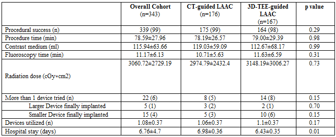
343 consecutive patients who underwent LAAC, from September 2009 to December 2019, were enrolled in this study. 176 patients received a preprocedural CT and a 3D-TEE, 167 patients received exclusively a preprocedural 3D-TEE. Baseline patient characteristics are shown in (Table 1).
Most patient characteristics did not differ significantly between the two groups. However, there was a significant difference in the HASBLED score with 4.1 ± 1.1 for the 3D-TEE group and 3.9 ± 1.01 for the CT/3D-TEE group (p = 0.05). Whereas, the CHA2DS2-VASc score was similar between the groups (4.57 ± 1.48 for 3D-TEE versus 4.72 ± 1.62 for CT/3D-TEE; p = 0.21).
Medications were well-balanced between the groups. However, 122 (73%) patients of the 3D-TEE group and 109 (62%) patients of the CT/3D-TEE group (p = 0.03) received diuretics. The corresponding values for ACE inhibitors were 48 (29%) patients for the 3D-TEE and 71 (40%) patients for the CT/3D-TEE (p = 0.02) group.
Furthermore, there were more smokers in the 3D-TEE guided group (n = 56 (34%) for 3D-TEE versus n = 37 (21%) for CT/3D-TEE, p = 0.01).
Not surprisingly, the 3D-TEE guided group had a higher prevalence of renal disease (n = 81 (49%) for 3D-TEE versus n = 47 (27%) for CT/3D-TEE; p < 0.001). This finding was also reflected by the creatinine levels (1.59 ± 1.0 mg/dl for 3D-TEE versus 1.21 ± 0.88 mg/dl for CT/3D-TEE; p < 0.001).
3.2 Indications for LAAC
In both groups, more than a half of all patients has had a previous major bleeding event (n = 93 (56%) for 3D-TEE versus n = 89 (51%) for CT/3D-TEE; p = 0.34), which was the most important indication for LAAC (listed in Table 2) in this study. Although, there were significantly more patients in the 3D-TEE guided group with previous gastrointestinal bleeding (n = 55 (33%) for 3D-TEE versus n = 41 (23%) for CT/3D-TEE; p = 0.047).


3.3 Data
339 of 343 LAAC procedures were successful, resulting in an overall success rate of 99%. There was no significant difference for the success rate between the CT and 3D-TEE guided group (n = 175 (99%) for CT/3D-TEE versus n = 164 (98%) for 3D-TEE; p = 0.29).
The failed attempt at device implantation in the CT/3D-TEE guided group was due to a complicated anatomy of the LAA. In the 3D-TEE guided group, the reasons for the failure of the LAAC procedure were:
1. periprocedural death due to pulseless electrical activity,
2. A complicated LAA anatomy, and
3. An unsuccessful puncture of the atrial septum as a result of a teflon patch of the interatrial septum after cardiac surgery.
There was no significant difference for both groups concerning the procedure time, the amount of contrast medium used, the fluoroscopy time, or the radiation dose.
Importantly, there was also no significant difference in the number of devices that needed to be resized in the 3D-TEE guided group (n = 14 (8%) versus n = 8 (5%) for CT/3D-TEE (p = 0.15).
However, patients who received a CCT before LAAC stayed longer in the hospital than those patients without CCT (6.43 ± 0.35 days for 3D-TEE versus 6.98 ± 0.36 days for CT/3D-TEE; p = 0.01).
3.4 Adverse events
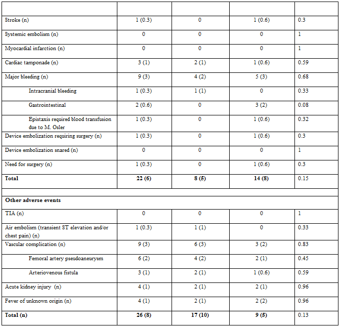
Twenty-two (6%) of 343 consecutive patients who underwent LAAC suffered from a major adverse event. Of these, 14 patients (8%) from the 3D-TEE guided group and 8 patients (5%) from the CT/3D-TEE guided group were affected (p = 0.15).
Three accumulated deaths were observed in both groups. One patient of the CT/3D-TEE guided group died after a cardiac tamponade following LAAC and a volume deficiency shock after cardiopulmonary resuscitation. Of the two reported deaths in the 3D-TEE group, one was caused by a pulseless electrical activity during the LAAC, and the other patient had a device dislocation after the intervention and died of a bleeding complication following the operation to remove the device. There was no statistical difference between the two groups regarding deaths (p = 0.53).
Major bleeding after LAAC appeared in five cases (3%) in the 3D-TEE group and four cases (2%) in the CT/3D-TEE group with no statistical difference (p = 0.68). Those events were divided into three (2%) gastrointestinal bleedings, one (0.6%) epistaxis due to M. Osler and one (0.6%) inguinal bleeding after puncture in the 3D-TEE group. In the CT/3D-TEE group, there was one (0.6%) intracranial bleeding, one (0.6%) hemorrhagic pericardial tamponade requiring one erythrocyte concentrate, one (0.6%) epistaxis, and one (0.6%) traumatic hard palate bleeding, that were defined as major bleeding events.
Two cases of acute kidney injuries were reported in each of the groups after LAAC, with no significant difference (p = 0.96).
3.5 Echocardiographic follow up
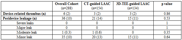
Of all the patients that underwent LAAC in this study, 288 (84%) patients received at least one follow up 3D-TEE, at the earliest, one month after the procedure. Those were divided in 154 (53%) patients for the CT/3D-TEE guided group and 134 (47%) patients for the 3D-TEE guided group (p = 0.09).
In each group, three cases (1.8% for 3D-TEE and 1.7% for CT/3D-TEE; p = 0.86) of device-related thrombi were detected (Table 5).
Furthermore, there were no significant differences in terms of minor leaks (n = 15 (11%) for 3D-TEE versus n = 20 (13%) for CT/3D-TEE; p = 0.64) or moderate leaks (n = 0 for 3D-TEE versus n = 1 (0.6 %) for CT/3D-TEE; p = 0.35). There were no major leaks in either group.
339 of 343 LAAC procedures were successful, resulting in an overall success rate of 99%. There was no significant difference for the success rate between the CT and 3D-TEE guided group (n = 175 (99%) for CT/3D-TEE versus n = 164 (98%) for 3D-TEE; p = 0.29).
The failed attempt at device implantation in the CT/3D-TEE guided group was due to a complicated anatomy of the LAA. In the 3D-TEE guided group, the reasons for the failure of the LAAC procedure were:
1. periprocedural death due to pulseless electrical activity,
2. A complicated LAA anatomy, and
3. An unsuccessful puncture of the atrial septum as a result of a teflon patch of the interatrial septum after cardiac surgery.
There was no significant difference for both groups concerning the procedure time, the amount of contrast medium used, the fluoroscopy time, or the radiation dose.
Importantly, there was also no significant difference in the number of devices that needed to be resized in the 3D-TEE guided group (n = 14 (8%) versus n = 8 (5%) for CT/3D-TEE (p = 0.15).
However, patients who received a CCT before LAAC stayed longer in the hospital than those patients without CCT (6.43 ± 0.35 days for 3D-TEE versus 6.98 ± 0.36 days for CT/3D-TEE; p = 0.01).
Twenty-two (6%) of 343 consecutive patients who underwent LAAC suffered from a major adverse event. Of these, 14 patients (8%) from the 3D-TEE guided group and 8 patients (5%) from the CT/3D-TEE guided group were affected (p = 0.15).
Three accumulated deaths were observed in both groups. One patient of the CT/3D-TEE guided group died after a cardiac tamponade following LAAC and a volume deficiency shock after cardiopulmonary resuscitation. Of the two reported deaths in the 3D-TEE group, one was caused by a pulseless electrical activity during the LAAC, and the other patient had a device dislocation after the intervention and died of a bleeding complication following the operation to remove the device. There was no statistical difference between the two groups regarding deaths (p = 0.53).
Major bleeding after LAAC appeared in five cases (3%) in the 3D-TEE group and four cases (2%) in the CT/3D-TEE group with no statistical difference (p = 0.68). Those events were divided into three (2%) gastrointestinal bleedings, one (0.6%) epistaxis due to M. Osler and one (0.6%) inguinal bleeding after puncture in the 3D-TEE group. In the CT/3D-TEE group, there was one (0.6%) intracranial bleeding, one (0.6%) hemorrhagic pericardial tamponade requiring one erythrocyte concentrate, one (0.6%) epistaxis, and one (0.6%) traumatic hard palate bleeding, that were defined as major bleeding events.
Two cases of acute kidney injuries were reported in each of the groups after LAAC, with no significant difference (p = 0.96).
3.5 Echocardiographic follow up
Of all the patients that underwent LAAC in this study, 288 (84%) patients received at least one follow up 3D-TEE, at the earliest, one month after the procedure. Those were divided in 154 (53%) patients for the CT/3D-TEE guided group and 134 (47%) patients for the 3D-TEE guided group (p = 0.09).
In each group, three cases (1.8% for 3D-TEE and 1.7% for CT/3D-TEE; p = 0.86) of device-related thrombi were detected (Table 5).
Furthermore, there were no significant differences in terms of minor leaks (n = 15 (11%) for 3D-TEE versus n = 20 (13%) for CT/3D-TEE; p = 0.64) or moderate leaks (n = 0 for 3D-TEE versus n = 1 (0.6 %) for CT/3D-TEE; p = 0.35). There were no major leaks in either group.

Table 5. Prevalence and severity of peridevice leaks at transesophageal echocardiographic follow up
LAAC is an established alternative to oral anticoagulation treatment in patients with AF and contraindications against OACs [46]. In order to achieve an effective procedure for each patient, in terms of present and long-term implantation success and a reduction of adverse events, we retrospectively investigated the influence of CCT as an additional preprocedural imaging modality besides the commonly utilized 3D-TEE.
In comparison to the ACP multicenter registry [41], we had a similar overall procedural success rate in our study (97.3% vs 98.8%) and periprocedural MAE rate (5% vs. 6.4%). In concordance with our results, Koskinas et al. [42] reported MAEs of 5.8% in 500 consecutive patients.
An important finding of our study was that we did not find a difference in the procedural success rate between patients who underwent a CCT-planned strategy for LAAC and patients with a 3D-TEE-only guided strategy (99% versus 98%). Furthermore, we did not detecthigher first-device accuracy in the CCT/3D-TEE group (94.9%) than in the 3D-TEE group (91%). In addition, the overall number of devices used was not different between the CCT/3D-TEE and 3D-TEE only groups (1.06 ± 0.37 for CCT vs. 1.1 ± 0.37 for 3D-TEE; p = 0.17).
In contrast, a much smaller study by Eng et al. [44], which randomized 24 patients to undergo LAAC using either 2D-TEE or 3D-CT for implanting WATCHMAN™ devices, found a higher procedural success rate in the 3D-CT than in the 2D-TEE group (100% vs 92%). Furthermore, this study showed that the accuracy for first device selection and the number of devices used was significantly better for 3D-CT than 2D-TEE (92% vs. 27% and 1.33 ± 0.7 vs. 2.5 ± 1.2, respectively). The difference between our findings and the study by Eng et al. may be explained by the smaller study group, different device used, and by the use of 2D-TEE, which is much less accurate than 3D-TEE [28, 32].
Dutcher et al. [31] compared a CT-guided and TEE-guided strategy in 154 consecutive patients (CT n = 76 vs. TEE n = 78) who received the WATCHMAN device. The authors of this study reported a significantly better accuracy rate concerning first device selection in the CT group with 86.7% for the CT-guided group vs 75.6% for the TEE group (p = 0.041). However, the difference between the groups was much smaller than in the study by Eng et al. [44]. Dutcher et al. also reported procedure times for the different groups. They observed significantly shorter procedure times in the CT group (33.6 min vs 46.5 min for TEE). In our study, the procedure time did not differ between the CCT/3D-TEE and 3D-TEE only groups.
We found in our study that fluoroscopy time and radiation doses did not differ between patients in the CCT/3D-TEE and in the 3D-TEE group. An interesting but small study of 24 patients receiving the WATCHMAN device by Obasare et al. [30] used a preprocedural CT to produce a latex model of the LAA using 3D printing (n = 14) and compared that to preprocedural 2D-TEE imaging. The authors of this study reported of a significantly reduced procedure time (70 ± 20 vs. 107 ± 53 min for 2D-TEE, p = 0.03) and fluoroscopy time (11 ± 4 vs. 20 ± 13 min for 2D-TEE, p = 0.02) for preprocedural CT. Unfortunately, there are some limitations concerning the comparisons of our study to Dutcher et al. and Obasare et al. as they both used the WATCHMAN device, were much smaller, and applied different imaging concepts, including 2D-TEE, which is inferior to 3D-TEE.
4.2 Long-term echocardiographic outcome
Landmesser et al. [43] showed in a large prospective Amplatzer Amulet observational study an adequate occlusion (< 3mm jet) of the appendage in 98.2% and detected a device thrombus in only 1.5% of 673 patients in the TEE follow-up, 67 ± 23 days after LAAC. Based on the definition of major leaks (jet-flow > 3 mm) by the Munich consensus document [40], we did not detect any major leaks, although we had one (0.6%) moderate and 35 (10%) minor leaks in the first follow-up TEE, at the earliest one month after LAAC in 288 available patients. However, our device-related thrombus rate (2.1%) was similar to the rate reported by Landmesser et al [43].
Importantly, in our study we could show that an absence of peri-device leaks was not statistically different between the 3D-TEE guided group and the CCT/3D-TEE group (88.8% vs. 86.4%). It is interesting that this finding is in concordance with the finding of Obasare et al., who reported an absence of peri-device leaks in 92% of patients who received a CT-based 3D-printed model.
Although patient characteristics were distributed predominantly equally in both groups, there were significantly more patients in the 3D-TEE guided group with a decreased renal function and a known renal disease. This is probably due to the selection bias of a non-randomized study, since the use of contrast agent during CCT can provoke acute renal failure or deteriorate renal function, in particular in patients with known renal disease, so it is likely that these patients were selected against for CCT.
Nevertheless, in our study, 27% of patients received a preprocedural CCT even though they had known renal disease. Interestingly, only two patients (1%) suffered from an acute kidney injury during their hospital stay in the CCT/3D-TEE arm, which was also the case in the 3D-TEE only guided group (two cases (1%)). All of these patients regained their original renal function during the hospital stay, under continued renal-protection measures. In this respect, it is important to note that in our study, patients in the CCT/3D-TEE arm did not need less contrast agent than in the 3D-TEE arm during the procedure (120 ml versus 112 ml). Hence, a CCT-guided strategy does not save contrast agent during the procedure.
Moreover, patients with an additional preprocedural CCT had a significantly longer hospital stay (6.4 ± 0.4 days for 3D-TEE alone versus 7 ± 0.4 days for CT/3D-TEE; p = 0.01) due to the fact that the CCT was performed one day before implantation of the device.
In conclusion, we did not find any difference between the preprocedural 3D-TEE and CCT/3D-TEE guided groups with respect to the primary endpoint, which was defined as the clinical outcome of the LAAC, or the secondary endpoint consisting of the echocardiographic follow-up.
In consideration of our results, we would suggest that a preprocedural CCT may be avoided before planning a LAAC, as we did not detect a decrease of MAEs or improved outcome. Moreover, the addition of a CCT leads to higher costs and a potentially longer hospital stay.
The most important limitation of our study is its retrospective design. However, we only included consecutive patients. Furthermore, it was regarded as potentially difficult to randomize patients with advanced renal dysfunction to the CCT/3D-TEE group.
In our study, 3D-TEE was used for LAAC implantation, which was performed by experienced operators. Hence our results cannot be applied to centers that use only 2D-TEE for LAAC implantation guidance.
Conflict of Interest Disclosures:
The authors have no conflicts of interest to declare.
Clearly Auctoresonline and particularly Psychology and Mental Health Care Journal is dedicated to improving health care services for individuals and populations. The editorial boards' ability to efficiently recognize and share the global importance of health literacy with a variety of stakeholders. Auctoresonline publishing platform can be used to facilitate of optimal client-based services and should be added to health care professionals' repertoire of evidence-based health care resources.

Journal of Clinical Cardiology and Cardiovascular Intervention The submission and review process was adequate. However I think that the publication total value should have been enlightened in early fases. Thank you for all.

Journal of Women Health Care and Issues By the present mail, I want to say thank to you and tour colleagues for facilitating my published article. Specially thank you for the peer review process, support from the editorial office. I appreciate positively the quality of your journal.

Journal of Clinical Research and Reports I would be very delighted to submit my testimonial regarding the reviewer board and the editorial office. The reviewer board were accurate and helpful regarding any modifications for my manuscript. And the editorial office were very helpful and supportive in contacting and monitoring with any update and offering help. It was my pleasure to contribute with your promising Journal and I am looking forward for more collaboration.

We would like to thank the Journal of Thoracic Disease and Cardiothoracic Surgery because of the services they provided us for our articles. The peer-review process was done in a very excellent time manner, and the opinions of the reviewers helped us to improve our manuscript further. The editorial office had an outstanding correspondence with us and guided us in many ways. During a hard time of the pandemic that is affecting every one of us tremendously, the editorial office helped us make everything easier for publishing scientific work. Hope for a more scientific relationship with your Journal.

The peer-review process which consisted high quality queries on the paper. I did answer six reviewers’ questions and comments before the paper was accepted. The support from the editorial office is excellent.

Journal of Neuroscience and Neurological Surgery. I had the experience of publishing a research article recently. The whole process was simple from submission to publication. The reviewers made specific and valuable recommendations and corrections that improved the quality of my publication. I strongly recommend this Journal.

Dr. Katarzyna Byczkowska My testimonial covering: "The peer review process is quick and effective. The support from the editorial office is very professional and friendly. Quality of the Clinical Cardiology and Cardiovascular Interventions is scientific and publishes ground-breaking research on cardiology that is useful for other professionals in the field.

Thank you most sincerely, with regard to the support you have given in relation to the reviewing process and the processing of my article entitled "Large Cell Neuroendocrine Carcinoma of The Prostate Gland: A Review and Update" for publication in your esteemed Journal, Journal of Cancer Research and Cellular Therapeutics". The editorial team has been very supportive.

Testimony of Journal of Clinical Otorhinolaryngology: work with your Reviews has been a educational and constructive experience. The editorial office were very helpful and supportive. It was a pleasure to contribute to your Journal.

Dr. Bernard Terkimbi Utoo, I am happy to publish my scientific work in Journal of Women Health Care and Issues (JWHCI). The manuscript submission was seamless and peer review process was top notch. I was amazed that 4 reviewers worked on the manuscript which made it a highly technical, standard and excellent quality paper. I appreciate the format and consideration for the APC as well as the speed of publication. It is my pleasure to continue with this scientific relationship with the esteem JWHCI.

This is an acknowledgment for peer reviewers, editorial board of Journal of Clinical Research and Reports. They show a lot of consideration for us as publishers for our research article “Evaluation of the different factors associated with side effects of COVID-19 vaccination on medical students, Mutah university, Al-Karak, Jordan”, in a very professional and easy way. This journal is one of outstanding medical journal.

Dear Hao Jiang, to Journal of Nutrition and Food Processing We greatly appreciate the efficient, professional and rapid processing of our paper by your team. If there is anything else we should do, please do not hesitate to let us know. On behalf of my co-authors, we would like to express our great appreciation to editor and reviewers.

As an author who has recently published in the journal "Brain and Neurological Disorders". I am delighted to provide a testimonial on the peer review process, editorial office support, and the overall quality of the journal. The peer review process at Brain and Neurological Disorders is rigorous and meticulous, ensuring that only high-quality, evidence-based research is published. The reviewers are experts in their fields, and their comments and suggestions were constructive and helped improve the quality of my manuscript. The review process was timely and efficient, with clear communication from the editorial office at each stage. The support from the editorial office was exceptional throughout the entire process. The editorial staff was responsive, professional, and always willing to help. They provided valuable guidance on formatting, structure, and ethical considerations, making the submission process seamless. Moreover, they kept me informed about the status of my manuscript and provided timely updates, which made the process less stressful. The journal Brain and Neurological Disorders is of the highest quality, with a strong focus on publishing cutting-edge research in the field of neurology. The articles published in this journal are well-researched, rigorously peer-reviewed, and written by experts in the field. The journal maintains high standards, ensuring that readers are provided with the most up-to-date and reliable information on brain and neurological disorders. In conclusion, I had a wonderful experience publishing in Brain and Neurological Disorders. The peer review process was thorough, the editorial office provided exceptional support, and the journal's quality is second to none. I would highly recommend this journal to any researcher working in the field of neurology and brain disorders.
Dear Agrippa Hilda, Journal of Neuroscience and Neurological Surgery, Editorial Coordinator, I trust this message finds you well. I want to extend my appreciation for considering my article for publication in your esteemed journal. I am pleased to provide a testimonial regarding the peer review process and the support received from your editorial office. The peer review process for my paper was carried out in a highly professional and thorough manner. The feedback and comments provided by the authors were constructive and very useful in improving the quality of the manuscript. This rigorous assessment process undoubtedly contributes to the high standards maintained by your journal.

International Journal of Clinical Case Reports and Reviews. I strongly recommend to consider submitting your work to this high-quality journal. The support and availability of the Editorial staff is outstanding and the review process was both efficient and rigorous.

Thank you very much for publishing my Research Article titled “Comparing Treatment Outcome Of Allergic Rhinitis Patients After Using Fluticasone Nasal Spray And Nasal Douching" in the Journal of Clinical Otorhinolaryngology. As Medical Professionals we are immensely benefited from study of various informative Articles and Papers published in this high quality Journal. I look forward to enriching my knowledge by regular study of the Journal and contribute my future work in the field of ENT through the Journal for use by the medical fraternity. The support from the Editorial office was excellent and very prompt. I also welcome the comments received from the readers of my Research Article.

Dear Erica Kelsey, Editorial Coordinator of Cancer Research and Cellular Therapeutics Our team is very satisfied with the processing of our paper by your journal. That was fast, efficient, rigorous, but without unnecessary complications. We appreciated the very short time between the submission of the paper and its publication on line on your site.

I am very glad to say that the peer review process is very successful and fast and support from the Editorial Office. Therefore, I would like to continue our scientific relationship for a long time. And I especially thank you for your kindly attention towards my article. Have a good day!

"We recently published an article entitled “Influence of beta-Cyclodextrins upon the Degradation of Carbofuran Derivatives under Alkaline Conditions" in the Journal of “Pesticides and Biofertilizers” to show that the cyclodextrins protect the carbamates increasing their half-life time in the presence of basic conditions This will be very helpful to understand carbofuran behaviour in the analytical, agro-environmental and food areas. We greatly appreciated the interaction with the editor and the editorial team; we were particularly well accompanied during the course of the revision process, since all various steps towards publication were short and without delay".

I would like to express my gratitude towards you process of article review and submission. I found this to be very fair and expedient. Your follow up has been excellent. I have many publications in national and international journal and your process has been one of the best so far. Keep up the great work.

We are grateful for this opportunity to provide a glowing recommendation to the Journal of Psychiatry and Psychotherapy. We found that the editorial team were very supportive, helpful, kept us abreast of timelines and over all very professional in nature. The peer review process was rigorous, efficient and constructive that really enhanced our article submission. The experience with this journal remains one of our best ever and we look forward to providing future submissions in the near future.

I am very pleased to serve as EBM of the journal, I hope many years of my experience in stem cells can help the journal from one way or another. As we know, stem cells hold great potential for regenerative medicine, which are mostly used to promote the repair response of diseased, dysfunctional or injured tissue using stem cells or their derivatives. I think Stem Cell Research and Therapeutics International is a great platform to publish and share the understanding towards the biology and translational or clinical application of stem cells.

I would like to give my testimony in the support I have got by the peer review process and to support the editorial office where they were of asset to support young author like me to be encouraged to publish their work in your respected journal and globalize and share knowledge across the globe. I really give my great gratitude to your journal and the peer review including the editorial office.

I am delighted to publish our manuscript entitled "A Perspective on Cocaine Induced Stroke - Its Mechanisms and Management" in the Journal of Neuroscience and Neurological Surgery. The peer review process, support from the editorial office, and quality of the journal are excellent. The manuscripts published are of high quality and of excellent scientific value. I recommend this journal very much to colleagues.

Dr.Tania Muñoz, My experience as researcher and author of a review article in The Journal Clinical Cardiology and Interventions has been very enriching and stimulating. The editorial team is excellent, performs its work with absolute responsibility and delivery. They are proactive, dynamic and receptive to all proposals. Supporting at all times the vast universe of authors who choose them as an option for publication. The team of review specialists, members of the editorial board, are brilliant professionals, with remarkable performance in medical research and scientific methodology. Together they form a frontline team that consolidates the JCCI as a magnificent option for the publication and review of high-level medical articles and broad collective interest. I am honored to be able to share my review article and open to receive all your comments.

“The peer review process of JPMHC is quick and effective. Authors are benefited by good and professional reviewers with huge experience in the field of psychology and mental health. The support from the editorial office is very professional. People to contact to are friendly and happy to help and assist any query authors might have. Quality of the Journal is scientific and publishes ground-breaking research on mental health that is useful for other professionals in the field”.

Dear editorial department: On behalf of our team, I hereby certify the reliability and superiority of the International Journal of Clinical Case Reports and Reviews in the peer review process, editorial support, and journal quality. Firstly, the peer review process of the International Journal of Clinical Case Reports and Reviews is rigorous, fair, transparent, fast, and of high quality. The editorial department invites experts from relevant fields as anonymous reviewers to review all submitted manuscripts. These experts have rich academic backgrounds and experience, and can accurately evaluate the academic quality, originality, and suitability of manuscripts. The editorial department is committed to ensuring the rigor of the peer review process, while also making every effort to ensure a fast review cycle to meet the needs of authors and the academic community. Secondly, the editorial team of the International Journal of Clinical Case Reports and Reviews is composed of a group of senior scholars and professionals with rich experience and professional knowledge in related fields. The editorial department is committed to assisting authors in improving their manuscripts, ensuring their academic accuracy, clarity, and completeness. Editors actively collaborate with authors, providing useful suggestions and feedback to promote the improvement and development of the manuscript. We believe that the support of the editorial department is one of the key factors in ensuring the quality of the journal. Finally, the International Journal of Clinical Case Reports and Reviews is renowned for its high- quality articles and strict academic standards. The editorial department is committed to publishing innovative and academically valuable research results to promote the development and progress of related fields. The International Journal of Clinical Case Reports and Reviews is reasonably priced and ensures excellent service and quality ratio, allowing authors to obtain high-level academic publishing opportunities in an affordable manner. I hereby solemnly declare that the International Journal of Clinical Case Reports and Reviews has a high level of credibility and superiority in terms of peer review process, editorial support, reasonable fees, and journal quality. Sincerely, Rui Tao.

Clinical Cardiology and Cardiovascular Interventions I testity the covering of the peer review process, support from the editorial office, and quality of the journal.

Clinical Cardiology and Cardiovascular Interventions, we deeply appreciate the interest shown in our work and its publication. It has been a true pleasure to collaborate with you. The peer review process, as well as the support provided by the editorial office, have been exceptional, and the quality of the journal is very high, which was a determining factor in our decision to publish with you.

The peer reviewers process is quick and effective, the supports from editorial office is excellent, the quality of journal is high. I would like to collabroate with Internatioanl journal of Clinical Case Reports and Reviews journal clinically in the future time.

Clinical Cardiology and Cardiovascular Interventions, I would like to express my sincerest gratitude for the trust placed in our team for the publication in your journal. It has been a true pleasure to collaborate with you on this project. I am pleased to inform you that both the peer review process and the attention from the editorial coordination have been excellent. Your team has worked with dedication and professionalism to ensure that your publication meets the highest standards of quality. We are confident that this collaboration will result in mutual success, and we are eager to see the fruits of this shared effort.

Dear Dr. Jessica Magne, Editorial Coordinator 0f Clinical Cardiology and Cardiovascular Interventions, I hope this message finds you well. I want to express my utmost gratitude for your excellent work and for the dedication and speed in the publication process of my article titled "Navigating Innovation: Qualitative Insights on Using Technology for Health Education in Acute Coronary Syndrome Patients." I am very satisfied with the peer review process, the support from the editorial office, and the quality of the journal. I hope we can maintain our scientific relationship in the long term.

Dear Monica Gissare, - Editorial Coordinator of Nutrition and Food Processing. ¨My testimony with you is truly professional, with a positive response regarding the follow-up of the article and its review, you took into account my qualities and the importance of the topic¨.

Dear Dr. Jessica Magne, Editorial Coordinator 0f Clinical Cardiology and Cardiovascular Interventions, The review process for the article “The Handling of Anti-aggregants and Anticoagulants in the Oncologic Heart Patient Submitted to Surgery” was extremely rigorous and detailed. From the initial submission to the final acceptance, the editorial team at the “Journal of Clinical Cardiology and Cardiovascular Interventions” demonstrated a high level of professionalism and dedication. The reviewers provided constructive and detailed feedback, which was essential for improving the quality of our work. Communication was always clear and efficient, ensuring that all our questions were promptly addressed. The quality of the “Journal of Clinical Cardiology and Cardiovascular Interventions” is undeniable. It is a peer-reviewed, open-access publication dedicated exclusively to disseminating high-quality research in the field of clinical cardiology and cardiovascular interventions. The journal's impact factor is currently under evaluation, and it is indexed in reputable databases, which further reinforces its credibility and relevance in the scientific field. I highly recommend this journal to researchers looking for a reputable platform to publish their studies.

Dear Editorial Coordinator of the Journal of Nutrition and Food Processing! "I would like to thank the Journal of Nutrition and Food Processing for including and publishing my article. The peer review process was very quick, movement and precise. The Editorial Board has done an extremely conscientious job with much help, valuable comments and advices. I find the journal very valuable from a professional point of view, thank you very much for allowing me to be part of it and I would like to participate in the future!”

Dealing with The Journal of Neurology and Neurological Surgery was very smooth and comprehensive. The office staff took time to address my needs and the response from editors and the office was prompt and fair. I certainly hope to publish with this journal again.Their professionalism is apparent and more than satisfactory. Susan Weiner

My Testimonial Covering as fellowing: Lin-Show Chin. The peer reviewers process is quick and effective, the supports from editorial office is excellent, the quality of journal is high. I would like to collabroate with Internatioanl journal of Clinical Case Reports and Reviews.

My experience publishing in Psychology and Mental Health Care was exceptional. The peer review process was rigorous and constructive, with reviewers providing valuable insights that helped enhance the quality of our work. The editorial team was highly supportive and responsive, making the submission process smooth and efficient. The journal's commitment to high standards and academic rigor makes it a respected platform for quality research. I am grateful for the opportunity to publish in such a reputable journal.

My experience publishing in International Journal of Clinical Case Reports and Reviews was exceptional. I Come forth to Provide a Testimonial Covering the Peer Review Process and the editorial office for the Professional and Impartial Evaluation of the Manuscript.

I would like to offer my testimony in the support. I have received through the peer review process and support the editorial office where they are to support young authors like me, encourage them to publish their work in your esteemed journals, and globalize and share knowledge globally. I really appreciate your journal, peer review, and editorial office.

Dear Agrippa Hilda- Editorial Coordinator of Journal of Neuroscience and Neurological Surgery, "The peer review process was very quick and of high quality, which can also be seen in the articles in the journal. The collaboration with the editorial office was very good."

I would like to express my sincere gratitude for the support and efficiency provided by the editorial office throughout the publication process of my article, “Delayed Vulvar Metastases from Rectal Carcinoma: A Case Report.” I greatly appreciate the assistance and guidance I received from your team, which made the entire process smooth and efficient. The peer review process was thorough and constructive, contributing to the overall quality of the final article. I am very grateful for the high level of professionalism and commitment shown by the editorial staff, and I look forward to maintaining a long-term collaboration with the International Journal of Clinical Case Reports and Reviews.

To Dear Erin Aust, I would like to express my heartfelt appreciation for the opportunity to have my work published in this esteemed journal. The entire publication process was smooth and well-organized, and I am extremely satisfied with the final result. The Editorial Team demonstrated the utmost professionalism, providing prompt and insightful feedback throughout the review process. Their clear communication and constructive suggestions were invaluable in enhancing my manuscript, and their meticulous attention to detail and dedication to quality are truly commendable. Additionally, the support from the Editorial Office was exceptional. From the initial submission to the final publication, I was guided through every step of the process with great care and professionalism. The team's responsiveness and assistance made the entire experience both easy and stress-free. I am also deeply impressed by the quality and reputation of the journal. It is an honor to have my research featured in such a respected publication, and I am confident that it will make a meaningful contribution to the field.

"I am grateful for the opportunity of contributing to [International Journal of Clinical Case Reports and Reviews] and for the rigorous review process that enhances the quality of research published in your esteemed journal. I sincerely appreciate the time and effort of your team who have dedicatedly helped me in improvising changes and modifying my manuscript. The insightful comments and constructive feedback provided have been invaluable in refining and strengthening my work".

I thank the ‘Journal of Clinical Research and Reports’ for accepting this article for publication. This is a rigorously peer reviewed journal which is on all major global scientific data bases. I note the review process was prompt, thorough and professionally critical. It gave us an insight into a number of important scientific/statistical issues. The review prompted us to review the relevant literature again and look at the limitations of the study. The peer reviewers were open, clear in the instructions and the editorial team was very prompt in their communication. This journal certainly publishes quality research articles. I would recommend the journal for any future publications.

Dear Jessica Magne, with gratitude for the joint work. Fast process of receiving and processing the submitted scientific materials in “Clinical Cardiology and Cardiovascular Interventions”. High level of competence of the editors with clear and correct recommendations and ideas for enriching the article.

We found the peer review process quick and positive in its input. The support from the editorial officer has been very agile, always with the intention of improving the article and taking into account our subsequent corrections.

My article, titled 'No Way Out of the Smartphone Epidemic Without Considering the Insights of Brain Research,' has been republished in the International Journal of Clinical Case Reports and Reviews. The review process was seamless and professional, with the editors being both friendly and supportive. I am deeply grateful for their efforts.

To Dear Erin Aust – Editorial Coordinator of Journal of General Medicine and Clinical Practice! I declare that I am absolutely satisfied with your work carried out with great competence in following the manuscript during the various stages from its receipt, during the revision process to the final acceptance for publication. Thank Prof. Elvira Farina

Dear Jessica, and the super professional team of the ‘Clinical Cardiology and Cardiovascular Interventions’ I am sincerely grateful to the coordinated work of the journal team for the no problem with the submission of my manuscript: “Cardiometabolic Disorders in A Pregnant Woman with Severe Preeclampsia on the Background of Morbid Obesity (Case Report).” The review process by 5 experts was fast, and the comments were professional, which made it more specific and academic, and the process of publication and presentation of the article was excellent. I recommend that my colleagues publish articles in this journal, and I am interested in further scientific cooperation. Sincerely and best wishes, Dr. Oleg Golyanovskiy.

Dear Ashley Rosa, Editorial Coordinator of the journal - Psychology and Mental Health Care. " The process of obtaining publication of my article in the Psychology and Mental Health Journal was positive in all areas. The peer review process resulted in a number of valuable comments, the editorial process was collaborative and timely, and the quality of this journal has been quickly noticed, resulting in alternative journals contacting me to publish with them." Warm regards, Susan Anne Smith, PhD. Australian Breastfeeding Association.

Dear Jessica Magne, Editorial Coordinator, Clinical Cardiology and Cardiovascular Interventions, Auctores Publishing LLC. I appreciate the journal (JCCI) editorial office support, the entire team leads were always ready to help, not only on technical front but also on thorough process. Also, I should thank dear reviewers’ attention to detail and creative approach to teach me and bring new insights by their comments. Surely, more discussions and introduction of other hemodynamic devices would provide better prevention and management of shock states. Your efforts and dedication in presenting educational materials in this journal are commendable. Best wishes from, Farahnaz Fallahian.

Dear Maria Emerson, Editorial Coordinator, International Journal of Clinical Case Reports and Reviews, Auctores Publishing LLC. I am delighted to have published our manuscript, "Acute Colonic Pseudo-Obstruction (ACPO): A rare but serious complication following caesarean section." I want to thank the editorial team, especially Maria Emerson, for their prompt review of the manuscript, quick responses to queries, and overall support. Yours sincerely Dr. Victor Olagundoye.

Dear Ashley Rosa, Editorial Coordinator, International Journal of Clinical Case Reports and Reviews. Many thanks for publishing this manuscript after I lost confidence the editors were most helpful, more than other journals Best wishes from, Susan Anne Smith, PhD. Australian Breastfeeding Association.

Dear Agrippa Hilda, Editorial Coordinator, Journal of Neuroscience and Neurological Surgery. The entire process including article submission, review, revision, and publication was extremely easy. The journal editor was prompt and helpful, and the reviewers contributed to the quality of the paper. Thank you so much! Eric Nussbaum, MD

Dr Hala Al Shaikh This is to acknowledge that the peer review process for the article ’ A Novel Gnrh1 Gene Mutation in Four Omani Male Siblings, Presentation and Management ’ sent to the International Journal of Clinical Case Reports and Reviews was quick and smooth. The editorial office was prompt with easy communication.

Dear Erin Aust, Editorial Coordinator, Journal of General Medicine and Clinical Practice. We are pleased to share our experience with the “Journal of General Medicine and Clinical Practice”, following the successful publication of our article. The peer review process was thorough and constructive, helping to improve the clarity and quality of the manuscript. We are especially thankful to Ms. Erin Aust, the Editorial Coordinator, for her prompt communication and continuous support throughout the process. Her professionalism ensured a smooth and efficient publication experience. The journal upholds high editorial standards, and we highly recommend it to fellow researchers seeking a credible platform for their work. Best wishes By, Dr. Rakhi Mishra.

Dear Jessica Magne, Editorial Coordinator, Clinical Cardiology and Cardiovascular Interventions, Auctores Publishing LLC. The peer review process of the journal of Clinical Cardiology and Cardiovascular Interventions was excellent and fast, as was the support of the editorial office and the quality of the journal. Kind regards Walter F. Riesen Prof. Dr. Dr. h.c. Walter F. Riesen.

Dear Ashley Rosa, Editorial Coordinator, International Journal of Clinical Case Reports and Reviews, Auctores Publishing LLC. Thank you for publishing our article, Exploring Clozapine's Efficacy in Managing Aggression: A Multiple Single-Case Study in Forensic Psychiatry in the international journal of clinical case reports and reviews. We found the peer review process very professional and efficient. The comments were constructive, and the whole process was efficient. On behalf of the co-authors, I would like to thank you for publishing this article. With regards, Dr. Jelle R. Lettinga.

Dear Clarissa Eric, Editorial Coordinator, Journal of Clinical Case Reports and Studies, I would like to express my deep admiration for the exceptional professionalism demonstrated by your journal. I am thoroughly impressed by the speed of the editorial process, the substantive and insightful reviews, and the meticulous preparation of the manuscript for publication. Additionally, I greatly appreciate the courteous and immediate responses from your editorial office to all my inquiries. Best Regards, Dariusz Ziora

Dear Chrystine Mejia, Editorial Coordinator, Journal of Neurodegeneration and Neurorehabilitation, Auctores Publishing LLC, We would like to thank the editorial team for the smooth and high-quality communication leading up to the publication of our article in the Journal of Neurodegeneration and Neurorehabilitation. The reviewers have extensive knowledge in the field, and their relevant questions helped to add value to our publication. Kind regards, Dr. Ravi Shrivastava.

Dear Clarissa Eric, Editorial Coordinator, Journal of Clinical Case Reports and Studies, Auctores Publishing LLC, USA Office: +1-(302)-520-2644. I would like to express my sincere appreciation for the efficient and professional handling of my case report by the ‘Journal of Clinical Case Reports and Studies’. The peer review process was not only fast but also highly constructive—the reviewers’ comments were clear, relevant, and greatly helped me improve the quality and clarity of my manuscript. I also received excellent support from the editorial office throughout the process. Communication was smooth and timely, and I felt well guided at every stage, from submission to publication. The overall quality and rigor of the journal are truly commendable. I am pleased to have published my work with Journal of Clinical Case Reports and Studies, and I look forward to future opportunities for collaboration. Sincerely, Aline Tollet, UCLouvain.

Dear Ms. Mayra Duenas, Editorial Coordinator, International Journal of Clinical Case Reports and Reviews. “The International Journal of Clinical Case Reports and Reviews represented the “ideal house” to share with the research community a first experience with the use of the Simeox device for speech rehabilitation. High scientific reputation and attractive website communication were first determinants for the selection of this Journal, and the following submission process exceeded expectations: fast but highly professional peer review, great support by the editorial office, elegant graphic layout. Exactly what a dynamic research team - also composed by allied professionals - needs!" From, Chiara Beccaluva, PT - Italy.

Dear Maria Emerson, Editorial Coordinator, we have deeply appreciated the professionalism demonstrated by the International Journal of Clinical Case Reports and Reviews. The reviewers have extensive knowledge of our field and have been very efficient and fast in supporting the process. I am really looking forward to further collaboration. Thanks. Best regards, Dr. Claudio Ligresti

Dear Chrystine Mejia, Editorial Coordinator, Journal of Neurodegeneration and Neurorehabilitation. “The peer review process was efficient and constructive, and the editorial office provided excellent communication and support throughout. The journal ensures scientific rigor and high editorial standards, while also offering a smooth and timely publication process. We sincerely appreciate the work of the editorial team in facilitating the dissemination of innovative approaches such as the Bonori Method.” Best regards, Dr. Giselle Pentón-Rol.
