AUCTORES
Globalize your Research

Review Article | DOI: https://doi.org/10.31579/2641-0419/073
*Corresponding Author: Jeremy William Docekal, Staff cardiologist Landstuhl Regional Military medical Center
Citation: Jeremy W. Docekal., (2020) A review of surgical Aortic Valve Repair. Indications and techniques for the General Cardiologist. J. Clinical Cardiology and Cardiovascular Interventions, 3(7);Doi:10.31579/2641-0419/073
Copyright: © 2020 Jeremy William Docekal, This is an open-access article distributed under the terms of the Creative Commons Attribution License, which permits unrestricted use, distribution, and reproduction in any medium, provided the original author and source are credited.
Received: 09 July 2020 | Accepted: 14 July 2020 | Published: 29 July 2020
Keywords: Aortic valve repair; aortic aneurysm; bicuspid aortic valve; prosthetic aortic valve
The management of surgical aortic valve disease is particularly challenging among young patients and patient’s employed in high risk occupations such as the military. Longer life expectancy predicts an increased probability for prosthetic-valve-related complications. Consequently, there is a recognized need for alternative therapies, and surgical aortic valve repair may offer an attractive option for selected patients. Furthermore, aortic valve repair surgical techniques have evolved, and data from contemporary studies show consistent, successful, and durable outcomes; particularly when performed in high volume centers. The purpose of this review is to discuss the indications for aortic valve repair, review basic operative techniques, and illustrate how pre-procedural echocardiographic imaging is contributing to surgical planning.
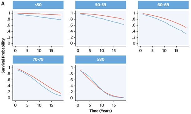
Young patients, particularly those employed in high-risk occupations such as the military or among competitive athletes, who have aortic valve pathology, present a well-documented challenge in the current era. For these patients, postoperative quality of life, occupational compatibility, and life expectancy are important considerations affecting choice of therapy. A commonly offered solution is aortic valve replacement (AVR) with either a mechanical or biologic prosthesis. However, AVR is associated with several disadvantages. In a recently published study on this topic, the authors conclude that their study found a “shorter life expectancy in patients after aortic valve replacement (AVR) compared with the general population. The estimated loss in life expectancy was substantial, and increased with younger age (Figure 1) [1].” Their observation may be explained, in part, by the fact that longer life expectancy predicts increased exposure to prosthetic-valve-related complications such as degeneration leading to reoperation, bleeding, and thromboembolism [2].
A retrospective review by Goldstone et al. [3] helps us to better understand the magnitude of the problem. Their study observed, among patients aged 45-54, a 15-year mortality rate of 30.6% in patients who received AVR with a biologic prosthesis and an estimated 15-year mortality rate of 26.4% following AVR with a mechanical prosthesis. Furthermore, a retrospective review performed in Ottawa, Canada concluded that the median interval to reoperation for contemporary stented aortic bioprosthesis was 7.74 years (95% CI 7.28 to 9.97 years,) in patients younger than 40 years of age [4].
With the disconcerting data emerging in regard to the long-term benefit of AVR, there is a need for alternative options. For this reason, there is a renewed interest in two surgical options in particular. Among young patients with aortic valve disease, the Ross procedure has several demonstrated advantages. Buratto et al. recently demonstrated that the Ross procedure is associated with a reduced risk of late mortality as compared with mechanical AVR, which may be partially explained by avoidance of anticoagulation and its associated complications. Furthermore, the Ross procedure achieves more favorable valve hemodynamics, whereby the effective orifice area is greater than that which can be achieved with a prosthesis [5]. The advantages of the Ross procedure have been recently and elegantly reviewed, and this is not the primary intent of this paper [6]. A second option is surgical aortic valve repair (AV repair), which is has long been regarded as a surgical alternative to prosthetic valve replacement among selected patients with aortic insufficiency (AI) or aortic aneurysm.
To help illustrate the advantage of AV repair, in a propensity score analysis matching patients who underwent surgical correction of severe AI by either surgical AVR or AV repair, it was determined that AV repair is associated with a better overall 9-year survival rate as compared to AVR (87% vs 60%; P = .007) [7]. In addition, a report published by the Society of Thoracic Surgeons confirms a low operative mortality among patients who underwent aortic valve sparing procedures (1.88%), which compares favorably with reported mortality rates of 5% among surgical AVR using a valved conduit (Bentall procedure) [8].
Yet, in spite of the published advantages of AV repair, this remains an underutilized surgical technique, particularly in the United States. In a report from the Society of Thoracic Surgeons database [8], it was determined that there is an increasing trend towards AVR using biostented valves, while AV repair remains an uncommon procedure for most surgical centers. According to this same report, currently only 5% of institutions participating in the Society of Thoracic Surgeons Adult Cardiac Surgery database (STS ACSD) performed >16 aortic valve sparing procedures in 2009.
Given the proposed advantages of AV repair and increasing familiarity of this procedure in several European institutions, it remains difficult to justify slow adoption of AV repair in the United States. One possible explanation is the historic wide range in reported surgical outcomes with AV repair. However, contemporary publications of AV repair results from specialized centers are now available and demonstrate excellent short- and long-term results. In addition, an industry generated bias towards valve replacement with current mechanical and bioprosthetic implants may possibly exist.
Advances in echocardiographic imaging have helped improve surgical planning. Thus, the cardiologist with training in 3-dimensional echocardiography now provides valuable insight and is an active contributor to successful AV repair.
The purpose of this review article is to discuss the indications for AV repair, discuss candidate patients, review current outcomes data, and help familiarize the reader in regard to basic surgical techniques used for AV repair. In addition, we review how advanced, non-invasive imaging is currently helping improve surgical planning and outcomes.
Anatomy of the aortic root
Prior to further discussion, a review of aortic root anatomy is instructive.
In general, the aortic root may be thought of as a bridge between the left ventricle and the ascending aorta, by which the aortic root acts as a native stent, surrounding and supporting the 3 aortic cusps [11].
In terms of function, valve integrity depends upon the cohesion of all structures forming the aortic root: the aortic valve leaflets and their attachments, Sinuses of Valsalva (SoV), interleaflet trigones, sinotubular junction (STJ), and the aortic annulus.
The aortic leaflets form the hemodynamic junction between the left ventricle and aorta and form the demarcation between structures subject to arterial pressures and those subjected to ventricular pressures [9,10].The aortic leaflets are attached to the aortic wall in a semilunar fashion and extend basally from the ventriculo-aortic junction (VAJ) to their distal attachment at the STJ. As the leaflet attachments insert into the wall of the aorta, they form a crown-shaped fibrous structure [11].
The anatomy of the valve leaflets is composed of the load bearing leaflet body, the free margin (otherwise known as the coapting surface) that contains a thickened circular node termed the nodule of Arantius [9].), and the leaflet attachment (otherwise known as the basal leaflet or hinge point) [9]. The free edge of the aortic leaflets are constructed such that when closed, the leaflets coapt over several millimeters. The margin of overlap has been defined as the lunula [12]. The term commissure refers to the zone of apposition between the two coronet-shaped, lateral attachments of adjacent leaflets to the aortic wall. A failure in the development of the commissural area results in the development of bicuspid aortic valves (BAV), as explained later in this text. The term “cusp” is used to describe the leaflet tip, but is commonly used synonymously with the term leaflet.
The exact definition of the aortic annulus is debated, and there is an absence of universally agreed upon criteria from either an anatomic or hemodynamic viewpoint. However, there exists a condensation of collagenous tissue at the leaflet hinge point, which follows the semi-lunar contour of the valvular attachment and is thickest at the nadir of the semilunar attachments [12]. This ring of collagenous tissue provides the substrate which anatomically defines the annulus. However, the functional annulus determines valve geometry and provides an important anatomical landmark for surgical repair. The functional annulus is determined by a horizontal plane connecting the cusp nadirs and is termed the basal ring (See Figure 2). This is discussed in more detail later in this text. The near universal desire to describe the structure of the aortic valve in terms of annulus has perhaps superseded the key fact that it is the semilunar arrangement of leaflet hinge points which is critical to normal valvular function [13].

Named after the Italian anatomist Antonio Valsalva, the SoV forms three distinct expanded portions of the aortic wall and is confined to the region bounded caudally by the valvular leaflets and cranially by the sinotubular ridge [12]. The function of the SoV is under investigation; however, it has been proposed that the area of the SoV creates flow turbulence, which may lead to a reduction in shear stress on the aortic leaflets and gradual valve closure, while supporting coronary flow [14].
Surgical technique
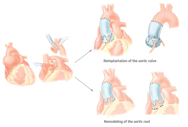
The most frequent mechanism for aortic regurgitation is dilation of the aortic root and ascending aorta. Therefore, we will begin by discussing the development of valve sparing root replacement. The aortic root remodeling procedure was initially described by Sarsam et al. In summary, in the aortic remodeling procedure, the aortic wall is excised to within approximately 3 mm of the leaflet attachments. Next, the coronary arteries are detached, and a Dacron graft is sized and sutured into the excised sinuses. Lastly, the coronary arteries are subsequently reimplanted [15].
A second option for accomplishing valve sparing aortic root replacement as originally described by David et al. [16]. is referred to as the reimplantation technique. This is a complex procedure, and a detailed description is beyond the scope of this review. In summary, the aneurysmal portion of the ascending aorta and SoV are excised, while leaving the aortic valve leaflets and portion of the arterial wall attached to the left ventricular outflow tract. Next, a carefully sized and constructed collagen-impregnated tubular Dacron graft is affixed proximally to the VAJ using pledgeted sutures placed, generally speaking, along the plane formed by the nadir of leaflet insertion [17]. The native valve is then implanted within the Dacron graft using a running suture. After valve reimplantation, identified leaflet pathology is corrected using techniques described below. The coronary arteries are subsequently re-attached, and the distal anastomosis between the graft and native aorta is performed (Figure 3). While technically more demanding, the reimplantation technique has the advantage of inherently stabilizing the aortic root at the level of the basal ring while the remodeling technique may be physiologically superior (18). Both the reimplantaton and the remodeling technique have been successfully performed among patients with Marfan syndrome and aortic root aneurysm [19,20].
Although the initial results of these two procedures was promising, follow-up studies demonstrated recurrent AI [21]. It was initially unclear as to the cause or mechanism of recurrent aortic regurgitation until Schäfers et al. [22, 23] published findings that the valve-preserving aortic root replacement procedure itself induced aortic valve leaflet prolapse. Their institution were early adopters of combining cusp prolapse repair in conjunction with valve sparing root replacement. Furthermore, Schäfers et al. [24] went on to describe the effective height concept into AV repair, which has been nearly universally adopted among surgeons performing AV repair.
The Effective Height Concept
Prior to summarizing techniques used to accomplish aortic leaflet repair, an understanding of the effective height (eH) concept is helpful. Various measurements are used to define aortic cusp geometry. These include dimensions of the SoV, annular dimensions, length of the free margin, and cusp height. Of the indices of cusp geometry, Schäfers et al. [24] determined that the distance between the basal plane (horizontal plane connecting the cusp nadirs) and central free margin is 8-10 mm in the normal aortic valve when measured in diastole; this measurement is termed the Effective Height (see Figures 4).
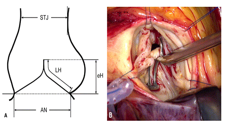
It was further observed that, when the distance between the aortic insertion lines and central free margins is low (typical cutoff is less than 8mm) following aortic remodeling, there is increased risk for recurrent aortic regurgitation. However, efforts to measure the effective height intraoperatively proved initially difficult. To overcome this obstacle, a unique caliper was invented for the purpose of easily, accurately, and reproducibly determining effective height (MSS-1, Fehling Instruments, Karlstein, Germany). With the aid of this tool, repair techniques discussed below are performed with the intention of restoring an effective height of 9 to 10 mm (See Figure 4).
In summary, when performing aortic valve sparing root replacement, assessing and correcting aortic cusp prolapse using the concept of effective height, in conjunction with root remodeling or reimplantation, has refined and improved long-term results.
Among patients with annular dilation as the primary mechanism causing AI, approximately 88% have additional leaflet prolapse, which further contributes to the severity of AI [25]. A proposed explanation for the association between aortic dilation and leaflet prolapse is the hypothesis that progressive annular dilatation increases stress on valve leaflets. Consequently, over time, this may result in leaflet stretching and prolapse [25]. Thus, aortic leaflet prolapse may coexist with aortic root dilation (mixed etiology). In addition, aortic leaflet prolapse may occur as a consequence of aortic valve preserving root replacement, or isolated prolapse may be the sole cause of aortic valve insufficiency. A successful surgical outcome may thus require a combination repair of aortic root dilation and additional leaflet pathology (most commonly aortic leaflet prolapse by mean of distention of the free margin) [26,27]. We will now shift our attention to surgical methods used for leaflet pathology. We will discuss the most common methods used for aortic leaflet repair: central plication, leaflet resuspension, leaflet resection, and pericardial patch.
In the tricuspid aortic valve, surgical repair techniques aim to elevate the level of coaptation, and restore effective height. Although several techniques have been proposed, due to associated technical difficulties, many have largely been abandoned. A technique termed central plication has emerged as the primary modality used to repair a prolapsing aortic leaflet [28, 29]. In this surgical method, simple 5‑0 or 6-0 polypropylene plication sutures are placed within the thickened leaflet free-edge cord, with the intent of shortening the prolapsing leaflet free-edge length adjacent to the nodule of Arantius. An advantage of this technique is its relative simplicity when compared to alternative techniques and the ability to adjust cusp geometry in a stepwise manner [29]. One significant disadvantage of central plication is that this procedure is not feasible in circumstances involving significant leaflet calcification.
The resuspension technique was developed, during which Gore-Tex sutures are passed in a running fashion over the entire length of the free margin. The free margin is shortened by applying tension on both Gore-Tex suture arms, which are subsequently locked. This technique may be a suitable option for closing aortic leaflet fenestrations and for reinforcing fragile free margins [30]. Additionally, it is further possible to combine both plication and resuspension, as demonstrated by Kerchove et al. [31].
A technique which is particularly well suited in the presence of leaflet perforation or fenestration is the use of pericardial patch repair. However, several publications [32,33] indicate that the use of pericardium for leaflet reconstruction may be associated with an increased rate of repair failure. In a retrospective review performed by Karliova et al., the authors observed that long-term stability following the use of pericardial patch repair is best suited for closure of fenestrations, followed by defect closure, and cusp augmentation. However, among bicuspid aortic valves, the use of pericardial patch was associated with particularly poor stability, regardless of technique [32]. The 10-year observed patch-related freedom from reoperation was only 78% among bicuspid AV repairs. Thus the durability after aortic repair using pericardium is dependent on both valve morphology and underlying cusp pathology.
In the presence of marked tissue redundancy (>10 mm) or dense fibrosis/calcification of the prolapsing cusp, triangular resection is a useful method. In this manner, a central resection of cusp tissue is created with the remaining tissue readapted using interrupted Prolene sutures [29]. If the resulting defect following triangular resection is too large to allow direct readaptation, then a pericardial patch is used for leaflet reconstruction.
Surgical Repair of the bicuspid valve
The anatomy of the bicuspid valve (BAV), as described by Sievers and Schmidtke (34), is divided into three general classifications. First, a Type 0 BAV consists of two symmetric aortic cusps without the presence of a central raphe (fused region of underdeveloped leaflets). The type 0 BAV occurs as the result of a complete failure in the development one commissure, resulting in two completely developed symmetric leaflets and commissures [33,34]. The mechanism of insufficiency is typically a result of excessive and redundant prolapsing cusp tissue. More common is the type 1 BAV, in which there are two fully developed commissures, one under-developed commissure, and a central raphe. The type 2 BAV occurs due to the full development of only one commissure, with two under-developed commissures, resulting in the presence of 2 raphe. The type 2 BAV may appear to be nearly tri-leaflet in configuration.
While the classification proposed by Sievers is the most widely adopted, there are recognized limitations with relevance to surgical planning for AV repair [35]. Thus, a novel repair-oriented classification scheme has recently been proposed by Kerchove et al. [36], which classifies bicuspid aortic valves in terms of the observed variability in degree of commissural orientation. In brief, the orientation of the two functional commissures has been observed to vary from 180 degrees (Kerchove Type A, symmetric such as described in a Sievers type 0) to 120-140 degrees (Kerchove Type C, very asymmetric, nearly tricuspid in configuration). Among asymmetric BAVs with a near tricuspid configuration, prolapse may preferentially involve the rudimentary right cusp.
The approach to repair of the BAV uses techniques similar to those described above. In the type 0 valve, with prolapse as the predominant mechanism of insufficiency, free margin plication or free margin resuspension with a goal for restoring effective height is the preferred approach. However, the repair technique is typically more complex in type 1 valves. If the raphe is relatively mobile and only mildly fibrosed, it may be preserved and shaved using a combination of a scalpel and scissors. However, if the raphe is found to be significantly calcified, then triangular resection is typically employed. Following triangular resection, the degree of remaining adequate cusp tissue is assessed. If deemed sufficient, then leaflets edges may be re-approximated using polypropylene sutures. However, in the absence of adequate tissue, the cusp may be restored with a bovine pericardial patch.
It is further observed that stability of BAV repair is affected by commissural orientation, as predicted by the Kerchove classification system described above. For example, one year freedom from reoperation was estimated to be less than 50% when preoperative commissural orientation was less than 160 degrees [37]. Schneider et al., demonstrated that plication of the fused sinus could effectively restore commissural orientation to greater than 160 degrees, and as a consequence could substantially improve outcomes [38] (See Figure 5).

n the presence of a dilated aortic root, the aortic root is typically replaced using the reimplantation technique. If the aortic root is not dilated, the aortic wall tissue is closely inspected. In circumstances where the tissue is found to be translucent or fragile, aortic root replacement may be indicated, even in the absence of aneurysm.
The anatomy of the unicuspid aortic valve (UAV), as defined by Novaro and colleagues [39], consists of the presence of an eccentric valvular orifice with either a single (typically posterior) or complete absence of commissural attachment, and one aortic leaflet with or without visualization of a raphe. The UAV is generally considered to be a rare finding and is estimated to occur with a frequency of 0.02 percent. However, due to under-recognition, UAV may be more common than previously estimated. The visualization of an obtuse angle of valvular orifice opening at the site of the commissure and an eccentric orifice are useful echocardiographic features for identifying a UAV.
It is furthermore controversial as to whether the UAV is a distinct entity or represents a continuum of BAV disease. However, Noly et al [40] demonstrate several distinguishing features which are unique to UAV, such as relatively more frequent dilation at the level of the aortic annulus, which argues that these represent separate and distinct entities.
In the absence of significant stenosis or calcification, the regurgitant UAV may be successfully repaired. The bicuspidization procedure was described initially by Schafers et al. [41], during which the UAV is reconstructed into a bicuspid configuration through the creation of a second commissure. In this procedure the fused cusp is incised toward the anterior commissure, and subsequently detached from the aortic insertion. A second commissure is then formed using autologous pericardium which is sutured within the gap formed by the excised tissue. A recently published case series demonstrated excellent 26 month follow-up outcomes data for the bicuspidization procedure, for both stenotic and regurgitant UAV, for a small population [42].
Currently, there are no head-to-head trials comparing surgical AV repair to that of valve replacement. However, several published reviews of AV repair are now available which provide contemporary outcomes for surgical aortic repair.
Results from a high-volume center over a 12-year period were published by Aicher et al. [43]. In their review of 640 AV repair cases over a 12-year period, the authors found a remarkably low overall incidence of valve-related complications. They report a hospital mortality rate of 3.4% in the total patient cohort. A higher mortality risk was found among patients over the age of 70 (6.7% mortality), among those who underwent concomitant coronary artery bypass graft (CABG) (8.1% mortality rate), and emergency operations (7.8%). However, after excluding these high-risk circumstances, mortality rate decreases to only 0.8% for cases involving isolated AV repair. Furthermore, freedom from reoperation at 5 and 10 years was 88% and 81% involving BAV, and 97% and 93% in tricuspid aortic valves ( p = 0.0013). For the 36 cases requiring reoperation, 13 out of 36 valves could be re-repaired, thus avoiding the need for valve replacement. Lastly, freedom from all valve-related complications (reoperation, thrombo-embolism, endocarditis, and hemorrhage) was 88% at 10 years (93% in tricuspid valves vs. 80% involving BAV).
Boodhwani et al. [44] published their review of 122 non-emergent AV repair procedures involving strictly BAVs. The authors found that overall survival at 8 years was determined to be 97% +/- 2%. During an approximate 5-year follow-up period, seven reoperations occurred (majority for recurrent AI), which corresponds to an overall freedom from reoperation of 94% +/- 2% at 5 years and 83% +/- 5% at 8 years. The authors further report four embolic events (one transient ischemic attack, three strokes) and no bleeding events over the same follow-up period.
Jasinski et al. [45] report their experience with AV repair for aortic regurgitation for 200 consecutive cases involving either tricuspid or bicuspid aortic valves over a 10-year period. They found an overall survival at a mean follow up of 48 months was 94% +/- 1.9%. In their cohort, the overall 6-year freedom from reoperation was 90%.
In a report authored by Schneider et al. (46), long-term outcomes among 852 patients with surgically repaired bicuspid aortic valve treated at Saarland University Medical Center between 1995 and December 2015 are published. The authors observe a cumulative incidence of reoperation of 12.3% at 10 years and 21.7% at 15 years. Furthermore, at 10 and 15 years, the cumulative incidence of aortic regurgitation grade II or higher was 12.3% and 17.1%, respectively. There was a statistically significant associations of aortic valve calcification (HR, 4.34; 95% confidence interval, 1.69-11.16; P = .002), and use of a pericardial patch for partial cusp replacement (HR, 4.00; 95% confidence interval, 1.65-9.66; P = .002) with respect to time to reoperation.
Over the prior two decades several technical refinements have led to improved durability of BAV repair. Examples include: measuring effective height intra-operatively, the use of sinus plication with intent to favorably change commissural orientation, and the incorporation of suture annuloplasty. Consequently, a demonstrated freedom from reoperation among patients with bicuspid aortic valve of 87.5 ± 2.8% and 80.1 ± 2.6% at 10 and 15 years is reported in a contemporary patient cohort who underwent surgery between 2000 and 2018. The authors conclude that with current technique, more than 90% of regurgitant bicuspid aortic valves are repairable [47].
Echocardiographic guided surgical planning
A classification system was proposed by El Khoury at al. (48) for purposes of classifying the mechanism of aortic regurgitation. In this scheme, Type 1 AI is described as AI due to abnormalities involving the aortic root. Type 1 AI is further divided into several sub classifications. First, Type 1a is AI determined to be due to combined effacement of the STJ and dilation of the ascending aorta. On the other hand, Type 1b is valvular regurgitation due to dilation of the SoV and STJ. Type 1c is AI that results from dilation of the VAJ. Type 1d is used to describe AI due to cusp perforation. Type 2 AI is due to leaflet prolapse as a result of excessive cusp tissue or disruption of the commissure. Lastly, Type 3 AI is due to leaflet restriction (as may be seen in cases involving bicuspid, degenerative, or rheumatic valve disease).
The above described classification system was shown by Boodwani et al. [49] to be relevant to preprocedural planning for surgical AV repair (Figure 6).

In their retrospective review, the authors found that, by classifying the mechanism of AI by echocardiography during the preprocedural evaluation, they were subsequently able to predict surgical repair technique. Thus, it was determined that accurate preoperative assessment of the mechanism of AI can help guide and standardize surgical technique. However, in spite of the findings demonstrated in this paper, adjunctive surgical techniques were necessary in as many as roughly 35% of patients on the basis of intraoperative findings, which were not anticipated during preoperative evaluation. Often, these adjunctive surgical techniques were required due to the finding of combined pathology (e.g., predominant Type 1c mechanism AI, with concomitant leaflet pathology, or induced leaflet prolapse following aortic root remodeling).
Recently, data have been published in regard to the use of 3-dimensional echocardiography in an attempt to improve preprocedural assessment of the mechanism of AI. First, 3-dimensional echocardiography helps to identify and classify the mechanism of AI into the classification system presented by Boodwani et al. This enables the fundamental framework with which operative planning may commence. However, 3-dimensional echocardiography further delineates morphology of the aortic valve leaflets, thereby improving preprocedural planning of intraoperative repair techniques by providing precise etiologic, morphologic, and functional assessments of aortic root pathology.
A detailed explanation regarding the 3-dimensional echocardiography protocol used to assess aortic cusp configuration is beyond the scope of this review. However, Hagendorff et al. [50] have published a proposed systematic approach with which to accomplish a detailed assessment of aortic cusp morphology pre- and post-surgical repair. In summary, multiplanar reconstruction allows adjustment of orthogonal imaging planes for optimal visualization of all AV coaptation lines [50,51]. After each coaptation point is identified, the effective height, geometric height, and coaptation length are determined. By determining the effective height preprocedurally, aortic cusp prolapse may be identified, thus helping to predict surgical strategies such as leaflet plication. Surgical strategy is further influenced by the potential findings of leaflet fenestrations or significant calcification of leaflet or aortic arch.
Transesophageal echocardiography is not only useful for preprocedural surgical planning and assessment but has been shown to identify patients at risk for developing recurrent severe AI following repair [52]. Several echocardiographic findings have been shown to predict recurrent aortic regurgitation. First, coaptation level below the aortic annulus is highly predictive for recurrence. Second, the absence of residual AI was shown to strongly favor long-term success. Coaptation length greater than 4 mm conferred a very low risk for AI recurrence. Last, Type 3 AI repair is associated with a high risk for recurrent AI. This is likely due to the frequent need to excise large areas of diseased tissue. As a consequence, the surgeon is frequently left with insufficient tissue to restore normal valve function.
There is growing awareness in regard to the limitations and shortcomings of surgical prosthetic valves. This is particularly true in the circumstance involving aortic valve surgery among our younger patient population. Consequently, there is a need for alternative surgical options for the treatment of aortic valve disease. Among properly selected patients, AV repair is one promising modality.
There has been a slow rate of adoption, particularly among institutions within the United States, for AV repair. One explanation for this is the historical lack of evidence and outcomes data for AV repair. However, new data show successful results for surgical AV repair, particularly in high-volume centers. Further surgical refinement has been accomplished with thoughtful preprocedural planning, with the aid of advanced echocardiographic imaging.
We hope that this review paper will encourage consideration for surgical AV repair among properly identified patients. In addition, we feel that AV repair may benefit from high-volume centers of excellence, with a Heart Team involving cardiologists with advanced imaging experience, who may provide valuable insight for purpose of surgical planning.
Clearly Auctoresonline and particularly Psychology and Mental Health Care Journal is dedicated to improving health care services for individuals and populations. The editorial boards' ability to efficiently recognize and share the global importance of health literacy with a variety of stakeholders. Auctoresonline publishing platform can be used to facilitate of optimal client-based services and should be added to health care professionals' repertoire of evidence-based health care resources.

Journal of Clinical Cardiology and Cardiovascular Intervention The submission and review process was adequate. However I think that the publication total value should have been enlightened in early fases. Thank you for all.

Journal of Women Health Care and Issues By the present mail, I want to say thank to you and tour colleagues for facilitating my published article. Specially thank you for the peer review process, support from the editorial office. I appreciate positively the quality of your journal.

Journal of Clinical Research and Reports I would be very delighted to submit my testimonial regarding the reviewer board and the editorial office. The reviewer board were accurate and helpful regarding any modifications for my manuscript. And the editorial office were very helpful and supportive in contacting and monitoring with any update and offering help. It was my pleasure to contribute with your promising Journal and I am looking forward for more collaboration.

We would like to thank the Journal of Thoracic Disease and Cardiothoracic Surgery because of the services they provided us for our articles. The peer-review process was done in a very excellent time manner, and the opinions of the reviewers helped us to improve our manuscript further. The editorial office had an outstanding correspondence with us and guided us in many ways. During a hard time of the pandemic that is affecting every one of us tremendously, the editorial office helped us make everything easier for publishing scientific work. Hope for a more scientific relationship with your Journal.

The peer-review process which consisted high quality queries on the paper. I did answer six reviewers’ questions and comments before the paper was accepted. The support from the editorial office is excellent.

Journal of Neuroscience and Neurological Surgery. I had the experience of publishing a research article recently. The whole process was simple from submission to publication. The reviewers made specific and valuable recommendations and corrections that improved the quality of my publication. I strongly recommend this Journal.

Dr. Katarzyna Byczkowska My testimonial covering: "The peer review process is quick and effective. The support from the editorial office is very professional and friendly. Quality of the Clinical Cardiology and Cardiovascular Interventions is scientific and publishes ground-breaking research on cardiology that is useful for other professionals in the field.

Thank you most sincerely, with regard to the support you have given in relation to the reviewing process and the processing of my article entitled "Large Cell Neuroendocrine Carcinoma of The Prostate Gland: A Review and Update" for publication in your esteemed Journal, Journal of Cancer Research and Cellular Therapeutics". The editorial team has been very supportive.

Testimony of Journal of Clinical Otorhinolaryngology: work with your Reviews has been a educational and constructive experience. The editorial office were very helpful and supportive. It was a pleasure to contribute to your Journal.

Dr. Bernard Terkimbi Utoo, I am happy to publish my scientific work in Journal of Women Health Care and Issues (JWHCI). The manuscript submission was seamless and peer review process was top notch. I was amazed that 4 reviewers worked on the manuscript which made it a highly technical, standard and excellent quality paper. I appreciate the format and consideration for the APC as well as the speed of publication. It is my pleasure to continue with this scientific relationship with the esteem JWHCI.

This is an acknowledgment for peer reviewers, editorial board of Journal of Clinical Research and Reports. They show a lot of consideration for us as publishers for our research article “Evaluation of the different factors associated with side effects of COVID-19 vaccination on medical students, Mutah university, Al-Karak, Jordan”, in a very professional and easy way. This journal is one of outstanding medical journal.

Dear Hao Jiang, to Journal of Nutrition and Food Processing We greatly appreciate the efficient, professional and rapid processing of our paper by your team. If there is anything else we should do, please do not hesitate to let us know. On behalf of my co-authors, we would like to express our great appreciation to editor and reviewers.

As an author who has recently published in the journal "Brain and Neurological Disorders". I am delighted to provide a testimonial on the peer review process, editorial office support, and the overall quality of the journal. The peer review process at Brain and Neurological Disorders is rigorous and meticulous, ensuring that only high-quality, evidence-based research is published. The reviewers are experts in their fields, and their comments and suggestions were constructive and helped improve the quality of my manuscript. The review process was timely and efficient, with clear communication from the editorial office at each stage. The support from the editorial office was exceptional throughout the entire process. The editorial staff was responsive, professional, and always willing to help. They provided valuable guidance on formatting, structure, and ethical considerations, making the submission process seamless. Moreover, they kept me informed about the status of my manuscript and provided timely updates, which made the process less stressful. The journal Brain and Neurological Disorders is of the highest quality, with a strong focus on publishing cutting-edge research in the field of neurology. The articles published in this journal are well-researched, rigorously peer-reviewed, and written by experts in the field. The journal maintains high standards, ensuring that readers are provided with the most up-to-date and reliable information on brain and neurological disorders. In conclusion, I had a wonderful experience publishing in Brain and Neurological Disorders. The peer review process was thorough, the editorial office provided exceptional support, and the journal's quality is second to none. I would highly recommend this journal to any researcher working in the field of neurology and brain disorders.
Dear Agrippa Hilda, Journal of Neuroscience and Neurological Surgery, Editorial Coordinator, I trust this message finds you well. I want to extend my appreciation for considering my article for publication in your esteemed journal. I am pleased to provide a testimonial regarding the peer review process and the support received from your editorial office. The peer review process for my paper was carried out in a highly professional and thorough manner. The feedback and comments provided by the authors were constructive and very useful in improving the quality of the manuscript. This rigorous assessment process undoubtedly contributes to the high standards maintained by your journal.

International Journal of Clinical Case Reports and Reviews. I strongly recommend to consider submitting your work to this high-quality journal. The support and availability of the Editorial staff is outstanding and the review process was both efficient and rigorous.

Thank you very much for publishing my Research Article titled “Comparing Treatment Outcome Of Allergic Rhinitis Patients After Using Fluticasone Nasal Spray And Nasal Douching" in the Journal of Clinical Otorhinolaryngology. As Medical Professionals we are immensely benefited from study of various informative Articles and Papers published in this high quality Journal. I look forward to enriching my knowledge by regular study of the Journal and contribute my future work in the field of ENT through the Journal for use by the medical fraternity. The support from the Editorial office was excellent and very prompt. I also welcome the comments received from the readers of my Research Article.

Dear Erica Kelsey, Editorial Coordinator of Cancer Research and Cellular Therapeutics Our team is very satisfied with the processing of our paper by your journal. That was fast, efficient, rigorous, but without unnecessary complications. We appreciated the very short time between the submission of the paper and its publication on line on your site.

I am very glad to say that the peer review process is very successful and fast and support from the Editorial Office. Therefore, I would like to continue our scientific relationship for a long time. And I especially thank you for your kindly attention towards my article. Have a good day!

"We recently published an article entitled “Influence of beta-Cyclodextrins upon the Degradation of Carbofuran Derivatives under Alkaline Conditions" in the Journal of “Pesticides and Biofertilizers” to show that the cyclodextrins protect the carbamates increasing their half-life time in the presence of basic conditions This will be very helpful to understand carbofuran behaviour in the analytical, agro-environmental and food areas. We greatly appreciated the interaction with the editor and the editorial team; we were particularly well accompanied during the course of the revision process, since all various steps towards publication were short and without delay".

I would like to express my gratitude towards you process of article review and submission. I found this to be very fair and expedient. Your follow up has been excellent. I have many publications in national and international journal and your process has been one of the best so far. Keep up the great work.

We are grateful for this opportunity to provide a glowing recommendation to the Journal of Psychiatry and Psychotherapy. We found that the editorial team were very supportive, helpful, kept us abreast of timelines and over all very professional in nature. The peer review process was rigorous, efficient and constructive that really enhanced our article submission. The experience with this journal remains one of our best ever and we look forward to providing future submissions in the near future.

I am very pleased to serve as EBM of the journal, I hope many years of my experience in stem cells can help the journal from one way or another. As we know, stem cells hold great potential for regenerative medicine, which are mostly used to promote the repair response of diseased, dysfunctional or injured tissue using stem cells or their derivatives. I think Stem Cell Research and Therapeutics International is a great platform to publish and share the understanding towards the biology and translational or clinical application of stem cells.

I would like to give my testimony in the support I have got by the peer review process and to support the editorial office where they were of asset to support young author like me to be encouraged to publish their work in your respected journal and globalize and share knowledge across the globe. I really give my great gratitude to your journal and the peer review including the editorial office.

I am delighted to publish our manuscript entitled "A Perspective on Cocaine Induced Stroke - Its Mechanisms and Management" in the Journal of Neuroscience and Neurological Surgery. The peer review process, support from the editorial office, and quality of the journal are excellent. The manuscripts published are of high quality and of excellent scientific value. I recommend this journal very much to colleagues.

Dr.Tania Muñoz, My experience as researcher and author of a review article in The Journal Clinical Cardiology and Interventions has been very enriching and stimulating. The editorial team is excellent, performs its work with absolute responsibility and delivery. They are proactive, dynamic and receptive to all proposals. Supporting at all times the vast universe of authors who choose them as an option for publication. The team of review specialists, members of the editorial board, are brilliant professionals, with remarkable performance in medical research and scientific methodology. Together they form a frontline team that consolidates the JCCI as a magnificent option for the publication and review of high-level medical articles and broad collective interest. I am honored to be able to share my review article and open to receive all your comments.

“The peer review process of JPMHC is quick and effective. Authors are benefited by good and professional reviewers with huge experience in the field of psychology and mental health. The support from the editorial office is very professional. People to contact to are friendly and happy to help and assist any query authors might have. Quality of the Journal is scientific and publishes ground-breaking research on mental health that is useful for other professionals in the field”.

Dear editorial department: On behalf of our team, I hereby certify the reliability and superiority of the International Journal of Clinical Case Reports and Reviews in the peer review process, editorial support, and journal quality. Firstly, the peer review process of the International Journal of Clinical Case Reports and Reviews is rigorous, fair, transparent, fast, and of high quality. The editorial department invites experts from relevant fields as anonymous reviewers to review all submitted manuscripts. These experts have rich academic backgrounds and experience, and can accurately evaluate the academic quality, originality, and suitability of manuscripts. The editorial department is committed to ensuring the rigor of the peer review process, while also making every effort to ensure a fast review cycle to meet the needs of authors and the academic community. Secondly, the editorial team of the International Journal of Clinical Case Reports and Reviews is composed of a group of senior scholars and professionals with rich experience and professional knowledge in related fields. The editorial department is committed to assisting authors in improving their manuscripts, ensuring their academic accuracy, clarity, and completeness. Editors actively collaborate with authors, providing useful suggestions and feedback to promote the improvement and development of the manuscript. We believe that the support of the editorial department is one of the key factors in ensuring the quality of the journal. Finally, the International Journal of Clinical Case Reports and Reviews is renowned for its high- quality articles and strict academic standards. The editorial department is committed to publishing innovative and academically valuable research results to promote the development and progress of related fields. The International Journal of Clinical Case Reports and Reviews is reasonably priced and ensures excellent service and quality ratio, allowing authors to obtain high-level academic publishing opportunities in an affordable manner. I hereby solemnly declare that the International Journal of Clinical Case Reports and Reviews has a high level of credibility and superiority in terms of peer review process, editorial support, reasonable fees, and journal quality. Sincerely, Rui Tao.

Clinical Cardiology and Cardiovascular Interventions I testity the covering of the peer review process, support from the editorial office, and quality of the journal.

Clinical Cardiology and Cardiovascular Interventions, we deeply appreciate the interest shown in our work and its publication. It has been a true pleasure to collaborate with you. The peer review process, as well as the support provided by the editorial office, have been exceptional, and the quality of the journal is very high, which was a determining factor in our decision to publish with you.

The peer reviewers process is quick and effective, the supports from editorial office is excellent, the quality of journal is high. I would like to collabroate with Internatioanl journal of Clinical Case Reports and Reviews journal clinically in the future time.

Clinical Cardiology and Cardiovascular Interventions, I would like to express my sincerest gratitude for the trust placed in our team for the publication in your journal. It has been a true pleasure to collaborate with you on this project. I am pleased to inform you that both the peer review process and the attention from the editorial coordination have been excellent. Your team has worked with dedication and professionalism to ensure that your publication meets the highest standards of quality. We are confident that this collaboration will result in mutual success, and we are eager to see the fruits of this shared effort.

Dear Dr. Jessica Magne, Editorial Coordinator 0f Clinical Cardiology and Cardiovascular Interventions, I hope this message finds you well. I want to express my utmost gratitude for your excellent work and for the dedication and speed in the publication process of my article titled "Navigating Innovation: Qualitative Insights on Using Technology for Health Education in Acute Coronary Syndrome Patients." I am very satisfied with the peer review process, the support from the editorial office, and the quality of the journal. I hope we can maintain our scientific relationship in the long term.

Dear Monica Gissare, - Editorial Coordinator of Nutrition and Food Processing. ¨My testimony with you is truly professional, with a positive response regarding the follow-up of the article and its review, you took into account my qualities and the importance of the topic¨.

Dear Dr. Jessica Magne, Editorial Coordinator 0f Clinical Cardiology and Cardiovascular Interventions, The review process for the article “The Handling of Anti-aggregants and Anticoagulants in the Oncologic Heart Patient Submitted to Surgery” was extremely rigorous and detailed. From the initial submission to the final acceptance, the editorial team at the “Journal of Clinical Cardiology and Cardiovascular Interventions” demonstrated a high level of professionalism and dedication. The reviewers provided constructive and detailed feedback, which was essential for improving the quality of our work. Communication was always clear and efficient, ensuring that all our questions were promptly addressed. The quality of the “Journal of Clinical Cardiology and Cardiovascular Interventions” is undeniable. It is a peer-reviewed, open-access publication dedicated exclusively to disseminating high-quality research in the field of clinical cardiology and cardiovascular interventions. The journal's impact factor is currently under evaluation, and it is indexed in reputable databases, which further reinforces its credibility and relevance in the scientific field. I highly recommend this journal to researchers looking for a reputable platform to publish their studies.

Dear Editorial Coordinator of the Journal of Nutrition and Food Processing! "I would like to thank the Journal of Nutrition and Food Processing for including and publishing my article. The peer review process was very quick, movement and precise. The Editorial Board has done an extremely conscientious job with much help, valuable comments and advices. I find the journal very valuable from a professional point of view, thank you very much for allowing me to be part of it and I would like to participate in the future!”

Dealing with The Journal of Neurology and Neurological Surgery was very smooth and comprehensive. The office staff took time to address my needs and the response from editors and the office was prompt and fair. I certainly hope to publish with this journal again.Their professionalism is apparent and more than satisfactory. Susan Weiner

My Testimonial Covering as fellowing: Lin-Show Chin. The peer reviewers process is quick and effective, the supports from editorial office is excellent, the quality of journal is high. I would like to collabroate with Internatioanl journal of Clinical Case Reports and Reviews.

My experience publishing in Psychology and Mental Health Care was exceptional. The peer review process was rigorous and constructive, with reviewers providing valuable insights that helped enhance the quality of our work. The editorial team was highly supportive and responsive, making the submission process smooth and efficient. The journal's commitment to high standards and academic rigor makes it a respected platform for quality research. I am grateful for the opportunity to publish in such a reputable journal.

My experience publishing in International Journal of Clinical Case Reports and Reviews was exceptional. I Come forth to Provide a Testimonial Covering the Peer Review Process and the editorial office for the Professional and Impartial Evaluation of the Manuscript.

I would like to offer my testimony in the support. I have received through the peer review process and support the editorial office where they are to support young authors like me, encourage them to publish their work in your esteemed journals, and globalize and share knowledge globally. I really appreciate your journal, peer review, and editorial office.

Dear Agrippa Hilda- Editorial Coordinator of Journal of Neuroscience and Neurological Surgery, "The peer review process was very quick and of high quality, which can also be seen in the articles in the journal. The collaboration with the editorial office was very good."

I would like to express my sincere gratitude for the support and efficiency provided by the editorial office throughout the publication process of my article, “Delayed Vulvar Metastases from Rectal Carcinoma: A Case Report.” I greatly appreciate the assistance and guidance I received from your team, which made the entire process smooth and efficient. The peer review process was thorough and constructive, contributing to the overall quality of the final article. I am very grateful for the high level of professionalism and commitment shown by the editorial staff, and I look forward to maintaining a long-term collaboration with the International Journal of Clinical Case Reports and Reviews.

To Dear Erin Aust, I would like to express my heartfelt appreciation for the opportunity to have my work published in this esteemed journal. The entire publication process was smooth and well-organized, and I am extremely satisfied with the final result. The Editorial Team demonstrated the utmost professionalism, providing prompt and insightful feedback throughout the review process. Their clear communication and constructive suggestions were invaluable in enhancing my manuscript, and their meticulous attention to detail and dedication to quality are truly commendable. Additionally, the support from the Editorial Office was exceptional. From the initial submission to the final publication, I was guided through every step of the process with great care and professionalism. The team's responsiveness and assistance made the entire experience both easy and stress-free. I am also deeply impressed by the quality and reputation of the journal. It is an honor to have my research featured in such a respected publication, and I am confident that it will make a meaningful contribution to the field.

"I am grateful for the opportunity of contributing to [International Journal of Clinical Case Reports and Reviews] and for the rigorous review process that enhances the quality of research published in your esteemed journal. I sincerely appreciate the time and effort of your team who have dedicatedly helped me in improvising changes and modifying my manuscript. The insightful comments and constructive feedback provided have been invaluable in refining and strengthening my work".

I thank the ‘Journal of Clinical Research and Reports’ for accepting this article for publication. This is a rigorously peer reviewed journal which is on all major global scientific data bases. I note the review process was prompt, thorough and professionally critical. It gave us an insight into a number of important scientific/statistical issues. The review prompted us to review the relevant literature again and look at the limitations of the study. The peer reviewers were open, clear in the instructions and the editorial team was very prompt in their communication. This journal certainly publishes quality research articles. I would recommend the journal for any future publications.

Dear Jessica Magne, with gratitude for the joint work. Fast process of receiving and processing the submitted scientific materials in “Clinical Cardiology and Cardiovascular Interventions”. High level of competence of the editors with clear and correct recommendations and ideas for enriching the article.

We found the peer review process quick and positive in its input. The support from the editorial officer has been very agile, always with the intention of improving the article and taking into account our subsequent corrections.

My article, titled 'No Way Out of the Smartphone Epidemic Without Considering the Insights of Brain Research,' has been republished in the International Journal of Clinical Case Reports and Reviews. The review process was seamless and professional, with the editors being both friendly and supportive. I am deeply grateful for their efforts.

To Dear Erin Aust – Editorial Coordinator of Journal of General Medicine and Clinical Practice! I declare that I am absolutely satisfied with your work carried out with great competence in following the manuscript during the various stages from its receipt, during the revision process to the final acceptance for publication. Thank Prof. Elvira Farina

Dear Jessica, and the super professional team of the ‘Clinical Cardiology and Cardiovascular Interventions’ I am sincerely grateful to the coordinated work of the journal team for the no problem with the submission of my manuscript: “Cardiometabolic Disorders in A Pregnant Woman with Severe Preeclampsia on the Background of Morbid Obesity (Case Report).” The review process by 5 experts was fast, and the comments were professional, which made it more specific and academic, and the process of publication and presentation of the article was excellent. I recommend that my colleagues publish articles in this journal, and I am interested in further scientific cooperation. Sincerely and best wishes, Dr. Oleg Golyanovskiy.

Dear Ashley Rosa, Editorial Coordinator of the journal - Psychology and Mental Health Care. " The process of obtaining publication of my article in the Psychology and Mental Health Journal was positive in all areas. The peer review process resulted in a number of valuable comments, the editorial process was collaborative and timely, and the quality of this journal has been quickly noticed, resulting in alternative journals contacting me to publish with them." Warm regards, Susan Anne Smith, PhD. Australian Breastfeeding Association.

Dear Jessica Magne, Editorial Coordinator, Clinical Cardiology and Cardiovascular Interventions, Auctores Publishing LLC. I appreciate the journal (JCCI) editorial office support, the entire team leads were always ready to help, not only on technical front but also on thorough process. Also, I should thank dear reviewers’ attention to detail and creative approach to teach me and bring new insights by their comments. Surely, more discussions and introduction of other hemodynamic devices would provide better prevention and management of shock states. Your efforts and dedication in presenting educational materials in this journal are commendable. Best wishes from, Farahnaz Fallahian.

Dear Maria Emerson, Editorial Coordinator, International Journal of Clinical Case Reports and Reviews, Auctores Publishing LLC. I am delighted to have published our manuscript, "Acute Colonic Pseudo-Obstruction (ACPO): A rare but serious complication following caesarean section." I want to thank the editorial team, especially Maria Emerson, for their prompt review of the manuscript, quick responses to queries, and overall support. Yours sincerely Dr. Victor Olagundoye.

Dear Ashley Rosa, Editorial Coordinator, International Journal of Clinical Case Reports and Reviews. Many thanks for publishing this manuscript after I lost confidence the editors were most helpful, more than other journals Best wishes from, Susan Anne Smith, PhD. Australian Breastfeeding Association.

Dear Agrippa Hilda, Editorial Coordinator, Journal of Neuroscience and Neurological Surgery. The entire process including article submission, review, revision, and publication was extremely easy. The journal editor was prompt and helpful, and the reviewers contributed to the quality of the paper. Thank you so much! Eric Nussbaum, MD

Dr Hala Al Shaikh This is to acknowledge that the peer review process for the article ’ A Novel Gnrh1 Gene Mutation in Four Omani Male Siblings, Presentation and Management ’ sent to the International Journal of Clinical Case Reports and Reviews was quick and smooth. The editorial office was prompt with easy communication.

Dear Erin Aust, Editorial Coordinator, Journal of General Medicine and Clinical Practice. We are pleased to share our experience with the “Journal of General Medicine and Clinical Practice”, following the successful publication of our article. The peer review process was thorough and constructive, helping to improve the clarity and quality of the manuscript. We are especially thankful to Ms. Erin Aust, the Editorial Coordinator, for her prompt communication and continuous support throughout the process. Her professionalism ensured a smooth and efficient publication experience. The journal upholds high editorial standards, and we highly recommend it to fellow researchers seeking a credible platform for their work. Best wishes By, Dr. Rakhi Mishra.

Dear Jessica Magne, Editorial Coordinator, Clinical Cardiology and Cardiovascular Interventions, Auctores Publishing LLC. The peer review process of the journal of Clinical Cardiology and Cardiovascular Interventions was excellent and fast, as was the support of the editorial office and the quality of the journal. Kind regards Walter F. Riesen Prof. Dr. Dr. h.c. Walter F. Riesen.

Dear Ashley Rosa, Editorial Coordinator, International Journal of Clinical Case Reports and Reviews, Auctores Publishing LLC. Thank you for publishing our article, Exploring Clozapine's Efficacy in Managing Aggression: A Multiple Single-Case Study in Forensic Psychiatry in the international journal of clinical case reports and reviews. We found the peer review process very professional and efficient. The comments were constructive, and the whole process was efficient. On behalf of the co-authors, I would like to thank you for publishing this article. With regards, Dr. Jelle R. Lettinga.

Dear Clarissa Eric, Editorial Coordinator, Journal of Clinical Case Reports and Studies, I would like to express my deep admiration for the exceptional professionalism demonstrated by your journal. I am thoroughly impressed by the speed of the editorial process, the substantive and insightful reviews, and the meticulous preparation of the manuscript for publication. Additionally, I greatly appreciate the courteous and immediate responses from your editorial office to all my inquiries. Best Regards, Dariusz Ziora

Dear Chrystine Mejia, Editorial Coordinator, Journal of Neurodegeneration and Neurorehabilitation, Auctores Publishing LLC, We would like to thank the editorial team for the smooth and high-quality communication leading up to the publication of our article in the Journal of Neurodegeneration and Neurorehabilitation. The reviewers have extensive knowledge in the field, and their relevant questions helped to add value to our publication. Kind regards, Dr. Ravi Shrivastava.

Dear Clarissa Eric, Editorial Coordinator, Journal of Clinical Case Reports and Studies, Auctores Publishing LLC, USA Office: +1-(302)-520-2644. I would like to express my sincere appreciation for the efficient and professional handling of my case report by the ‘Journal of Clinical Case Reports and Studies’. The peer review process was not only fast but also highly constructive—the reviewers’ comments were clear, relevant, and greatly helped me improve the quality and clarity of my manuscript. I also received excellent support from the editorial office throughout the process. Communication was smooth and timely, and I felt well guided at every stage, from submission to publication. The overall quality and rigor of the journal are truly commendable. I am pleased to have published my work with Journal of Clinical Case Reports and Studies, and I look forward to future opportunities for collaboration. Sincerely, Aline Tollet, UCLouvain.

Dear Ms. Mayra Duenas, Editorial Coordinator, International Journal of Clinical Case Reports and Reviews. “The International Journal of Clinical Case Reports and Reviews represented the “ideal house” to share with the research community a first experience with the use of the Simeox device for speech rehabilitation. High scientific reputation and attractive website communication were first determinants for the selection of this Journal, and the following submission process exceeded expectations: fast but highly professional peer review, great support by the editorial office, elegant graphic layout. Exactly what a dynamic research team - also composed by allied professionals - needs!" From, Chiara Beccaluva, PT - Italy.

Dear Maria Emerson, Editorial Coordinator, we have deeply appreciated the professionalism demonstrated by the International Journal of Clinical Case Reports and Reviews. The reviewers have extensive knowledge of our field and have been very efficient and fast in supporting the process. I am really looking forward to further collaboration. Thanks. Best regards, Dr. Claudio Ligresti

Dear Chrystine Mejia, Editorial Coordinator, Journal of Neurodegeneration and Neurorehabilitation. “The peer review process was efficient and constructive, and the editorial office provided excellent communication and support throughout. The journal ensures scientific rigor and high editorial standards, while also offering a smooth and timely publication process. We sincerely appreciate the work of the editorial team in facilitating the dissemination of innovative approaches such as the Bonori Method.” Best regards, Dr. Giselle Pentón-Rol.
