AUCTORES
Globalize your Research

Review Article | DOI: https://doi.org/10.31579/NPCP.2021/011
*Corresponding Author: Abdulwahab Alahmari, Radiology Department, Al-Namas General Hospital, Ministry of Health, Al-Namas City, Saudi Arabia.
Citation: Abdulwahab Alahmari (2021) Neuroimaging Role in Mental Illnesses,Neural Plasticity and Clinical Practice 4 (1); DOI: 10.31579/NPCP.2021/011
Copyright: © Abdulwahab Alahmari , This is an open access article distributed under the Creative Commons Attribution License, which permits unrestricted use, distribution, and reproduction in any medium, provided the original work is properly cited.
Received: 16 March 2021 | Accepted: 31 March 2021 | Published: 08 April 2021
Keywords: neuroimaging; mental illnesses; psychiatry; psychology; psych radiology
The role of neuroimaging in diagnosing, evaluating, and staging mental illnesses is underestimated globally. Most of the Psychiatrists do not even know that neuroimaging can be used to help them. If there is any neurological issue, the Psychiatrist will refer their patient to a Neurologist and the Neurologist can request any neuroimaging scan, but the Psychiatrist will not even think about using medical imaging for their patients. This misunderstanding is caused by majority of medical associations and societies who have made negative statements about using of medical imaging in mental illnesses. Until the day of publishing of this paper, all medical associations do not endorse using of medical imaging in evaluating mental illnesses as what will be mentioned later in this paper. The aim of this paper is to present astonishing scientific facts that prove the usefulness of medical imaging in diagnosing mental illnesses.
Here are three famous stories that were published in different newspapers worldwide that support this paper argument. In 1992, a 65 ̶ year ̶ old male named Herbert Weinstin strangled his wife. Due to presence of a cyst in his brain, Wintein’s Lawyer argued his mental functions and ability to differentiate between rights and wrong because of the cyst that was compressing his brain and the court reduced the charge from a murder to a manslaughter [1].
In 2000, an American male acquired paedophiliac behaviours. He was a school teacher in his 40s and he started collecting child pornography. He started making sexual harassment to his stepdaughter. The police arrested him and charged him with child molestation. Inside prison, he started experiencing severe headaches and he was taken to the hospital for a check. An MRI scan revealed a tumour in the right orbitofrontal cortex which is responsible for social behaviours and decision making. In the hospital, they removed the tumour and the patient did not have any paedophilic interests. After one year, the man start having paedophilic sexual desire again and another MRI was done which revealed a new grow back of the tumour [2].
In 2009, the famous Indian case of integration with a suspected wife who killed her husband which was done on EEG and it was the first case to use an EEG to convict a suspect. This EEG technique called brain electrical oscillation signature profile which can detect the criminals. By using an EEG which shows a specific brain wave pattern called P300 during integration. Using of medical imaging in forensic science to analysis the psychology of the suspects’ is a new trend now. An American company made an fMRI-based-lie-detector which can have too many psychological and psychiatric applications. These techniques can be used for forensic psychology with serial killers and psychopaths. As well, these techniques raise too many questions about human memory privacy [3].
Neuroimaging and Psychiatry
Many psychiatric theories over the years, have been tested and proven to be inaccurate like; the extraction of all teeth to heal madness “surgical bacteriology” [4] or the masturbation theory of insanity in the 18th century which connect masturbation with mental illnesses which has been tested and showed no relation [5]. The chemical imbalance theory which connect low levels of serotonin to depression and high levels of dopamine to schizophrenia (hallucinations, paranoid, and voices). The depression is treated with antidepressants to increase the level of serotonin on the synaptic level and the schizophrenia is treated with antipsychotics which reduced the level of dopamine in presynaptic neurons or some neurons have high density of dopamine receptors. Serotonin metabolizing to become 5−hydroxyindole acetic acid (5−HIAA) while dopamine becomes a homovanillic acid (HVA) which can detect their levels in cerebrospinal fluid (CSF). This theory have been tested and there are many claims that the chemical imbalance theory is a wishful thinking [6]. This theory was introduced and supported, so that pharmaceutical companies can sale more medications with no scientific evidence that prove and support the chemical imbalance theory in the brain [6].
Similar to all the previous mentioned psychiatric theories, the idea of using medical imaging to diagnose mental illnesses has been criticized heavily. One example is Daniel Amen the director of Amen Clinics where he uses SPECT scan to evaluate mental illnesses to compare the activity in healthy individual to ill patients [7]. Amen criticized by the Society of Nuclear Medicine and Molecular Imaging (SNMMI) by stating that SPECT has no value in diagnosing psychological disorders [8]. The American Psychiatric Association (APA) claimed “there is no sufficient evidence for the usefulness of neuroimaging in psychiatry” which is responsible for making all Psychiatrists ovoid using medical imaging with mentally ill patients to diagnose these illnesses [8]. Other researchers like Martha Farah and Seth Gillihan, condemned Amen’s approach in employing medical imaging in diagnosing psychiatric illnesses [9, 10]. In addition, they claimed that he is exposing his patients to radiation and he is making financial profit of unproved science which is ethical issues associated with Amen’s work. Irving Kirsch as well, condemned Amen’s work and he asked to publish the scientific benefits of using a SPECT scan for mentally ill patients in a peer−reviewed journal [11]. Anjan Chatterjee published a paper about one of Amen’s cases discussing how Amen broke the standards of care [12]. Amen claim that SPECT does not only show blood’s flow instead it shows the brain’s function [13]. Amen prefers SPECT due to it’s availability in many hospitals [13]. In addition, he claimed that researchers are biased to PET scanners [13], while SPECT is better which is not accurate scientifically as it’s well known that SPECT gives poor contrast and spatial resolution compared to PET scan. On the other hand, SPECT is generally known to be cheap and widely used due to the long half−lives tracers (6 hours) which allow more imaging time, while PET tracers have have−lives (75 seconds).
Amen had published a book titled “images of human behavior” showing the different patterns in different mental disorders like post-traumatic stress disorder (PTSD), road rage, obsessive−compulsive disorder (OCD), attention deficit hyperactivity disorder (ADHD) and a long list of mental disorders [13]. As well, Amen did many press interviews, one segment with Dr. Oz to explain the usefulness of SPECT scan in mental illnesses, TED talks, and he published many information on his website. Some of Amen’s papers are not published in a peer review journals with a high impact factors. Amen was in charge of the National Football League Kicker Tom Dempsey’s case, where Amen found 3 holes in the frontal lobes of Dempsey’s brain. In 2012, Tom was diagnosed with dementia [14]. After all the denying from the Society of Nuclear Medicine & Molecular Imaging and the APA, the Neuroscientist James Fallon found a SPECT pattern in psychopaths (i.e. serial killers) [7]. The prefrontal cortex is damaged in psychopath which makes them show a lack of empathy [15]. Both areas (i.e. orbital cortex and anterior temporal cortex), are damaged. As well, Fallon found a genetic pattern in serial killers (all they have the MAO−A gene which makes the person violent and aggressive) which support the SPECT findings in psychopaths [15]. Fallon’s findings support the usefulness of SPECT and medical imaging in mental disorder [15].
Dopaminergic Pathways
There are three major dopaminergic pathways that started from two points; either from the substantia nigra or ventral tegmentem. Both points will project their axons to the frontal lobes through (mesocortical system), limbic region (mesolimbic system), or basal ganglia (nigrostriatal system). This circle is extending from the ventral tegmentam of the midbrain & substantia nigra in the midbrain which passes through nigrostriatal pathway to corpus straitum to nucleus accumbens, stritatum, cingulate gyrus, and prefrontal cortex. There are two type of dopamine receptors which are D1 and D2. The antipsychotic medication can block 90% of the D2 receptors [6]. Steven Yemen in molecular biology book stated that, if there is a lesion in the dopamine system, it will not be the primary cause of schizophrenia and there is no a compelling evidence to prove it [6]. The dopaminergic pathways is associated with addiction and schizophrenia as what will be mentioned subsequently in this paper.
Addiction & Gambling
During participation in using an addictive drug or in gambling, both will cause a release of dopamine in high amounts which will lead to euphoria. Cocaine stops nucleus accambenous from removing of the dopamine in synapses. In both situation (i.e. gambling or cocaine use) the blood flow will increase to the nucleus accambenous which can be detected by fMRI [16]. Similarly, nicotine connect Actylecholine to nucleus accumbenous’ recptors which allow dopamine release. Over time, drugs suppress the reward circuitry by inducing synaptic plasticity of the ventral tegmentem which increase the craving with withdrawal that requires an increase in the amount of the drugs to have the same euphoric effect which can lead to a drug overdose which could cause death eventually [16].
Déjà Vu
is when someone feels too had lived through the current situation before. This psychological phenomena can be normal or related to temporal lobe epilepsy which called ictal déjà vu. On any 18 FDG PET scan combined with an MRI scan for the temporal lobe in ictal déjà vu patient which can show the metabolism in region of interest (ROIs) in the temporal lobe [17]. The result showed a marked hypometabolism in the ROIs in temporal lobe in epilepsy patients with déjà vu [17].
Emotional Engagement (Sympathy and Empathy)
Dubbed mirror systems “mirror neurons” in inferior parietal cortex of the brain found to be activated on fMRI associated with empathy feeling [18]. Sympathy and empathy vary based on the level of engagement in other people suffering. As well, mirror neurons cause imitation of people like; when yawning upon seeing others yawn, crying when others cry, feeling happy when other laughing, etc. Mirror neuron has a relation with personality disorder (psychopath) and autism [18].
Infarctions
Many papers claimed that patients who had a stroke could develop depression [19, 20, 21]. By using medical imaging, Psychiatrists can identify the cause of depression in patients who had stroke in the past. By neglecting the use of medical imaging, Psychiatrists will waste their time trying to treat a patient without knowing the cause of this patient’s depression. Furthermore, not all strokes are symptomatic. Some of the strokes are asymptomatic which called “silent strokes”. Requesting a CT or an MRI scan can help in finding those silent strokes.

Neuroleptic Drug-Induced Brain Damage
The effect on a brain’s volume caused by anti-psychotic medications had been studied by using an MRI which revealed a frontal lobe atrophy with thalamic and basal ganglia swelling which caused by anti-psychotic medications [22]. This phenomena is known as “drug-induced brain damage” which occurs in tardive dyskinesia patients. The neuroleptic drugs can causes drug-related shrinkage which can be evaluated by MRI [22].
Meditation
The effect of meditation on the human brain can be seen in the amygdala by using fMRI. Amygdala is responsible for emotional responses, while cortex is responsible for logical response. Both PET and fMRI can show the blood distribution in the brain during meditation. Meditation is proven therapy for many illnesses (i.e. medical and mental like; gastric ulcer or anxiety respectively) and medical imaging can help in showing that effect of meditation on the brain which support the usefulness of medical imaging in psychiatry and psychology.
The famous TV show called “Khawatir” presented by Ahmed Al-Shugairi on different channels in the Arab world about meditation during prayer showed a huge difference between focusing and meditating during a prayer on the brain. Ahmed did this experiment and it was reordered for the TV show “Khawatir”. Basically, Ahmed experiment was to undertake a SPECT scan then pray very fast without focusing, meditating, or connecting with God during the prayer then undertake a SPECT scan. After that, Ahmed went for a second prayer, but this time he was focusing, mediating, connecting with God, and thinking about nothing. The result on SPECT scans was astonishing. The SPECT scans showed no different in the pre and post praying the first time (the fast prayer), but the second prayer (meditating prayer) showed a change in the brain in three regions: the frontal lobe, basal ganglia, and parietal lobe which was confirmed by Dr. Andrew B. Newberg who is a specialist in neurological study of spiritual and religious experiences.

Suicide
In a systematic review of 33 neuroimaging studies by using MRI, it was found that grey matter volume (GMV) reduction with cortical thinning of frontal and temporal lobes were found in and associated with suicidal patients [23]. As well, hippocampus is responsible for emotional responses and some of the published studies found that the hippocampus showed size reduction in suicidal patients compared to normal individuals. A simple brain MRI scan, can reveal the reasons behind patients’ suicidal symptoms by measuring the gray matter volume, checking for any the cortical thinning, and measuring the hippocampal’s volume which can help in determine the biological factors in suicide [23].
Sexual disorders
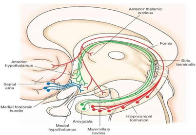
Gender identity disorder (GID) or gender dysphoria and homosexuality are classified according to Diagnostic and Statistical Manual of Mental Disorders (DMS) as mental disorders. Homosexuality has been proven that has no genetic basis [24]. Based on the brain sex theory, the GID patients have brain structure that is incompatible with same sex. There is a structure that has been associated with GID; stria terminalis which is a central subdivision of bed nucleus (BSTc) which in males is the twice size of the ones in females [25] (Zhou et al., 1997) and twice number of neurons [26] (Kruijver et al., 2000). As well, Kruijver et al (2000) found in BSTc of transsexuals contain the same number of neurons in females [26]. Both previously mentioned studies are cadaveric –post mortem− studies, but with advances in technologies of medical imaging, it could help in measuring the stria terminalis.

Dementia
Dementia is a decline of mental functions like; memory, thinking, problem solving, perception, and concentration because of Alzahimer’s disease, vascular dementia, etc. The neuroimaging role in diagnosing, staging, and evaluating dementia is very important. An article published in Scientific America magazine by “ a science writer” where the writer claimed that brain scans can’t help in psychiatry [28] and the author finished his article by stating list of diseases that can be diagnosed by brain scans including Alzheimer’s disease which the primary cause of dementia! If there is a decline in the mental abilities, it’s considered a mental illness. If medical imaging can help in diagnosing, staging, and evaluating a mental illness like dementia, but such arrogant, broad, and generalizing statements by the APA or Scientific America magazine should not be made. Not to forget what Amen and others stated that the big failure of the APA in the last 40 years and how the APA made mental illnesses in America worse [6, 14, 29].
In Vivo Imaging of Neurotransmitters & Neuromodulators
Neurotransmitter is a chemical that transfers to a nerve, control muscle cell, or other structure, while neuromodulator is a chemical that control another neuron’s function.
Based on the brain chemical imbalance theory, MRI and PET in vivo imaging have been developed to study the dynamic of the neuroreciptors and the levels of both neurotransmitters and neuromodulators [16].


Related Disease to Imbalance of Brain Chemical
Tourette’s syndrome (TS) is a neurodevelopmental psychiatric disorders which characterized by chronic tics [30]. Even though, motion artefact is an issue in such cases, but the usefulness of neuroimaging wither by MRI or PET scans’ both are useful. On MRI, subcortical regions, white matter, grey matter, brain connectivity in general were smaller in Tourette’s patients compared to normal individuals [30].
Medical Conditions Associated With Mental Illnesses
Corpus callosum agenesis is associated with many mental illnesses, but the most common one is schizophrenia [31]. It appears as “Moose head” sign on sagittal section and as “racing car” sign on coronal section of MRI scan (both are classic signs which easy to identify). It’s known that corpus callosum agenesis is association with depression, learning problems, epilepsy, Asperger's syndrome, conduct disorder, conversion symptoms, etc.
Schizophrenia can be caused by the following biological factors: cerebral lupus, Traumatic Brain Injury (TBI), brain tumor, subdural hematoma, pheochromocytoma, metachromatic leukodystrophy, HIV infection, Wilson disease, vitamin B12 deficiency, vitamin D deficiency, neurosyphilis, and dementia of any etiology, temporal lobe epilepsy, and multiple sclerosis [31]. Most of these etiologies can be diagnosed on neuroimaging scans [31].
Chronic Traumatic Encephalopathy (CTE) is associated with cognitive decline, dementia, suicide, low life quality due to chronic headache. CTE is caused by chronic TBI which appears as micro−bleeds in the brain which could be detected by MRI [32].

Parkinson ’s disease (PD) is characterized by decrease the dopamine levels only from substintia nigra to corpus striatum. The psychiatric disorders that are associated with PD include; delusion, apathy, depression, anxiety, anhedonia, impulsive & compulsive behavior, hallucination, and cognitive dysfunction. Parkinson can be diagnosed by neuroimaging scans [33] see (Figure 6)

Huntington’s disease (HD) is characterized by decreasing levels of dopamine, GABA and P substance from corpus striatum to substintia nigra. The psychiatric disorders that are associated with HD include; irritability disorder, cognitive impairment, psychiatric disturbance, affective disorder, psychosis, and apathy [34].
Multiple Sclerosis (MS) is characterized by attacking the myelin sheet of the neurons in the central nervous system by the immune system (i.e. macrophages). The psychiatric disorders that are associated with MS include; anxiety, agitation, irritability, and dysphoria [35].
Neuroimaging Applications in Mental Illnesses
It has been proven that psychiatric disorders can affect the brain’s morphology, neural circuits, and their activities. The following are some of the applications of neuroimaging in mental illnesses:
Schizophrenia
Bilateral ventricular enlargement is a classic sign for schizophrenia that were identified in the 70s [31]. Voxel−based morphometric changes usually found in the left medial lobe and superior temporal gyrus [31].
Depression
MRI voxel−based morphometric changes was detected in subgenual cingulate cortex and hippocampal’s volume in depression patients who were treated with ElectroConvulsion Therapy (ECT) [36].
Bipolar I Disorder (Manic Depression)
Diffusion Tensor Imaging (DTI) of the whole brain showed white matter abnormalities in the corpus callosum, fornix, stria terminalis, and tapetum [37].
Borderline personality disorder
It is associated with decrease of the white matter’s integrity in the cingulum and fornix [38, 39]. Abandonment avoidance and affective instability are associated with fractional anisotropy in the fornix [38, 39]. Anger emotion is associated with patients who have a fractional anisotropy in the cingulum [39].
Connectome and Childhood Development
Connectome (i.e. cortical connectivity networks) can help in monitoring of childhood development in the future to detect any change caused by mental illness.
Autism
Many sources classify this disease as neurological or developmental disorder due to its biological origin, but its still needs Psychiatrists, Psychologists, and Speech Therapists intervention in treating autism patients. The mental illnesses on the other hand, originated from the surrounding environment to acquire a mental illness where the autism is caused by biological factors. However, autism required cognitive therapy. Autism can be diagnosed on MRI scan [40].
Drug Abuse
The famous chicken or egg dilemma which presents the question which one came first? The chicken or the egg? The same scenario in psychiatry when ask the famous question “is mental illness cause drug abuse or drug abuse lead to mental illness?” A pregnant women who abuse drugs can cause neonatal abstinence syndrome which affect the mental health of the baby, but the mother abuse or dependence on a substance is due to her addiction which classified as mental illness by the DMS-5. In vivo imaging of neurotransmitters & neuromodulators are affected by using drugs which include illegal drugs, medications, other substances (i.e. nicotine and alcohol). Furthermore, there are many papers claim that smoking marijuana can cause schizophrenia [13].
The Mind of a Dictator
There are two famous psychological experiments Stanford prison experiment and Milgram’s experiment which demonstrates authority misuse and blind following of order. But yet nobody focused on the merciless dictators and horrible tyrants. A new theory mentioned by James Fallon that dictators probably have a prefrontal cortex damage that causes a lack of apathy. This can be detected on brain scans, but unfortunately no dictator has been undergo brain neuroimaging study before and shared the scan with Scientists.
Are mental illnesses biological in their kind?
The APA is claiming that mental illnesses are biological in their kind, but in the same time, they refuse to indicate the usefulness of medical imaging in detecting those illnesses. The APA is the one who supports the chemical imbalance theory and oppose the Freudian method (psychoanalysis) [29]. They encourage using the medications “magic bullets” instead of evaluating and exposing patients to their subconscious minds to find the reasons behind their mental illnesses [6].
The Author of “The Myth of Mental Illness” argue that mental illnesses are not biological in their kind [41]. Thomas Szasz (1961) in his book where he inspired the start of the antipsychiatry movement which oppose all of medications, electrical shocks, and lobotomy (i.e. psychosurgery). Szasz’s friend (David Rosenhan), did a famous experiment that shocked the entire psychiatric field by sending 8 individuals to pretend to be sick (i.e. they should present one symptoms to the mental healthcare provider by saying they hear only the word “empty” which is “hearing hallucination”) [42]. Then after they have been hospitalized they acted normal and showed no sickness, but they did not release them [42]. The number of pseudopatients is 8 and the number of mental facilities that were deceived is 12 hospitals [42]. The APA panicked because they can’t differentiate between the mad and sane [42] by a scan, blood test, etc. The APA stood for David’s published paper (on being sane in insane places which was published in the nature journal in that time) and the APA published a new manual that uses the tick−the−box approach which was introduced in 18th−century. If someone has 6 symptoms out of 10, this person will be diagnosed with the disorder that fits with the disorder’s criteria [43]. One of the tricked hospital responded to Rosenhan and they challenged him to send the next month any pseudopatients he wants and the hospital will identify them. By the end of the month, the mental hospital declared that 41 pseudopatients were sent by Rosenhan and they were identified by the hospital. Rosenhan responded “I sent 0 pseudopatients which means the mental hospital let 41 sick patients out because they have no scientific criteria”. Similar to what Rosenhan said, the Psychiatrists until today, they do not know the reasons that causes mental illnesses after 50 years from that time.
Mental illness’ etiology could be a result of chemical imbalance, genetic, hormonal imbalance, electrical imbalance, altered brain temperature, metabolism imbalance, hormonal imbalance, environmental, prior negative experience, abnormal development, TBI, associated with other medical conditions, substance use, sole related spirituality (parapsychology), etc.
Pornography vs. Neuroplasticity
It is well known that addiction to watch pornography will cause change to the brain plasticity (neuroplasticity), gray matter to become thinner which will affect brain structures like the striatum which lead to the dysfunction reward system, low IQ, bad memory, unacceptable social behaviors, unsuccessful relationships, sexual dysfunction, etc. Brain plasticity can be evaluated by MRI [44].
Brain Calcification
Basal ganglia calcifications can be diagnosed on a CT scan and the calcification have been reported to be associated with psychosis and schizophrenia [45, 46]. Basal ganglia can be calcified in Fahr disease patients and other conditions.
Brain Morphology
There are neurological conditions that cause changes of the brain morphology. These neurological conditions have been reported to be associated with mental illnesses in the literature like; psychosis, schizophrenia, developmental language disorder, intellectual developmental disorder, mental disability (i.e. previously known as mental retardation), etc. These mental illnesses have been associated with a huge list of neurological conditions including; polymicrogyria, Dany−Walker malformation, schizencephaly, microcephaly, megalencephaly, macrocephaly, hemimegalencephaly, syntelencephaly, colpocephaly, holoprosencephaly, porencephaly, dysgenesis or agenesis of corpus callosum or vermis, etc.
Neuropsychiatric As Specialty
Psychiatry should become more involved with neurology and psychoanalysis. All the psychiatric graduate programs should be renamed neuropsychiatry and it must be changed to study the neurological causes of mental illnesses. As well, it must focus on psychoanalysis to understand the emotional reasons behind any mental illness. All Psychiatrists must wait for 1 month period before prescribing any psychiatric medications for any patients which will affect the patient’s live forever. Talking more with patients is the key to understand the reasons behind their sickness, to know their medical history, and to know their family’s medical history. Focusing on treating the reasons is important than immediate treating of the symptoms by psychiatric medications and experimental cocktails which could make psychiatric patients more sick.
Neuroimaging can help in diagnosing and evaluating of mental illnesses. As well, neuroimaging can help in evaluating psychiatric medications’ effects on the brain, and the finding associated medical illnesses with mental illnesses. Neuroimaging can bring more scientific approach to identifying a mental illness more than tick−the−box approach
Clearly Auctoresonline and particularly Psychology and Mental Health Care Journal is dedicated to improving health care services for individuals and populations. The editorial boards' ability to efficiently recognize and share the global importance of health literacy with a variety of stakeholders. Auctoresonline publishing platform can be used to facilitate of optimal client-based services and should be added to health care professionals' repertoire of evidence-based health care resources.

Journal of Clinical Cardiology and Cardiovascular Intervention The submission and review process was adequate. However I think that the publication total value should have been enlightened in early fases. Thank you for all.

Journal of Women Health Care and Issues By the present mail, I want to say thank to you and tour colleagues for facilitating my published article. Specially thank you for the peer review process, support from the editorial office. I appreciate positively the quality of your journal.

Journal of Clinical Research and Reports I would be very delighted to submit my testimonial regarding the reviewer board and the editorial office. The reviewer board were accurate and helpful regarding any modifications for my manuscript. And the editorial office were very helpful and supportive in contacting and monitoring with any update and offering help. It was my pleasure to contribute with your promising Journal and I am looking forward for more collaboration.

We would like to thank the Journal of Thoracic Disease and Cardiothoracic Surgery because of the services they provided us for our articles. The peer-review process was done in a very excellent time manner, and the opinions of the reviewers helped us to improve our manuscript further. The editorial office had an outstanding correspondence with us and guided us in many ways. During a hard time of the pandemic that is affecting every one of us tremendously, the editorial office helped us make everything easier for publishing scientific work. Hope for a more scientific relationship with your Journal.

The peer-review process which consisted high quality queries on the paper. I did answer six reviewers’ questions and comments before the paper was accepted. The support from the editorial office is excellent.

Journal of Neuroscience and Neurological Surgery. I had the experience of publishing a research article recently. The whole process was simple from submission to publication. The reviewers made specific and valuable recommendations and corrections that improved the quality of my publication. I strongly recommend this Journal.

Dr. Katarzyna Byczkowska My testimonial covering: "The peer review process is quick and effective. The support from the editorial office is very professional and friendly. Quality of the Clinical Cardiology and Cardiovascular Interventions is scientific and publishes ground-breaking research on cardiology that is useful for other professionals in the field.

Thank you most sincerely, with regard to the support you have given in relation to the reviewing process and the processing of my article entitled "Large Cell Neuroendocrine Carcinoma of The Prostate Gland: A Review and Update" for publication in your esteemed Journal, Journal of Cancer Research and Cellular Therapeutics". The editorial team has been very supportive.

Testimony of Journal of Clinical Otorhinolaryngology: work with your Reviews has been a educational and constructive experience. The editorial office were very helpful and supportive. It was a pleasure to contribute to your Journal.

Dr. Bernard Terkimbi Utoo, I am happy to publish my scientific work in Journal of Women Health Care and Issues (JWHCI). The manuscript submission was seamless and peer review process was top notch. I was amazed that 4 reviewers worked on the manuscript which made it a highly technical, standard and excellent quality paper. I appreciate the format and consideration for the APC as well as the speed of publication. It is my pleasure to continue with this scientific relationship with the esteem JWHCI.

This is an acknowledgment for peer reviewers, editorial board of Journal of Clinical Research and Reports. They show a lot of consideration for us as publishers for our research article “Evaluation of the different factors associated with side effects of COVID-19 vaccination on medical students, Mutah university, Al-Karak, Jordan”, in a very professional and easy way. This journal is one of outstanding medical journal.

Dear Hao Jiang, to Journal of Nutrition and Food Processing We greatly appreciate the efficient, professional and rapid processing of our paper by your team. If there is anything else we should do, please do not hesitate to let us know. On behalf of my co-authors, we would like to express our great appreciation to editor and reviewers.

As an author who has recently published in the journal "Brain and Neurological Disorders". I am delighted to provide a testimonial on the peer review process, editorial office support, and the overall quality of the journal. The peer review process at Brain and Neurological Disorders is rigorous and meticulous, ensuring that only high-quality, evidence-based research is published. The reviewers are experts in their fields, and their comments and suggestions were constructive and helped improve the quality of my manuscript. The review process was timely and efficient, with clear communication from the editorial office at each stage. The support from the editorial office was exceptional throughout the entire process. The editorial staff was responsive, professional, and always willing to help. They provided valuable guidance on formatting, structure, and ethical considerations, making the submission process seamless. Moreover, they kept me informed about the status of my manuscript and provided timely updates, which made the process less stressful. The journal Brain and Neurological Disorders is of the highest quality, with a strong focus on publishing cutting-edge research in the field of neurology. The articles published in this journal are well-researched, rigorously peer-reviewed, and written by experts in the field. The journal maintains high standards, ensuring that readers are provided with the most up-to-date and reliable information on brain and neurological disorders. In conclusion, I had a wonderful experience publishing in Brain and Neurological Disorders. The peer review process was thorough, the editorial office provided exceptional support, and the journal's quality is second to none. I would highly recommend this journal to any researcher working in the field of neurology and brain disorders.
Dear Agrippa Hilda, Journal of Neuroscience and Neurological Surgery, Editorial Coordinator, I trust this message finds you well. I want to extend my appreciation for considering my article for publication in your esteemed journal. I am pleased to provide a testimonial regarding the peer review process and the support received from your editorial office. The peer review process for my paper was carried out in a highly professional and thorough manner. The feedback and comments provided by the authors were constructive and very useful in improving the quality of the manuscript. This rigorous assessment process undoubtedly contributes to the high standards maintained by your journal.

International Journal of Clinical Case Reports and Reviews. I strongly recommend to consider submitting your work to this high-quality journal. The support and availability of the Editorial staff is outstanding and the review process was both efficient and rigorous.

Thank you very much for publishing my Research Article titled “Comparing Treatment Outcome Of Allergic Rhinitis Patients After Using Fluticasone Nasal Spray And Nasal Douching" in the Journal of Clinical Otorhinolaryngology. As Medical Professionals we are immensely benefited from study of various informative Articles and Papers published in this high quality Journal. I look forward to enriching my knowledge by regular study of the Journal and contribute my future work in the field of ENT through the Journal for use by the medical fraternity. The support from the Editorial office was excellent and very prompt. I also welcome the comments received from the readers of my Research Article.

Dear Erica Kelsey, Editorial Coordinator of Cancer Research and Cellular Therapeutics Our team is very satisfied with the processing of our paper by your journal. That was fast, efficient, rigorous, but without unnecessary complications. We appreciated the very short time between the submission of the paper and its publication on line on your site.

I am very glad to say that the peer review process is very successful and fast and support from the Editorial Office. Therefore, I would like to continue our scientific relationship for a long time. And I especially thank you for your kindly attention towards my article. Have a good day!

"We recently published an article entitled “Influence of beta-Cyclodextrins upon the Degradation of Carbofuran Derivatives under Alkaline Conditions" in the Journal of “Pesticides and Biofertilizers” to show that the cyclodextrins protect the carbamates increasing their half-life time in the presence of basic conditions This will be very helpful to understand carbofuran behaviour in the analytical, agro-environmental and food areas. We greatly appreciated the interaction with the editor and the editorial team; we were particularly well accompanied during the course of the revision process, since all various steps towards publication were short and without delay".

I would like to express my gratitude towards you process of article review and submission. I found this to be very fair and expedient. Your follow up has been excellent. I have many publications in national and international journal and your process has been one of the best so far. Keep up the great work.

We are grateful for this opportunity to provide a glowing recommendation to the Journal of Psychiatry and Psychotherapy. We found that the editorial team were very supportive, helpful, kept us abreast of timelines and over all very professional in nature. The peer review process was rigorous, efficient and constructive that really enhanced our article submission. The experience with this journal remains one of our best ever and we look forward to providing future submissions in the near future.

I am very pleased to serve as EBM of the journal, I hope many years of my experience in stem cells can help the journal from one way or another. As we know, stem cells hold great potential for regenerative medicine, which are mostly used to promote the repair response of diseased, dysfunctional or injured tissue using stem cells or their derivatives. I think Stem Cell Research and Therapeutics International is a great platform to publish and share the understanding towards the biology and translational or clinical application of stem cells.

I would like to give my testimony in the support I have got by the peer review process and to support the editorial office where they were of asset to support young author like me to be encouraged to publish their work in your respected journal and globalize and share knowledge across the globe. I really give my great gratitude to your journal and the peer review including the editorial office.

I am delighted to publish our manuscript entitled "A Perspective on Cocaine Induced Stroke - Its Mechanisms and Management" in the Journal of Neuroscience and Neurological Surgery. The peer review process, support from the editorial office, and quality of the journal are excellent. The manuscripts published are of high quality and of excellent scientific value. I recommend this journal very much to colleagues.

Dr.Tania Muñoz, My experience as researcher and author of a review article in The Journal Clinical Cardiology and Interventions has been very enriching and stimulating. The editorial team is excellent, performs its work with absolute responsibility and delivery. They are proactive, dynamic and receptive to all proposals. Supporting at all times the vast universe of authors who choose them as an option for publication. The team of review specialists, members of the editorial board, are brilliant professionals, with remarkable performance in medical research and scientific methodology. Together they form a frontline team that consolidates the JCCI as a magnificent option for the publication and review of high-level medical articles and broad collective interest. I am honored to be able to share my review article and open to receive all your comments.

“The peer review process of JPMHC is quick and effective. Authors are benefited by good and professional reviewers with huge experience in the field of psychology and mental health. The support from the editorial office is very professional. People to contact to are friendly and happy to help and assist any query authors might have. Quality of the Journal is scientific and publishes ground-breaking research on mental health that is useful for other professionals in the field”.

Dear editorial department: On behalf of our team, I hereby certify the reliability and superiority of the International Journal of Clinical Case Reports and Reviews in the peer review process, editorial support, and journal quality. Firstly, the peer review process of the International Journal of Clinical Case Reports and Reviews is rigorous, fair, transparent, fast, and of high quality. The editorial department invites experts from relevant fields as anonymous reviewers to review all submitted manuscripts. These experts have rich academic backgrounds and experience, and can accurately evaluate the academic quality, originality, and suitability of manuscripts. The editorial department is committed to ensuring the rigor of the peer review process, while also making every effort to ensure a fast review cycle to meet the needs of authors and the academic community. Secondly, the editorial team of the International Journal of Clinical Case Reports and Reviews is composed of a group of senior scholars and professionals with rich experience and professional knowledge in related fields. The editorial department is committed to assisting authors in improving their manuscripts, ensuring their academic accuracy, clarity, and completeness. Editors actively collaborate with authors, providing useful suggestions and feedback to promote the improvement and development of the manuscript. We believe that the support of the editorial department is one of the key factors in ensuring the quality of the journal. Finally, the International Journal of Clinical Case Reports and Reviews is renowned for its high- quality articles and strict academic standards. The editorial department is committed to publishing innovative and academically valuable research results to promote the development and progress of related fields. The International Journal of Clinical Case Reports and Reviews is reasonably priced and ensures excellent service and quality ratio, allowing authors to obtain high-level academic publishing opportunities in an affordable manner. I hereby solemnly declare that the International Journal of Clinical Case Reports and Reviews has a high level of credibility and superiority in terms of peer review process, editorial support, reasonable fees, and journal quality. Sincerely, Rui Tao.

Clinical Cardiology and Cardiovascular Interventions I testity the covering of the peer review process, support from the editorial office, and quality of the journal.
