AUCTORES
Globalize your Research

Research Article | DOI: https://doi.org/10.31579/2643-1912/002
1 Deportment of Pharmaceutics, Jangaon Institute of Pharmaceutical Sciences, India.
*Corresponding Author: Stalin Reddy Challa, Deportment of Pharmaceutics, Jangaon Institute of Pharmaceutical Sciences, India.
Citation: Stalin Reddy Challa and Swathi Goli, Differentiation of Human Embryonic Stem Cells into Engrafting Myogenic Precursor Cells. J. Stem cell Research and Therapeutics International, DOI: 10.31579/2643-1912/002
Copyright: © 2019 Stalin Reddy Challa, This is an open-access article distributed under the terms of the Creative Commons Attribution License, which permits unrestricted use, distribution, and reproduction in any medium, provided the original author and source are credited.
Received: 16 March 2019 | Accepted: 09 April 2019 | Published: 11 April 2019
Keywords: muscle tissue; human embryonic stem cells; myogenic precursor; immunodeficient
Degenerative muscle diseases affect muscle tissue integrity and function. Human embryonic stem cells (hESC) are an attractive source of cells to use in regenerative therapies due to their unlimited capacity to divide and ability to specialize into a wide variety of cell types. A practical way to derive therapeutic myogenic stem cells from hESC is lacking. In this study, we demonstrate the development of two serum-free conditions to direct the differentiation of hESC towards a myogenic precursor state. Using TGFß and PI3Kinase inhibitors in combination with bFGF we showed that one week of differentiation is sufficient for hESC to specialize into PAX3+/PAX7+ myogenic precursor cells. These cells also possess the capacity to further differentiate in vitro into more specialized myogenic cells that express MYOD, Myogenin, Desmin and MYHC, and showed engraftment in vivo upon transplantation in immunodeficient mice. Ex vivo myomechanical studies of dystrophic mouse hindlimb muscle showed functional improvement one month post-transplantation. In summary, this study describes a promising system to derive engrafting muscle precursor cells solely using chemical substances in serum-free conditions and without genetic manipulation.
Muscular dystrophies are debilitating degenerative muscle diseases caused by defective muscle proteins that disrupt normal homeostasis [1,2]. During embryogenesis Pax3 and Pax7-expressing cells drive muscle formation, growth and repair [3-8]. In the postnatal period, these cells are termed satellite cells –stem cells that remain quiescent beneath the basal lamina of muscle fibers under normal conditions. Tissue damage triggers these cells to proliferate and differentiate into myoblasts, leading to the formation of multinucleated myotubes and fibers [9-12]. However, dystrophic tissue induces repeated activation of satellite cells, ultimately leading to stem cell exhaustion, limiting the ability for further restorative myogenesis. Given their role in muscle formation, growth and repair, satellite cells, or other myogenic precursors with similar characteristics, would be attractive candidates for cell-based therapies for muscular dystrophies. Successful cell therapy would require myogenic precursor cells to: i. repopulate damaged muscle, ii. contribute to the formation of muscle fibers, and iii. lead to expression of normal muscle proteins in host tissue [13]. In addition, an important therapeutic benefit would be repopulation of the muscle stem cell niche to allow for ongoing homeostasis.
Embryonic stem cells (ESC) have the capacity to expand indefinitely, while maintaining the ability to differentiate into a wide variety of cell types. As such ESC may be useful for cell transplantation therapy or tissue engineering. However, a non-genetics method to obtain hESC derivatives with muscle regenerative abilities is lacking. Promising attempts to derive human myogenic precursors from pluripotent stem cells have relied on viral–mediated MYOD-expression [14-18], serum-containing media [14,15], use of demethylating agents [15], or selective multi-step cell culture approaches and cell sorting procedures that may be difficult to implement on a large-scale [16]. The most efficient methods described to date used genetic manipulation of human ESC (hESC) to overexpress the myogenic transcription factors, PAX3 and PAX7 [19]. Importantly, the use of genetically manipulated or virally induced cells, as well as cells exposed to serum that contains non-human components, may be difficult to use in human trials due to safety concerns.
Therefore, in order to establish alternative ways of driving early myogenic specification, we leveraged previous studies describing chemical compounds that stimulate and facilitate hESC differentiation toward paraxial mesoderm, the lineage responsible for limb and limb-associated skeletal muscle development. hESC can be chemically stimulated by activation of the bone morphogenetic protein (BMP) pathway, which is a key inducer of mesoderm formation during embryogenesis [14,20-24]. Treatment with the phosphatidylinositol 3-kinase (PI3K) inhibitor, Ly294002 hydrochloride (Ly), facilitates the differentiation of hESC by reducing self-renewal ability [25,26]. Additionally, paraxial mesoderm, the lineage responsible for limb muscle development, can be formed by brief stimulation with BMP and inhibition of the TGFb/Activin pathway using the small molecule, SB431542 (SB) [27]. This compound specifically inhibits the Activin pathway without affecting BMP signaling. It has been shown that inhibition of the TGFb/Activin pathway using SB up-regulates myogenic genes during hESC differentiation in defined culture conditions [14]. Another important factor in mesoderm formation is fibroblast growth factor (FGF), which also determines myogenic fate during development and plays a role in satellite cell activation [28-31]. Therefore, it is plausible that these components could be used to drive myogenesis in hESC cultures.
Based on these observations, we hypothesized that engrafting myogenic precursors could be derived from hESC by the sequential and coordinated manipulation of the BMP, TGFß and FGF pathways. Importantly, we sought to prove that myogenic specification of hESC could be achieved without the use of allogeneic components in serum or genomic alteration. To this end, we tested whether myogenesis could be induced through embryoid body (EB) differentiation in medium containing basic fibroblast growth factor (bFGF), Ly and low levels of FGF, in combination with short exposure to BMP4 (referred to as KFLy(B)S medium). Separately, a combination of FGF and SB alone was tested for the ability to drive myogenesis (referred to as KFS medium). We determined the capacity of hESC-derived myogenic precursors to engraft in skeletal muscle upon transplantation in hindlimb muscles of SCID mice. In addition, we determined functional changes in transplanted muscle of mdx mice using ex vivo myomechanical analysis. The novel differentiation approach described in this study circumvents many of the limitations encountered in the transition of pluripotent stem cells into myogenic precursor cells, while enhancing our understanding of the mechanisms that govern stem cell differentiation and skeletal myoge
Human ESC culture and myogenic differentiation
All work with hESC was done with the approval of UCSF Stem Cell Research Oversight Committee. The H9 hESC (WA09; WiCell) were maintained on irradiated CF1 mouse embryonic fibroblasts (MEF; Millipore, Temecula, CA) in KSR medium [Knockout Dulbecco’s Modified Eagle Medium containing 20% Knockout Serum Replacement, 2mM glutamine, 0.1 mM nonessential amino acids, 0.1 mM β-mercaptoethanol (all from Invitrogen, Carlsbad, CA)] supplemented with 12.5 ng/ml recombinant human fibroblast growth factor basic (bFGF; R&D Systems, Minneapolis, MN) as previously described [32].
hESC colonies were detached after brief exposure to Collagenase IV (1 mg/ml; Sigma-Aldrich, St. Lois, MO), washed and collected by settlement. Differentiation was initiated by embryoid body (EB) formation in pre-differentiation medium KFLy(B)S: KSR medium freshly supplemented with 20 ng/ml bFGF, 10 μM Ly294,002 hydrochloride (Ly; Sigma-Aldrich), 10 μM SB431542 hydrate (SB; Sigma-Aldrich), and for the first 36 hours supplementing with recombinant human bone morphogenetic protein-4 (BMP-4) (10 ng/ml; HumanZyme, Chicago, IL), or pre-differentiation medium KFS: KSR medium freshly supplemented with 40 ng/ml bFGF and 10 μM SB. Both pre-differentiation media were supplemented one time with 10 μM Rho-associated protein kinase inhibitor (ROCKi) at the day of EB formation. SB and Ly were reconstituted in dimethylsulfoxide (DMSO) at 20 mM, and diluted to the final concentration in pre-differentiation medium. Human EBs were kept in suspension in ultra-low cluster 6-well plates (Costar, Corning, NY) and placed onto an orbital shaker (Daigger, Vernon Hills, IL) at a speed of 90 rpm. After 36 hours, medium was refreshed, which was repeated every other day until day six.
To assess the myocyte-forming potential of EB-derived myogenic precursor cells derived in KFLy(B)S and KFS (differentiation day 6) media, EBs were plated onto 12.5 μg/cm2 fibronectin (from human plasma; Sigma-Aldrich) in 0.1% gelatin-coated culture surfaces in pre-differentiation medium to stimulate cell outgrowth. After two days, medium was replaced with muscle differentiation medium [DMEM-high glucose, 10% fetal bovine serum (FBS; Hyclone, Thermo Scientific, South Logan, UT), 5% horse serum (HS; also from Hyclone) supplemented with 5 ng/ml bFGF] and cultured for an additional 4 days.
Quantitative real-time PCR
RNA was extracted from cells using the RNeasy Mini Kit including DNase treatment (Qiagen, Valencia, CA) according to the manufacturer's instruction. RNA concentration was measured using a Nanodrop ND-1000 Spectrophotometer (Nanodrop Technologies) to convert equal quantities of mRNA into cDNA using the Superscript™ III First-Strand Synthesis System (Invitrogen). Relative gene expression was determined using TaqMan Assay (Applied Biosytems) on an ABI 7300 Real-Time PCR system with the following human primer pairs (ABI): OCT4 (Hs00742896_s1); Brachyury (T) (HS00610080_m1); PAX3 (Hs00240950_m1); PAX7 (Hs00242962m1); MYOD (Hs00159528_m1); Desmin (Hs01090875_m1); Myogenin (Hs002311167_m1); MYHC (Hs00430042_m1) and GAPDH (4326317E). Cycle threshold (Ct) value for detection of gene of interest was normalized against Ct value of the housekeeping gene, human GAPDH, and relative changes were calculated according the ΔΔCt method. Results of relative fold expression were analyzed using GraphPad prism software in a two-way ANOVA with Bonferroni post-test.
Immunostaining
Immunofluorescence staining was performed on EB-outgrowths cultured in of KFLy(B)S or KFS with mouse anti-human Pax7 (undiluted), Myogenin (F5D; undiluted) and MyHC (MF20; 1:20) antibodies (all three supernatants from Developmental Studies Hybridoma Bank); mouse anti-human MyoD (5.8A; 20 μg/ml; R&D systems); or mouse anti-human Desmin (D33; 10 μg/ml; Dako, Glostrup, Denmark). Cells were fixed in 4% paraformaldehyde (PFA; EMS, Fort Washington, PA) for 15 min. and permeabilized in 0.4% Triton-X100 for 10 min. After blocking for 30 min in blocking solution [2% normal goat serum (NGS; Dako), 1% bovine serum albumin (BSA; Sigma-Aldrich), 0.1% cold fish gelatin (Sigma-Aldrich), 0.1% Triton-X100, 0.05% Tween-20 in phosphate buffered saline (PBS)], cells were subsequently incubated with the primary antibodies diluted in blocking solution for 1 hr. After washing (three times; 5 min in PBS with 0.05% Tween-20), primary antibodies were detected using Alexa Fluor® 488-labeled goat anti-mouse (1:200; Life Technologies, Grand Island, NY) for 30 min. All steps were performed at room temperature (RT). Fluorescence microscopy was performed using a Nikon Eclipse TE300 microscope and Zeiss AxioScope Imager Z.2 with Apotome (Zeiss, Inc, Thornwood, NY).
Intramuscular cell transplantation
All experiments involving animals were done with the approval of the UCSF Institutional Animal Care and Use Committee. Hindlimbs of CB17.B6-PrkdcscidLyst bg/Crl (8-12 wk-old) and C57BL/10ScSn-Dmdmdx/J (X-linked muscular dystrophy; aged 6-8 weeks) mice (Jackson Laboratories, Bar Harbor, ME) were exposed to gamma radiation (5 Gy and 18 Gy, respectively) three days before cell transplantation to inhibit muscle regeneration by host muscle cells. Mice cages were supplied with a water bottle containing an antibacterial oral suspension (200 mg sulfamethoxazole-40 mg trimethoprim/ 200 ml water) from the day of irradiation through euthanasia. For intramuscular injection, EB-derived in KFLy(B)S or KFS were trypsinized and filtered through 40 μm nylon cell strainer (Gibco) to obtain a single cell suspension. One million cells cultured in KFLy(B)S or KFS were suspended in 50 μl PBS (supplemented with 10 μM ROCKi) and injected into the tibialis anterior (TA) muscle at two sites. As control, the contra-lateral muscle was injected with 50 μl PBS with 10 μM ROCKi. TA muscles were analyzed by immunohistochemistry one month post-transplantation, as described above.
Immunohistochemistry
TA muscles were embedded in OCT, snap-frozen in liquid nitrogen and stored at -80°C. Cryosections (5 mm) were fixed in acetone (ice cold, 10 min) and blocked in blocking solution [5% NGS, 5% Normal Donkey Serum (Dako), 5% BSA, 0.1% cold fish gelatin, 0.1% Triton-X100, 0.05% Tween-20 in PBS]. Sections were incubated with rabbit anti-human Dystrophin (1:2500; Novus Biologicals, Littleton, CO), chicken anti-mouse Laminin (2 mg/ml; Abcam), mouse anti-human Desmin (10 μg/ml; D33; Dako), or mouse anti-human HLA-1 (W6/32; antigen determinant common to HLA-A, B and C; 4 mg/ml; provided by Frances Brodsky, Dept. of Biopharmaceutical Sciences, UCSF) in blocking buffer for 1 hour. Alexa488/594-conjugated goat anti-mouse IgG, Alexa594-Donkey anti-Rabbit IgG, or Alexa488-Goat anti-Chicken IgG (1:250; all from Life Technologies) were used to detect primary antibodies. Sections were then washed and dehydrated in ethanol (70%, 90%, 100%; 5 min each), air-dried, and mounted in Vectashield with DAPI (Vector, Burlingame, CA). For immunohistochemical detection, sections were additionally blocked for endogenous peroxidase (0.3% H2O2 in PBS, 20min). Primary antibody HLA-1 was detected by peroxidase labeled goat anti-mouse. Visualization was performed by a 3-amino-9-ethyl-carbazole (AEC; Sigma) substrate incubation for 4 minutes. Nuclei were counterstained with Mayer’s haematoxyline (2 min, Fluka, Buchs, Zwitserland). All steps were performed at RT. Fluorescence microscopy was performed using a Nikon Eclipse TE300 microscope and Zeiss AxioScope Imager Z.2 with Apotome (Zeiss, Inc, Thornwood, NY).
Ex vivo myomechanical analysis of transplanted EDL muscle in mdx mouse model
Ex vivo myography was performed to determine the increase in force production and to determine muscle fatigue level after cell transplantation into the extensor digitorum longus (EDL) muscle of mdx mouse. Myomechanical analysis was performed as previously described [33,34]. Force-frequency fatigue was measured by exposing muscle to a supramaximal stimulus train of 3-5 pulses (300 msec duration separated by 3 sec) at successive frequencies (30, 60, 100 and 140 Hz), with 5 minute intervals between stimulations. Low-frequency time-fatigue was measured by supramaximal tetanic muscle stimulation at low frequency (60 Hz) for duration of 300 msec, repeated every 3 seconds for a period of 10 minutes. For myo-mechanical analysis, the left and right leg variables of an animal were averaged in each group and muscle mass, cross-sectional area, and length was measured. Significant differences were present between the means of each group compared to the PBS group. Multiple t-tests were performed with the number of comparisons adjusted by Bonferroni correction. A value of p <0.05 was considered significant.
nhibition of TGFβ pathway directs myogenic commitment
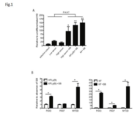
To compare previously described, serum-containing methods for their capacity to induce the formation of myogenic precursors, we differentiated hESC by a selective culture approach using serum-containing (10% FBS) medium to enrich for CD73+ mesodermal stem cells followed by a second cell sort for expression of neural cell adhesion molecule (NCAM), a molecule also expressed by myogenic cells [16]. Furthermore, we differentiated hESC-EB in low serum conditions (2% HS) [35-37] and high serum conditions (10% FBS) during 2-4 weeks in culture [15]. We observed expression of PAX7 in cells derived through the selective multi-step methods (preCD73/NCAM sort) and when exposed to low and high serum conditions (Figure 1A). Addition of SB, a compound that inhibits the TGFb pathway, to cells differentiated in 10% FBS culture conditions resulted in markedly increased PAX7 levels (Figure 1A) *p<0.01. This indicated the myogenic stimulatory effect of TGFb inhibition in hESC. Next, we transiently stimulated the BMP pathway with BMP4 during EB differentiation combined with Ly and high levels of bFGF in serum-free conditions (KFLy(B)). This resulted in high levels of PAX7 expression (Figure 1A) p<0.001. This was also seen when EB were exposed solely to high concentrations of FGF (KF) (Figure 1A) p<0.001.
Myogenic differentiation in EB culture
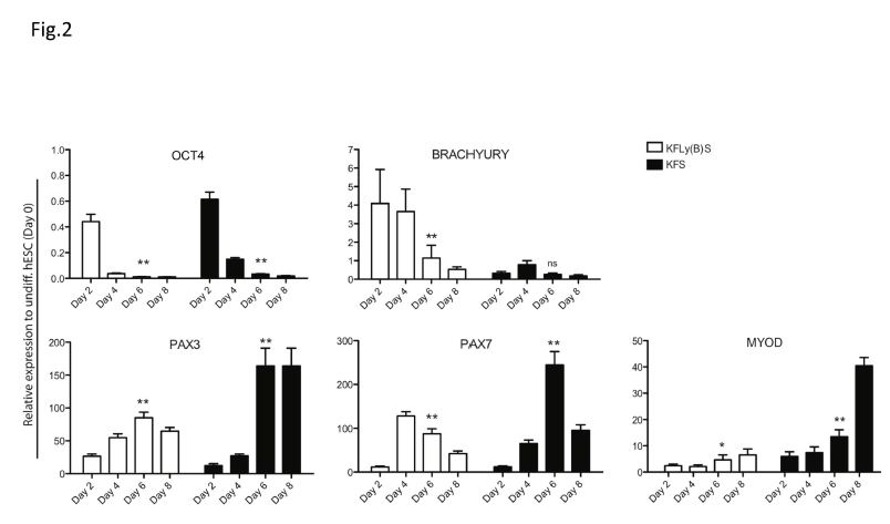
We hypothesized that myogenic precursor cells would be more likely to engraft in host muscle, and have a greater chance of repopulating the stem cell niche, than fully differentiated myoblasts. To monitor the stages of myogenic differentiation, we cultured hESC-EB in serum-free KFLy(B)S or KFS and examined myogenic gene expression by qRT-PCR at days 2, 4, 6 and 8, relative to undifferentiated hESC (day 0). OCT4 expression was used as an indicator of residual pluripotency after initiation of hESC differentiation. In both differentiation conditions, OCT4 expression fell dramatically by day 2 and further diminished through day 6 of differentiation (Figure 2) p<0.001.
KFLy(B)S- and KFS-derived precursors differentiate into myoblasts in vitro
Directed differentiation of stem cells can be stimulated with exposure to specific extracellular matrix (ECM) components they would likely encounter in the stem cell niche [42]. This has specifically been shown for stem cell differentiation toward the myogenic lineage [43,44]. EB differentiated for 6 days in KFLy(B)S or KFS were plated in dishes coated with Matrigel, laminin or fibronectin to stimulate myogenic cell outgrowth. We observed that attachment to fibronectin-coated dishes, compared to Matrigel and laminin, more effectively promoted myogenic differentiation (data not shown). Therefore, fibronectin coating was used to evaluate the myogenic potential of EB-outgrowth cells (EB-O) during the subsequent 6 days in muscle differentiation medium.
PAX3 and PAX7 are critical factors in embryonic skeletal myogenesis and participate after birth in muscle homeostasis. They are, therefore, important indicators of a myogenic precursor stage that harbors regenerative capacity. Several methods have been published describing the myogenic differentiation of hESC, however, limitations of published methods include the use of non-human serum and genetic manipulation that may hamper therapeutic application due to safety concerns [48,49]. This study underscores that myogenesis can be achieved in hESC cultures in a short time frame when exposed to appropriate developmental cues. We have demonstrated two approaches to derive PAX3- and PAX7-expressing myogenic precursors from hESC using a novel sequence of chemical compounds that direct a myogenic cell fate. Upon transplantation, these muscle precursor cells engrafted into muscle, formed new fibers, and improved muscle strength.
In the course of this study a publication revealed the success of a chemically defined culture system in transgenic zebrafish that directs skeletal myogenesis of human induced pluripotent stem cells [50]. Therefore, the combination of pathway inhibitors and stimulators described in this study provide an effective platform to advances approaches to muscle differentiation from hESC. Among the benefits are the avoidance of the use of serum and genetic alteration to promote gene expression. Furthermore, we showed that hESC entered the myogenic program in less then one-week making this approach faster than other published methods applying hESC differentiation during 2-4 weeks. Additionally, lacking selective cell culture approaches contributed to a method that is costs effective and easy.
In mdx mice, transplantation with hESC-derived precursors derived using these methods resulted in improved mechanical force generation and decreased fatigue. Both muscles injected with KFLy(B)S and KFS-derived cells demonstrated higher maximal tension than muscle injected with PBS or EDL that was not injected with cells, indicating improved functional capacity. Interestingly, EDL from mice injected with KFLy(B)S-derived cells demonstrated greater maximal tetanic tension, compared to both EDL from mice injected with PBS and KFS-derived cells and EDL that was not injected. However, force-frequency analysis demonstrated decreased fatigue in muscle from mice injected with KFS-derived cells compared to the other groups. The mechanisms that might account for these differences are unclear, but it is intriguing that the ESC-derived precursors could alter functional capacity by affecting different muscle properties (e.g strength vs endurance). Most importantly, however, the findings of the myomechanical analysis indicate preliminary proof-of-concept for functional myogenic potential for these hESC-derived precursor cells.
We have shown that a short exposure of hESC to chemically defined media promotes differentiation of hESC into engrafting myogenic precursor cells that can contribute to the formation of new fibers in vivo and improve muscle strength as analyzed ex vivo. These techniques are less laborious than previously published methods, and the avoidance of xenobiotic agents and serum may mitigate safety concerns. Further studies to assess durability of transplanted cells and newly formed fibers, as well as their contribution to muscle homeostasis over time are warranted.
Clearly Auctoresonline and particularly Psychology and Mental Health Care Journal is dedicated to improving health care services for individuals and populations. The editorial boards' ability to efficiently recognize and share the global importance of health literacy with a variety of stakeholders. Auctoresonline publishing platform can be used to facilitate of optimal client-based services and should be added to health care professionals' repertoire of evidence-based health care resources.

Journal of Clinical Cardiology and Cardiovascular Intervention The submission and review process was adequate. However I think that the publication total value should have been enlightened in early fases. Thank you for all.

Journal of Women Health Care and Issues By the present mail, I want to say thank to you and tour colleagues for facilitating my published article. Specially thank you for the peer review process, support from the editorial office. I appreciate positively the quality of your journal.

Journal of Clinical Research and Reports I would be very delighted to submit my testimonial regarding the reviewer board and the editorial office. The reviewer board were accurate and helpful regarding any modifications for my manuscript. And the editorial office were very helpful and supportive in contacting and monitoring with any update and offering help. It was my pleasure to contribute with your promising Journal and I am looking forward for more collaboration.

We would like to thank the Journal of Thoracic Disease and Cardiothoracic Surgery because of the services they provided us for our articles. The peer-review process was done in a very excellent time manner, and the opinions of the reviewers helped us to improve our manuscript further. The editorial office had an outstanding correspondence with us and guided us in many ways. During a hard time of the pandemic that is affecting every one of us tremendously, the editorial office helped us make everything easier for publishing scientific work. Hope for a more scientific relationship with your Journal.

The peer-review process which consisted high quality queries on the paper. I did answer six reviewers’ questions and comments before the paper was accepted. The support from the editorial office is excellent.

Journal of Neuroscience and Neurological Surgery. I had the experience of publishing a research article recently. The whole process was simple from submission to publication. The reviewers made specific and valuable recommendations and corrections that improved the quality of my publication. I strongly recommend this Journal.

Dr. Katarzyna Byczkowska My testimonial covering: "The peer review process is quick and effective. The support from the editorial office is very professional and friendly. Quality of the Clinical Cardiology and Cardiovascular Interventions is scientific and publishes ground-breaking research on cardiology that is useful for other professionals in the field.

Thank you most sincerely, with regard to the support you have given in relation to the reviewing process and the processing of my article entitled "Large Cell Neuroendocrine Carcinoma of The Prostate Gland: A Review and Update" for publication in your esteemed Journal, Journal of Cancer Research and Cellular Therapeutics". The editorial team has been very supportive.

Testimony of Journal of Clinical Otorhinolaryngology: work with your Reviews has been a educational and constructive experience. The editorial office were very helpful and supportive. It was a pleasure to contribute to your Journal.

Dr. Bernard Terkimbi Utoo, I am happy to publish my scientific work in Journal of Women Health Care and Issues (JWHCI). The manuscript submission was seamless and peer review process was top notch. I was amazed that 4 reviewers worked on the manuscript which made it a highly technical, standard and excellent quality paper. I appreciate the format and consideration for the APC as well as the speed of publication. It is my pleasure to continue with this scientific relationship with the esteem JWHCI.

This is an acknowledgment for peer reviewers, editorial board of Journal of Clinical Research and Reports. They show a lot of consideration for us as publishers for our research article “Evaluation of the different factors associated with side effects of COVID-19 vaccination on medical students, Mutah university, Al-Karak, Jordan”, in a very professional and easy way. This journal is one of outstanding medical journal.

Dear Hao Jiang, to Journal of Nutrition and Food Processing We greatly appreciate the efficient, professional and rapid processing of our paper by your team. If there is anything else we should do, please do not hesitate to let us know. On behalf of my co-authors, we would like to express our great appreciation to editor and reviewers.

As an author who has recently published in the journal "Brain and Neurological Disorders". I am delighted to provide a testimonial on the peer review process, editorial office support, and the overall quality of the journal. The peer review process at Brain and Neurological Disorders is rigorous and meticulous, ensuring that only high-quality, evidence-based research is published. The reviewers are experts in their fields, and their comments and suggestions were constructive and helped improve the quality of my manuscript. The review process was timely and efficient, with clear communication from the editorial office at each stage. The support from the editorial office was exceptional throughout the entire process. The editorial staff was responsive, professional, and always willing to help. They provided valuable guidance on formatting, structure, and ethical considerations, making the submission process seamless. Moreover, they kept me informed about the status of my manuscript and provided timely updates, which made the process less stressful. The journal Brain and Neurological Disorders is of the highest quality, with a strong focus on publishing cutting-edge research in the field of neurology. The articles published in this journal are well-researched, rigorously peer-reviewed, and written by experts in the field. The journal maintains high standards, ensuring that readers are provided with the most up-to-date and reliable information on brain and neurological disorders. In conclusion, I had a wonderful experience publishing in Brain and Neurological Disorders. The peer review process was thorough, the editorial office provided exceptional support, and the journal's quality is second to none. I would highly recommend this journal to any researcher working in the field of neurology and brain disorders.
Dear Agrippa Hilda, Journal of Neuroscience and Neurological Surgery, Editorial Coordinator, I trust this message finds you well. I want to extend my appreciation for considering my article for publication in your esteemed journal. I am pleased to provide a testimonial regarding the peer review process and the support received from your editorial office. The peer review process for my paper was carried out in a highly professional and thorough manner. The feedback and comments provided by the authors were constructive and very useful in improving the quality of the manuscript. This rigorous assessment process undoubtedly contributes to the high standards maintained by your journal.

International Journal of Clinical Case Reports and Reviews. I strongly recommend to consider submitting your work to this high-quality journal. The support and availability of the Editorial staff is outstanding and the review process was both efficient and rigorous.

Thank you very much for publishing my Research Article titled “Comparing Treatment Outcome Of Allergic Rhinitis Patients After Using Fluticasone Nasal Spray And Nasal Douching" in the Journal of Clinical Otorhinolaryngology. As Medical Professionals we are immensely benefited from study of various informative Articles and Papers published in this high quality Journal. I look forward to enriching my knowledge by regular study of the Journal and contribute my future work in the field of ENT through the Journal for use by the medical fraternity. The support from the Editorial office was excellent and very prompt. I also welcome the comments received from the readers of my Research Article.

Dear Erica Kelsey, Editorial Coordinator of Cancer Research and Cellular Therapeutics Our team is very satisfied with the processing of our paper by your journal. That was fast, efficient, rigorous, but without unnecessary complications. We appreciated the very short time between the submission of the paper and its publication on line on your site.

I am very glad to say that the peer review process is very successful and fast and support from the Editorial Office. Therefore, I would like to continue our scientific relationship for a long time. And I especially thank you for your kindly attention towards my article. Have a good day!

"We recently published an article entitled “Influence of beta-Cyclodextrins upon the Degradation of Carbofuran Derivatives under Alkaline Conditions" in the Journal of “Pesticides and Biofertilizers” to show that the cyclodextrins protect the carbamates increasing their half-life time in the presence of basic conditions This will be very helpful to understand carbofuran behaviour in the analytical, agro-environmental and food areas. We greatly appreciated the interaction with the editor and the editorial team; we were particularly well accompanied during the course of the revision process, since all various steps towards publication were short and without delay".

I would like to express my gratitude towards you process of article review and submission. I found this to be very fair and expedient. Your follow up has been excellent. I have many publications in national and international journal and your process has been one of the best so far. Keep up the great work.

We are grateful for this opportunity to provide a glowing recommendation to the Journal of Psychiatry and Psychotherapy. We found that the editorial team were very supportive, helpful, kept us abreast of timelines and over all very professional in nature. The peer review process was rigorous, efficient and constructive that really enhanced our article submission. The experience with this journal remains one of our best ever and we look forward to providing future submissions in the near future.

I am very pleased to serve as EBM of the journal, I hope many years of my experience in stem cells can help the journal from one way or another. As we know, stem cells hold great potential for regenerative medicine, which are mostly used to promote the repair response of diseased, dysfunctional or injured tissue using stem cells or their derivatives. I think Stem Cell Research and Therapeutics International is a great platform to publish and share the understanding towards the biology and translational or clinical application of stem cells.

I would like to give my testimony in the support I have got by the peer review process and to support the editorial office where they were of asset to support young author like me to be encouraged to publish their work in your respected journal and globalize and share knowledge across the globe. I really give my great gratitude to your journal and the peer review including the editorial office.

I am delighted to publish our manuscript entitled "A Perspective on Cocaine Induced Stroke - Its Mechanisms and Management" in the Journal of Neuroscience and Neurological Surgery. The peer review process, support from the editorial office, and quality of the journal are excellent. The manuscripts published are of high quality and of excellent scientific value. I recommend this journal very much to colleagues.

Dr.Tania Muñoz, My experience as researcher and author of a review article in The Journal Clinical Cardiology and Interventions has been very enriching and stimulating. The editorial team is excellent, performs its work with absolute responsibility and delivery. They are proactive, dynamic and receptive to all proposals. Supporting at all times the vast universe of authors who choose them as an option for publication. The team of review specialists, members of the editorial board, are brilliant professionals, with remarkable performance in medical research and scientific methodology. Together they form a frontline team that consolidates the JCCI as a magnificent option for the publication and review of high-level medical articles and broad collective interest. I am honored to be able to share my review article and open to receive all your comments.

“The peer review process of JPMHC is quick and effective. Authors are benefited by good and professional reviewers with huge experience in the field of psychology and mental health. The support from the editorial office is very professional. People to contact to are friendly and happy to help and assist any query authors might have. Quality of the Journal is scientific and publishes ground-breaking research on mental health that is useful for other professionals in the field”.

Dear editorial department: On behalf of our team, I hereby certify the reliability and superiority of the International Journal of Clinical Case Reports and Reviews in the peer review process, editorial support, and journal quality. Firstly, the peer review process of the International Journal of Clinical Case Reports and Reviews is rigorous, fair, transparent, fast, and of high quality. The editorial department invites experts from relevant fields as anonymous reviewers to review all submitted manuscripts. These experts have rich academic backgrounds and experience, and can accurately evaluate the academic quality, originality, and suitability of manuscripts. The editorial department is committed to ensuring the rigor of the peer review process, while also making every effort to ensure a fast review cycle to meet the needs of authors and the academic community. Secondly, the editorial team of the International Journal of Clinical Case Reports and Reviews is composed of a group of senior scholars and professionals with rich experience and professional knowledge in related fields. The editorial department is committed to assisting authors in improving their manuscripts, ensuring their academic accuracy, clarity, and completeness. Editors actively collaborate with authors, providing useful suggestions and feedback to promote the improvement and development of the manuscript. We believe that the support of the editorial department is one of the key factors in ensuring the quality of the journal. Finally, the International Journal of Clinical Case Reports and Reviews is renowned for its high- quality articles and strict academic standards. The editorial department is committed to publishing innovative and academically valuable research results to promote the development and progress of related fields. The International Journal of Clinical Case Reports and Reviews is reasonably priced and ensures excellent service and quality ratio, allowing authors to obtain high-level academic publishing opportunities in an affordable manner. I hereby solemnly declare that the International Journal of Clinical Case Reports and Reviews has a high level of credibility and superiority in terms of peer review process, editorial support, reasonable fees, and journal quality. Sincerely, Rui Tao.

Clinical Cardiology and Cardiovascular Interventions I testity the covering of the peer review process, support from the editorial office, and quality of the journal.

Clinical Cardiology and Cardiovascular Interventions, we deeply appreciate the interest shown in our work and its publication. It has been a true pleasure to collaborate with you. The peer review process, as well as the support provided by the editorial office, have been exceptional, and the quality of the journal is very high, which was a determining factor in our decision to publish with you.

The peer reviewers process is quick and effective, the supports from editorial office is excellent, the quality of journal is high. I would like to collabroate with Internatioanl journal of Clinical Case Reports and Reviews journal clinically in the future time.

Clinical Cardiology and Cardiovascular Interventions, I would like to express my sincerest gratitude for the trust placed in our team for the publication in your journal. It has been a true pleasure to collaborate with you on this project. I am pleased to inform you that both the peer review process and the attention from the editorial coordination have been excellent. Your team has worked with dedication and professionalism to ensure that your publication meets the highest standards of quality. We are confident that this collaboration will result in mutual success, and we are eager to see the fruits of this shared effort.

Dear Dr. Jessica Magne, Editorial Coordinator 0f Clinical Cardiology and Cardiovascular Interventions, I hope this message finds you well. I want to express my utmost gratitude for your excellent work and for the dedication and speed in the publication process of my article titled "Navigating Innovation: Qualitative Insights on Using Technology for Health Education in Acute Coronary Syndrome Patients." I am very satisfied with the peer review process, the support from the editorial office, and the quality of the journal. I hope we can maintain our scientific relationship in the long term.

Dear Monica Gissare, - Editorial Coordinator of Nutrition and Food Processing. ¨My testimony with you is truly professional, with a positive response regarding the follow-up of the article and its review, you took into account my qualities and the importance of the topic¨.

Dear Dr. Jessica Magne, Editorial Coordinator 0f Clinical Cardiology and Cardiovascular Interventions, The review process for the article “The Handling of Anti-aggregants and Anticoagulants in the Oncologic Heart Patient Submitted to Surgery” was extremely rigorous and detailed. From the initial submission to the final acceptance, the editorial team at the “Journal of Clinical Cardiology and Cardiovascular Interventions” demonstrated a high level of professionalism and dedication. The reviewers provided constructive and detailed feedback, which was essential for improving the quality of our work. Communication was always clear and efficient, ensuring that all our questions were promptly addressed. The quality of the “Journal of Clinical Cardiology and Cardiovascular Interventions” is undeniable. It is a peer-reviewed, open-access publication dedicated exclusively to disseminating high-quality research in the field of clinical cardiology and cardiovascular interventions. The journal's impact factor is currently under evaluation, and it is indexed in reputable databases, which further reinforces its credibility and relevance in the scientific field. I highly recommend this journal to researchers looking for a reputable platform to publish their studies.

Dear Editorial Coordinator of the Journal of Nutrition and Food Processing! "I would like to thank the Journal of Nutrition and Food Processing for including and publishing my article. The peer review process was very quick, movement and precise. The Editorial Board has done an extremely conscientious job with much help, valuable comments and advices. I find the journal very valuable from a professional point of view, thank you very much for allowing me to be part of it and I would like to participate in the future!”

Dealing with The Journal of Neurology and Neurological Surgery was very smooth and comprehensive. The office staff took time to address my needs and the response from editors and the office was prompt and fair. I certainly hope to publish with this journal again.Their professionalism is apparent and more than satisfactory. Susan Weiner

My Testimonial Covering as fellowing: Lin-Show Chin. The peer reviewers process is quick and effective, the supports from editorial office is excellent, the quality of journal is high. I would like to collabroate with Internatioanl journal of Clinical Case Reports and Reviews.

My experience publishing in Psychology and Mental Health Care was exceptional. The peer review process was rigorous and constructive, with reviewers providing valuable insights that helped enhance the quality of our work. The editorial team was highly supportive and responsive, making the submission process smooth and efficient. The journal's commitment to high standards and academic rigor makes it a respected platform for quality research. I am grateful for the opportunity to publish in such a reputable journal.

My experience publishing in International Journal of Clinical Case Reports and Reviews was exceptional. I Come forth to Provide a Testimonial Covering the Peer Review Process and the editorial office for the Professional and Impartial Evaluation of the Manuscript.

I would like to offer my testimony in the support. I have received through the peer review process and support the editorial office where they are to support young authors like me, encourage them to publish their work in your esteemed journals, and globalize and share knowledge globally. I really appreciate your journal, peer review, and editorial office.

Dear Agrippa Hilda- Editorial Coordinator of Journal of Neuroscience and Neurological Surgery, "The peer review process was very quick and of high quality, which can also be seen in the articles in the journal. The collaboration with the editorial office was very good."

I would like to express my sincere gratitude for the support and efficiency provided by the editorial office throughout the publication process of my article, “Delayed Vulvar Metastases from Rectal Carcinoma: A Case Report.” I greatly appreciate the assistance and guidance I received from your team, which made the entire process smooth and efficient. The peer review process was thorough and constructive, contributing to the overall quality of the final article. I am very grateful for the high level of professionalism and commitment shown by the editorial staff, and I look forward to maintaining a long-term collaboration with the International Journal of Clinical Case Reports and Reviews.

To Dear Erin Aust, I would like to express my heartfelt appreciation for the opportunity to have my work published in this esteemed journal. The entire publication process was smooth and well-organized, and I am extremely satisfied with the final result. The Editorial Team demonstrated the utmost professionalism, providing prompt and insightful feedback throughout the review process. Their clear communication and constructive suggestions were invaluable in enhancing my manuscript, and their meticulous attention to detail and dedication to quality are truly commendable. Additionally, the support from the Editorial Office was exceptional. From the initial submission to the final publication, I was guided through every step of the process with great care and professionalism. The team's responsiveness and assistance made the entire experience both easy and stress-free. I am also deeply impressed by the quality and reputation of the journal. It is an honor to have my research featured in such a respected publication, and I am confident that it will make a meaningful contribution to the field.

"I am grateful for the opportunity of contributing to [International Journal of Clinical Case Reports and Reviews] and for the rigorous review process that enhances the quality of research published in your esteemed journal. I sincerely appreciate the time and effort of your team who have dedicatedly helped me in improvising changes and modifying my manuscript. The insightful comments and constructive feedback provided have been invaluable in refining and strengthening my work".

I thank the ‘Journal of Clinical Research and Reports’ for accepting this article for publication. This is a rigorously peer reviewed journal which is on all major global scientific data bases. I note the review process was prompt, thorough and professionally critical. It gave us an insight into a number of important scientific/statistical issues. The review prompted us to review the relevant literature again and look at the limitations of the study. The peer reviewers were open, clear in the instructions and the editorial team was very prompt in their communication. This journal certainly publishes quality research articles. I would recommend the journal for any future publications.

Dear Jessica Magne, with gratitude for the joint work. Fast process of receiving and processing the submitted scientific materials in “Clinical Cardiology and Cardiovascular Interventions”. High level of competence of the editors with clear and correct recommendations and ideas for enriching the article.

We found the peer review process quick and positive in its input. The support from the editorial officer has been very agile, always with the intention of improving the article and taking into account our subsequent corrections.

My article, titled 'No Way Out of the Smartphone Epidemic Without Considering the Insights of Brain Research,' has been republished in the International Journal of Clinical Case Reports and Reviews. The review process was seamless and professional, with the editors being both friendly and supportive. I am deeply grateful for their efforts.

To Dear Erin Aust – Editorial Coordinator of Journal of General Medicine and Clinical Practice! I declare that I am absolutely satisfied with your work carried out with great competence in following the manuscript during the various stages from its receipt, during the revision process to the final acceptance for publication. Thank Prof. Elvira Farina

Dear Jessica, and the super professional team of the ‘Clinical Cardiology and Cardiovascular Interventions’ I am sincerely grateful to the coordinated work of the journal team for the no problem with the submission of my manuscript: “Cardiometabolic Disorders in A Pregnant Woman with Severe Preeclampsia on the Background of Morbid Obesity (Case Report).” The review process by 5 experts was fast, and the comments were professional, which made it more specific and academic, and the process of publication and presentation of the article was excellent. I recommend that my colleagues publish articles in this journal, and I am interested in further scientific cooperation. Sincerely and best wishes, Dr. Oleg Golyanovskiy.

Dear Ashley Rosa, Editorial Coordinator of the journal - Psychology and Mental Health Care. " The process of obtaining publication of my article in the Psychology and Mental Health Journal was positive in all areas. The peer review process resulted in a number of valuable comments, the editorial process was collaborative and timely, and the quality of this journal has been quickly noticed, resulting in alternative journals contacting me to publish with them." Warm regards, Susan Anne Smith, PhD. Australian Breastfeeding Association.

Dear Jessica Magne, Editorial Coordinator, Clinical Cardiology and Cardiovascular Interventions, Auctores Publishing LLC. I appreciate the journal (JCCI) editorial office support, the entire team leads were always ready to help, not only on technical front but also on thorough process. Also, I should thank dear reviewers’ attention to detail and creative approach to teach me and bring new insights by their comments. Surely, more discussions and introduction of other hemodynamic devices would provide better prevention and management of shock states. Your efforts and dedication in presenting educational materials in this journal are commendable. Best wishes from, Farahnaz Fallahian.

Dear Maria Emerson, Editorial Coordinator, International Journal of Clinical Case Reports and Reviews, Auctores Publishing LLC. I am delighted to have published our manuscript, "Acute Colonic Pseudo-Obstruction (ACPO): A rare but serious complication following caesarean section." I want to thank the editorial team, especially Maria Emerson, for their prompt review of the manuscript, quick responses to queries, and overall support. Yours sincerely Dr. Victor Olagundoye.

Dear Ashley Rosa, Editorial Coordinator, International Journal of Clinical Case Reports and Reviews. Many thanks for publishing this manuscript after I lost confidence the editors were most helpful, more than other journals Best wishes from, Susan Anne Smith, PhD. Australian Breastfeeding Association.

Dear Agrippa Hilda, Editorial Coordinator, Journal of Neuroscience and Neurological Surgery. The entire process including article submission, review, revision, and publication was extremely easy. The journal editor was prompt and helpful, and the reviewers contributed to the quality of the paper. Thank you so much! Eric Nussbaum, MD

Dr Hala Al Shaikh This is to acknowledge that the peer review process for the article ’ A Novel Gnrh1 Gene Mutation in Four Omani Male Siblings, Presentation and Management ’ sent to the International Journal of Clinical Case Reports and Reviews was quick and smooth. The editorial office was prompt with easy communication.

Dear Erin Aust, Editorial Coordinator, Journal of General Medicine and Clinical Practice. We are pleased to share our experience with the “Journal of General Medicine and Clinical Practice”, following the successful publication of our article. The peer review process was thorough and constructive, helping to improve the clarity and quality of the manuscript. We are especially thankful to Ms. Erin Aust, the Editorial Coordinator, for her prompt communication and continuous support throughout the process. Her professionalism ensured a smooth and efficient publication experience. The journal upholds high editorial standards, and we highly recommend it to fellow researchers seeking a credible platform for their work. Best wishes By, Dr. Rakhi Mishra.

Dear Jessica Magne, Editorial Coordinator, Clinical Cardiology and Cardiovascular Interventions, Auctores Publishing LLC. The peer review process of the journal of Clinical Cardiology and Cardiovascular Interventions was excellent and fast, as was the support of the editorial office and the quality of the journal. Kind regards Walter F. Riesen Prof. Dr. Dr. h.c. Walter F. Riesen.

Dear Ashley Rosa, Editorial Coordinator, International Journal of Clinical Case Reports and Reviews, Auctores Publishing LLC. Thank you for publishing our article, Exploring Clozapine's Efficacy in Managing Aggression: A Multiple Single-Case Study in Forensic Psychiatry in the international journal of clinical case reports and reviews. We found the peer review process very professional and efficient. The comments were constructive, and the whole process was efficient. On behalf of the co-authors, I would like to thank you for publishing this article. With regards, Dr. Jelle R. Lettinga.

Dear Clarissa Eric, Editorial Coordinator, Journal of Clinical Case Reports and Studies, I would like to express my deep admiration for the exceptional professionalism demonstrated by your journal. I am thoroughly impressed by the speed of the editorial process, the substantive and insightful reviews, and the meticulous preparation of the manuscript for publication. Additionally, I greatly appreciate the courteous and immediate responses from your editorial office to all my inquiries. Best Regards, Dariusz Ziora

Dear Chrystine Mejia, Editorial Coordinator, Journal of Neurodegeneration and Neurorehabilitation, Auctores Publishing LLC, We would like to thank the editorial team for the smooth and high-quality communication leading up to the publication of our article in the Journal of Neurodegeneration and Neurorehabilitation. The reviewers have extensive knowledge in the field, and their relevant questions helped to add value to our publication. Kind regards, Dr. Ravi Shrivastava.

Dear Clarissa Eric, Editorial Coordinator, Journal of Clinical Case Reports and Studies, Auctores Publishing LLC, USA Office: +1-(302)-520-2644. I would like to express my sincere appreciation for the efficient and professional handling of my case report by the ‘Journal of Clinical Case Reports and Studies’. The peer review process was not only fast but also highly constructive—the reviewers’ comments were clear, relevant, and greatly helped me improve the quality and clarity of my manuscript. I also received excellent support from the editorial office throughout the process. Communication was smooth and timely, and I felt well guided at every stage, from submission to publication. The overall quality and rigor of the journal are truly commendable. I am pleased to have published my work with Journal of Clinical Case Reports and Studies, and I look forward to future opportunities for collaboration. Sincerely, Aline Tollet, UCLouvain.

Dear Ms. Mayra Duenas, Editorial Coordinator, International Journal of Clinical Case Reports and Reviews. “The International Journal of Clinical Case Reports and Reviews represented the “ideal house” to share with the research community a first experience with the use of the Simeox device for speech rehabilitation. High scientific reputation and attractive website communication were first determinants for the selection of this Journal, and the following submission process exceeded expectations: fast but highly professional peer review, great support by the editorial office, elegant graphic layout. Exactly what a dynamic research team - also composed by allied professionals - needs!" From, Chiara Beccaluva, PT - Italy.

Dear Maria Emerson, Editorial Coordinator, we have deeply appreciated the professionalism demonstrated by the International Journal of Clinical Case Reports and Reviews. The reviewers have extensive knowledge of our field and have been very efficient and fast in supporting the process. I am really looking forward to further collaboration. Thanks. Best regards, Dr. Claudio Ligresti

Dear Chrystine Mejia, Editorial Coordinator, Journal of Neurodegeneration and Neurorehabilitation. “The peer review process was efficient and constructive, and the editorial office provided excellent communication and support throughout. The journal ensures scientific rigor and high editorial standards, while also offering a smooth and timely publication process. We sincerely appreciate the work of the editorial team in facilitating the dissemination of innovative approaches such as the Bonori Method.” Best regards, Dr. Matteo Bonori.

I recommend without hesitation submitting relevant papers on medical decision making to the International Journal of Clinical Case Reports and Reviews. I am very grateful to the editorial staff. Maria Emerson was a pleasure to communicate with. The time from submission to publication was an extremely short 3 weeks. The editorial staff submitted the paper to three reviewers. Two of the reviewers commented positively on the value of publishing the paper. The editorial staff quickly recognized the third reviewer’s comments as an unjust attempt to reject the paper. I revised the paper as recommended by the first two reviewers.

Dear Maria Emerson, Editorial Coordinator, Journal of Clinical Research and Reports. Thank you for publishing our case report: "Clinical Case of Effective Fetal Stem Cells Treatment in a Patient with Autism Spectrum Disorder" within the "Journal of Clinical Research and Reports" being submitted by the team of EmCell doctors from Kyiv, Ukraine. We much appreciate a professional and transparent peer-review process from Auctores. All research Doctors are so grateful to your Editorial Office and Auctores Publishing support! I amiably wish our article publication maintained a top quality of your International Scientific Journal. My best wishes for a prosperity of the Journal of Clinical Research and Reports. Hope our scientific relationship and cooperation will remain long lasting. Thank you very much indeed. Kind regards, Dr. Andriy Sinelnyk Cell Therapy Center EmCell

Dear Editorial Team, Clinical Cardiology and Cardiovascular Interventions. It was truly a rewarding experience to work with the journal “Clinical Cardiology and Cardiovascular Interventions”. The peer review process was insightful and encouraging, helping us refine our work to a higher standard. The editorial office offered exceptional support with prompt and thoughtful communication. I highly value the journal’s role in promoting scientific advancement and am honored to be part of it. Best regards, Meng-Jou Lee, MD, Department of Anesthesiology, National Taiwan University Hospital.

Dear Editorial Team, Journal-Clinical Cardiology and Cardiovascular Interventions, “Publishing my article with Clinical Cardiology and Cardiovascular Interventions has been a highly positive experience. The peer-review process was rigorous yet supportive, offering valuable feedback that strengthened my work. The editorial team demonstrated exceptional professionalism, prompt communication, and a genuine commitment to maintaining the highest scientific standards. I am very pleased with the publication quality and proud to be associated with such a reputable journal.” Warm regards, Dr. Mahmoud Kamal Moustafa Ahmed

Dear Maria Emerson, Editorial Coordinator of ‘International Journal of Clinical Case Reports and Reviews’, I appreciate the opportunity to publish my article with your journal. The editorial office provided clear communication during the submission and review process, and I found the overall experience professional and constructive. Best regards, Elena Salvatore.

Dear Mayra Duenas, Editorial Coordinator of ‘International Journal of Clinical Case Reports and Reviews Herewith I confirm an optimal peer review process and a great support of the editorial office of the present journal

Dear Editorial Team, Clinical Cardiology and Cardiovascular Interventions. I am really grateful for the peers review; their feedback gave me the opportunity to reflect on the message and impact of my work and to ameliorate the article. The editors did a great job in addition by encouraging me to continue with the process of publishing.

Dear Cecilia Lilly, Editorial Coordinator, Endocrinology and Disorders, Thank you so much for your quick response regarding reviewing and all process till publishing our manuscript entitled: Prevalence of Pre-Diabetes and its Associated Risk Factors Among Nile College Students, Sudan. Best regards, Dr Mamoun Magzoub.

International Journal of Clinical Case Reports and Reviews is a high quality journal that has a clear and concise submission process. The peer review process was comprehensive and constructive. Support from the editorial office was excellent, since the administrative staff were responsive. The journal provides a fast and timely publication timeline.

Dear Maria Emerson, Editorial Coordinator of International Journal of Clinical Case Reports and Reviews, What distinguishes International Journal of Clinical Case Report and Review is not only the scientific rigor of its publications, but the intellectual climate in which research is evaluated. The submission process is refreshingly free of unnecessary formal barriers and bureaucratic rituals that often complicate academic publishing without adding real value. The peer-review system is demanding yet constructive, guided by genuine scientific dialogue rather than hierarchical or authoritarian attitudes. Reviewers act as collaborators in improving the manuscript, not as gatekeepers imposing arbitrary standards. This journal offers a rare balance: high methodological standards combined with a respectful, transparent, and supportive editorial approach. In an era where publishing can feel more burdensome than research itself, this platform restores the original purpose of peer review — to refine ideas, not to obstruct them Prof. Perlat Kapisyzi, FCCP PULMONOLOGIST AND THORACIC IMAGING.

Dear Grace Pierce, International Journal of Clinical Case Reports and Reviews I appreciate the opportunity to review for Auctore Journal, as the overall editorial process was smooth, transparent and professionally managed. This journal maintains high scientific standards and ensures timely communications with authors, which is truly commendable. I would like to express my special thanks to editor Grace Pierce for his constant guidance, promt responses, and supportive coordination throughout the review process. I am also greatful to Eleanor Bailey from the finance department for her clear communication and efficient handling of all administrative matters. Overall, my experience with Auctore Journal has been highly positive and rewarding. Best regards, Sabita sinha

Dear Mayra Duenas, Editorial Coordinator of the journal IJCCR, I write here a little on my experience as an author submitting to the International Journal of Clinical Case Reports and Reviews (IJCCR). This was my first submission to IJCCR and my manuscript was inherently an outsider’s effort. It attempted to broadly identify and then make some sense of life’s under-appreciated mysteries. I initially had responded to a request for possible submissions. I then contacted IJCCR with a tentative topic for a manuscript. They quickly got back with an approval for the submission, but with a particular requirement that it be medically relevant. I then put together a manuscript and submitted it. After the usual back-and-forth over forms and formality, the manuscript was sent off for reviews. Within 2 weeks I got back 4 reviews which were both helpful and also surprising. Surprising in that the topic was somewhat foreign to medical literature. My subsequent updates in response to the reviewer comments went smoothly and in short order I had a series of proofs to evaluate. All in all, the whole publication process seemed outstanding. It was both helpful in terms of the paper’s content and also in terms of its efficient and friendly communications. Thank you all very much. Sincerely, Ted Christopher, Rochester, NY.
