AUCTORES
Globalize your Research

Review Article | DOI: https://doi.org/10.31579/2637-8914/189
Addis Ababa University College of Veterinary Medicine and Agriculture P. O. Box 34, Bishoftu, Ethiopia.
*Corresponding Author: Samson Terefe Kassa, Addis Ababa University College of Veterinary Medicine and Agriculture P. O. Box 34, Bishoftu, Ethiopia.
Citation: Samson T. Kassa, (2024), Review on the Economic Impacts of Lumpy Skin Disease of Cattle and its Prevention and Control Strategies in Ethiopia, J. Nutrition and Food Processing, 7(2); DOI:10.31579/2637-8914/189
Copyright: © 2024, Samson Terefe Kassa. This is an open access article distributed under the Creative Commons Attribution License, which permits unrestricted use, distribution, and reproduction in any medium, provided the original work is properly cited.
Received: 15 January 2024 | Accepted: 16 February 2024 | Published: 29 February 2024
Keywords: cattle; economic impact; Ethiopia; lumpy skin disease; prevention and control
Lumpy skin disease (LSD) is an infectious disease of cattle, caused by a Lumpy Skin Disease Virus. It is an economically important viral disease of cattle affecting all ages and breeds that can be prevented by vaccination in endemic regions including Ethiopia. LSD is an OIE listed disease because of considerable financial losses and in Ethiopia due to the endemic nature of LSD; the country is facing serious difficulties in exporting live cattle and their products. In addition, this situation contributes a negative impact on the national economic growth through the loss of meat and milk production and poor quality of skin and hides. Serious economic losses can follow outbreaks that have a high morbidity and high mortality rates of 40 percent or more have been encountered. There is no specific antiviral treatment available for LSD-infected cattle. The disease is now the problem of almost all the regions and agro ecological zones of Ethiopia. Major outbreaks of LSD have been occurred in different regions of Ethiopia. Two vaccines, however, Neethling and Kenya sheep and goat pox virus, have been used widely in Africa with success. Preventing movement the diseased animals along with vector control, regular annual vaccinations and awareness creation for cattle owners play great role in disease prevention and control. The disease outbreak was observed in the cattle regardless of previous vaccination with Kenyan sheep- and goat pox vaccine the occurrence of LSD outbreak scale, despite the use of a vaccination regime, is suggestive of vaccine failure. Apparent emerging vaccine failure is a serious problem for efficient control of LSD, as the disease has been manifested by high morbidity and mortality rates, regardless of vaccination status. Consequently continuous surveillance on the status of the disease and genetic information on circulating field viruses is mandatory in order to take effective measures for the control of the disease in the country.
Ethiopia is believed to have the largest livestock population in Africa and livestock production constitutes a vital part of the agricultural system and it accounts about 40% of the agricultural gross domestic product (GDP) (CSA, 2017). In Ethiopia, the livestock sector has been contributing considerable portion to the country’s economy, and still promising to bring together the economic development of the country. In relation to this, 85% of country’s population economy depends on farming and animal husbandry that rely on this population (Ayelet et al., 2014). However, livestock diseases in the country pose a huge problem on fundamental sector that plays a crucial role to lift the country from poverty. Lumpy skin disease (LSD) is one of the most common viral diseases mentioned in hampering livestock production and productivity with its higher morbidity rate (Tuppurainen and Oura, 2012; Molla et al., 2018).
The disease is now the problem of almost all the regions and agro ecological zones of Ethiopia. Major outbreaks of LSD have been occurred in different regions of Ethiopia like Amhara and west Oromia Regions in 2000/2001, Oromia and Southern nations nationalities and people (SNNP) regions in 2003/2004 and Tigray, Amhara and Benishangul regions in 2006/2007 (Ayelet et al., 2013). LSD is an OIE listed disease because of considerable financial losses and in Ethiopia due to the endemic nature of LSD; the country is facing serious difficulties in exporting live cattle and their products. In addition, this situation contributes a negative impact on the national economic growth through the loss of meat and milk production and poor quality of skin and hides (Gelaye et al., 2015). The disease outbreak was observed in the cattle regardless of previous vaccination with Kenyan sheep- and goat pox vaccine the occurrence of LSD outbreak scale, despite the use of a vaccination regime, is suggestive of vaccine failure. Apparent emerging vaccine failure is a serious problem for efficient control of LSD, as the disease has been manifested by high morbidity and mortality rates, regardless of vaccination status (Ayelet et al., 2013). Consequently continuous surveillance on the status of the disease and genetic information on circulating field viruses is mandatory in order to take effective measures for the control disease in the country (Body et al., 2011).
Control and prevention of LSD in endemic countries like Ethiopia is mainly by vaccination. In Africa and the Middle East countries several live attenuated CaPV vaccine strains are currently used for cattle and small ruminants. These include LSDV Neethling strain, Kenyan sheep and goat pox virus (KSGPV) O-240 and O-180 strains, Yugoslavian RM65 SPP strain, Romanian SPP and Gorgan GTP strains (Gari et al., 2015). In Ethiopia there are problems related to lack of vaccine efficacy and continuous outbreaks in vaccinated animals (Tilahun et al., 2014).
Immunization of the susceptible animals is the effective methods to control the disease. The most commonly used live LSDV vaccines are derived either from the South-African LSDV Neethling strain or an attenuated LSDV field strain and are manufactured in South Africa (Ben et al., 2015). In Ethiopia vaccine against LSD has been producing at the National Veterinary Institute (NVI), Bishoftu. The attenuated SPPV Kenya O-180 vaccine strain (referred to as KS-1) of Kenyan origin is used for the production of capripoxvirus vaccine, for small ruminants and cattle, by the NVI (Ayelet et al., 2013).
Ethiopia has been striving to control LSD using mass vaccination at a specified season as well as following a report of the case. Most research finding also characterise LSD virus following an outbreak. Yet few have checked for immune response of cattle after vaccination aiming mainly on different capripox vaccine comparison. Even the reported one mostly focused on stationed farm of crossbred cattle (Zenebe et al., 2014). Additionally, different report has been coming out from animals’ owner and in LSD outbreak areas of Ethiopia about a suspected vaccine failure (Ayelet et al., 2013; Ayelet et al., 2014; Molla et al., 2017).
The first description of the clinical signs of LSD was reported in 1929 in Zambia (Morris 1931). Same clinical signs were occurred in Botswana, Zimbabwe and the Republic of South Africa between 1943 and 1945, where the infectious nature of the disease was recognized in these outbreaks. In 1949 South Africa, LSD occurred as a panzootic, which affected eight million cattle. In1957, LSD was identified in East Africa in Kenya. In 1972, the disease was reported in Sudan and West Africa in 1974. Nowadays, LSD occurs in most of African continent (Tuppurainen and Oura 2012). In the Middle East, the outbreaks of the LSD were reported in Oman in 1984 and 2009 (Kumar 2011; Tageldin 2014). Kuwait in 1986 and 1991, Egypt in 1988 and 2006 (Ali and Amina 2013), Israel in 1989 and 2006 (APHIS 2006), Bahrain in 1993 and 2002-2003, Yemen, United Arab Emirates in 2000 and the West Bank also reported LSD invasion (Kumar 2011; Sherrylin et al., 2013). LSD is exotic to the European Union (EU), but incursions of LSD have occurred in EU neighboring areas (EFSA Journal, 2015).
Initially, it was considered to be the result either of poisoning or a hypersensitivity to insect bites. Between 1943 and 1945, cases occurred in Botswana (Bechuanaland), Zimbabwe (Southern Rhodesia) and the Republic of South Africa. The infectious nature of the disease was recognized at this time (OIE 2008). Until 1988 LSD was confined to sub-Saharan Africa, but then spread into Egypt. There have been only two laboratory-confirmed outbreaks of LSD outside Africa: in Israel in 1989, which was eliminated by slaughter of all infected and in-contact cattle, and vaccination and in Bahrain in 1993. There was an outbreak in 2000 in cattle imported into Mauritius; the diagnosis was confirmed by electron microscopy (OIE 2008). And more recent outbreaks of LSD outside Africa have been reported in Israel (2006 and 2007), Palestine (2007 and 2008) and Bahrain (2006-2009) (OIE, 2010; Body et al., 2012).
LSD virus (LSDV) belongs to the family Poxviridae that is divided into two subfamilies, poxviruses affecting insects (Entomopoxvirinae) and vertebrates (Chordopoxvirinae). CaPVs represent one of eight genera within the Chordopox virus (ChPV) subfamily. The capripox virus genus consists of Lumpy skin Disease Virus, as well as sheeppox virus, and goatpox virus. The prototype of LSDV, Neethling strain, was isolated in South Africa. CaPV infections are usually host specific within specific geographic distributions even though they are serologically indistinguishable from one another (Tulman et al., 2001).
LSDV as a member of CaPVs, it has a single serotype, do not cause persistent infection, have a limited host range and vaccines are available that may provide long term immunity. These attributes increase the prospect of successfully implementing regional control programs, leading to the elimination of the virus and conceivably global eradication. Control and prevention of LSD in endemic countries like Ethiopia is mainly by vaccination. In Africa and the Middle East countries several live attenuated CaPV vaccine strains are currently used for cattle and small ruminants. These include LSDV Neethling strain, Kenyan sheep and goat pox virus (KSGPV) O-240 and O-180 strains, Yugoslavian RM65 SPP strain, Romanian SPP and Gorgan GTP strains (Gari et al., 2015; Babuik et al., 2008). In Ethiopia there are problems related to lack of vaccine efficacy and continuous outbreaks in vaccinated animals (Tilahun et al., 2014).
4.1. Geographic distribution
Until the 1980s, LSD was only found south of the Sahara desert and in Madagascar, but in 1988, it spread into Egypt. It also occurs in other Middle Eastern countries. In 1989, an outbreak in Israel was eradicated by slaughter and vaccination (OIE, 2008) and more recent outbreaks of LSD outside Africa have been reported in Israel (2006 and 2007), Palestine (2007 and 2008) and Bahrain (2006-2009). Some field outbreaks are associated with severe and generalized infections and a high mortality, while with others there are few obviously affected animals and no deaths but in general outbreaks are more severe with the initial introduction of the infection to a region and then abate, probably associated with the development of widespread immunity (OIE, 2010; Body et al., 2012).
Lumpy skin disease (LSD) was first seen in Zambia in 1929, spreading into Botswana by 1943, and then into South Africa, where it affected over eight million cattle causing major economic loss. In1957, it entered Kenya, associated with an outbreak of sheep pox. In1970, LSD spread north into the Sudan; by 1974 it had spread west as far as Nigeria, and in 1977 was reported from Mauritania, Mali, Ghana and Liberia. Another epizootic of LSD between 1981 and 1986 affected Tanzania, Kenya, Zimbabwe, Somalia and the Cameroon, with reported mortality rates in affected cattle of 20%. The occurrence of LSD north of the Sahara desert and outside the African continent was confirmed for the first time in Egypt and Israel between1988 and 1989, and was reported again in 2006 (Brenner et al.,2006). In the past decade, LSD occurrences have been reported in the Middle Eastern, European and west Asian regions (OIE, 2016). Lumpy skin disease outbreaks tend to be sporadic, depending upon animal movements, immune status, and wind and rainfall patterns affecting vector populations. The principal method of transmission is thought to be mechanical by arthropod vector (Tuppurainen et al., 2015).
4.2. Occurrence of the disease
LSD is an endemic disease of most African countries particularly in those of the sub Saharan region. After 2012 it has spread rapidly through the Middle East, south-east Europe, the Balkans, Caucasus, Russia and Kazakhstan (OIE, 2017; Coezer and Tuppurainen, 2004). Mostly, field outbreaks can be severe and generalized infection with high morbidity and mortality rates, while in others there may be few affected animals and few or no deaths recorded but in general outbreaks are more severe with the initial introduction of the infection to a region and then will decrease, probably associated with the development of widespread immunity. Morbidity rates reach 80% during epizootics, but are nearer 20% in endemic areas (Radostits et al., 2006).
4.3. Hosts and susceptibility
Domestic cattle and Asian water buffalo are the animals affected by LSDV naturally during field outbreaks (El-Nahas et al., 2011; Al-Salihi, 2014). Some strains may replicate in sheep and goats but to date no epidemiological studies have evidenced small ruminants as reservoirs for the virus (Tuppurainen, 2017).Very little is known about the susceptibility of wild ruminants to LSDV. The susceptibility of host animals mostly depends on immune status, age and breed rather than the virulence of the virus. European cattle breeds are generally more susceptible than indigenous African and Asian breeds (Tageldin et al., 2014).
4.4. Sources of the virus
Lumpy skin diseases virus is present in cutaneous lesions and crusts. Virus is also present in blood, nasal and lachrymal secretions, milk, semen and saliva, which may be sources for transmission. All secretions contain LSD virus when nodules on the mucous membranes of the eyes, nose, mouth, rectum udder and genitalia ulcerate. Shedding in semen may be prolonged since viral DNA has been found in the semen of some bulls for at least 5 months after infection. Approximately 50% of infected animals are likely to show clinical signs; the majority of experimentally infected animals become viraemic and source of the virus. In experimentally infected cattle LSD virus was demonstrated in saliva for 11 days, semen for 22 days and in skin nodules for 33 days, but not in urine or faces (Tuppurainen et al., 2012).
4.5. Transmission
Different types of biting and blood feeding arthropods (including mosquitoes and other flies such astabanids, Culicoides, biting midges and Glossinaspecies) are likely responsible for the mechanical spread of the LSD virus (Chihota et al., 2003). Disease incidence is highest in wet/warm weather. Incidence decreases during the dry season, which is possibly linked to decreases in insect vector occurrence/numbers. Minor sources of infection could include direct and indirect contact (e.g. through infective-saliva contaminated feed and water). Pox viruses are highly resistant and can remain viable in infected tissue for more than 120 days or probably longer time. The virus is also found in blood, nasal discharge, lachrymal secretion, semen and saliva, which considered as main sources for LSD transmission (FAO, 2011). Other potential transmission routes include the milk of lactating cows and the semen of infected bulls, since the LSD virus can persist for extended periods of time in both ( Irons et al., 2005 ).
The actual incubation period of LSD under field conditions has not been reported, but following experimental inoculation of the virus is 6–9 days until the onset of fever. LSDV replicates inside the host cells such as macrophages, fibroblasts, pericytes and endothelial cells in the lymphatics and blood vessels walls leads to vasculitis and lymphangitis, in severe cases thrombosis and infarction may also develop (Al Salhi, 2014). In the acutely infected animal, there is initial pyrexia, which may exceed 41°C and can persist for 1 week. The superficial lymph nodes become enlarged and lesions may develop over the body, particularly on the head, neck, udder, scrotum, vulva and perineum between 7 and 19 days and the first ones usually appearing in the perineum. In lactating cattle there is a marked reduction in milk yield (OIE, 2017).
In experimental studies, the intravenous route develops severe generalized infection, while in the intradermal inoculation only 40-50% of animals may develop localized lesions or no apparent disease at all. A localized swelling at the site of inoculation after four to seven days and enlargement of the regional lymph nodes, develop after subcutaneous or intradermal inoculation of cattle with LSDV (Al-Salihi, 2014; Abdulqa et al., 2016).
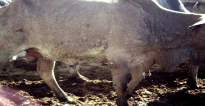
Figure 1: Progression of lumpy skin disease; necrotic nodules and formation of deep scab
Lesions of LSD are round and firm, 1 to 4 cm in diameter, and are flattened and the hair on them stands on end. They vary in number from a few to hundreds; they are intradermal and, mostly confined to the skin area. Lacrimation, nasal discharge, salivation, and lameness can also be observed in association with the pyrexia. Lesions in the nostrils and on the turbinates, causing mucopurulent nasal discharge, respiratory obstruction and snoring; plaques and ulcers in the mouth causing salivation, nodules on the conjunctiva, causing severe lacrimation can be observed in severe cases. Lymph nodes draining the affected area become enlarged and cause local edema (Radostits et al., 2006; Maclanchilan and Dubovi, 2011).
Diagnosis of CaPV is based upon clinical signs with laboratory confirmation by virus isolation, polymerase chain reaction (PCR) and electron microscopy. Field diagnosis of LSD is often based on characteristic clinical signs of the disease. However, mild and subclinical forms require rapid and reliable laboratory testing to confirm diagnosis (Alaa et al., 2008; Knopvelsiekte, 2008). Isolation of virus can be made from collected biopsy or at post-mortem from skin nodules, lung lesions or lymph nodes within the first week of the occurrence of clinical signs, before the development of neutralizing antibodies (OIE, 2010; CFSPH, 2008).
Laboratory diagnosis of LSD comprised either identification of the virus using: electron microscopy, egg inoculation, isolation in cell cultures, fluorescent antibody test; or detection of its specific antibody using serological tests. Several polymerase chain reaction (PCR) assays have been developed for more accurate and rapid detection of LSDV in suitable specimens (Stram et al., 2008). PCR for the diagnosis of LSD is with a greater sensitivity and good specificity and it is most appropriate technique (Kholy et al., 2008; OIE, 2010).
Samples submitted for laboratory diagnosis of LSD includes; take biopsy specimen at least two early lesions (for viral isolation), clipped and cleansed with a none-disinfectant soap; if a punch biopsy is used, specimens must be collected at the lesions edge. An enlarged LN can be aspirated aseptically with a syringe and 16- gage needle or a biopsy can be taken. Organ samples should be sealed in screw-caped vials and taped shut. Tissue specimens should include all organs with emphasis on those showing lesions i.e., skin turbinate‟s, trachea, lung and lymph nodes. specimens should arriving to laboratory within 24 hours ship with wet ice; if more than one day shipment is required dry ice should be used (Tuppurainen et al., 2011).
Generally, LSDV diagnostic tests can be grouped into 3 categories (1) direct detection, (2) indirect examination (virus isolation), and (3) serology. In direct examination, the clinical specimen is examined directly for the presence of virus particles, virus antigen or viral nucleic acids. In indirect examination, the specimen into cell culture, eggs or animals in an attempt to grow the virus: this is called virus isolation. Serology actually constitute far the bulk of the work of any virology laboratory to demonstrate the presence antibody against the virus infection (Vorster and Mapham, 2008; OIE, 2010).
The polymerase chain reaction (PCR) is a scientific technique in molecular biology to amplify a single or a few copies of a piece of DNA across several orders of magnitude, generating thousands to millions of copies of a particular DNA sequence. The conventional gel-based PCR method is a simple, fast and sensitive method for the detection of capripoxvirus genome. In EDTA blood, biopsy, semen or tissue culture samples. However, it does not allow differentiation between LSD and sheep and goat pox viruses. Primers for the viral attachment protein gene and the viral fusion protein gene are specific for all the strains within the genus Capripoxvirus. By the use of sequence and phylogenetic analysis; strains of virus can be identified (Le Goff et al., 2009).
LSD was first reported in 1983 in the northwestern part of the country near Lake Tana (Mebratu et al., 1984). There were frequent outbreak reports of LSD in the county that are highly associated with seasonal peak of mechanical vectors in wet and warm weather conditions (Getachew et al., 2010).The disease has spread to almost all regions and agro-ecological zones of the country. Because of the wide distribution of the disease and the size and structure of the cattle population in Ethiopia, it is likely that LSD is one of the most economically important livestock diseases in the country. One of the recent outbreaks of LSD was occurred in central Ethiopia in 2007 to 2011. These outbreaks were described as active. It was investigated in four districts: Adama, Wenji, Mojo and Welenchiti. The totally 1,675 outbreaks were reported over 5 years period from 2007 to 2011, with 62,176 cases and 4,372 deaths. The Oromia represented the highest numbers of outbreaks (1,066), followed by Amhara (365) and the Southern Nations, Nationalities and People’s Region (123). The 2010 were reported the highest number of outbreaks that were frequently seen between September and December. The morbidity and mortality rates were 13.61% (296) and 4.97% respectively (Ayelet et al., 2014).
In Ethiopia LSD is the one of the most economically important livestock diseases. After the first occurrence in 1983 it has spread widely throughout the country and now it is the problem of almost all the regions and agro ecological zones (Gari et al., 2010; Mebratu et al., 1984). Its spread was mainly enhanced by cattle movements, communal grazing and watering, and pastoralist ways of life (Tuppurainen and Oura, 2011; Gari et al., 2012).
In Ethiopia from 2007-2011 totals of 1352 disease outbreaks of LSD have been reported and highest frequency was documented in Oromia region and the least in Afar region (Gumbe, 2018). Another study also showed that a total 3811 LSD outbreaks reported in Ethiopia between 2000 and 2015. Most of these outbreaks were from Oromia (54.5%), Amhara (27.9%), SNNP (10.1%) and Tigray regional states (3.6%) No out breaks were reported from Harari and Dire dawa. It also shows that LSD affects districts for one or two years and then spreads to other nearby areas with a susceptible cattle population with a trend of LSD outbreaks increased over time (Molla et al., 2017a).
Since the country has no well-designed control strategy for this disease it is continuing to be a great problem. Even if the animal health authorities undertake vaccination campaigns when outbreak is reported, researches have shown that the different vaccines used in Ethiopia are not fully effective (Molla et al., 2017b; Ayelet et al., 2013). There have been repeated concerns reported to NVI on the insufficient protection provided by the vaccine, for cattle against LSDV. In addition to this, lack of genetic information on the circulating isolates in the field and their relation to the vaccine strain in use, which is essential for better vaccine matching, is also a great problem in the country (Gelaye et al., 2015).
LSD is an economically important disease of cattle, serious economic losses from outbreaks that have a high morbidity and can produce a chronic debility in infected cattle. LSD is usually considered economically important diseases due to its impact on livestock productivity. This is usually related with the prolonged effect on productivity of dairy and beef cattle through decreased body weight, mastitis, infertility (can be temporary or permanent) and abortion. Additionally, the lesions on animal skin can bring a permanent damage to the hides which affect the leather. There is a great loss of milk production since the disease is more severe in cows in the peak of lactation and causes a sharp drop in milk yield because of high fever caused by the viral infection itself and secondary bacterial mastitis predisposed by the development of lesions on the teats (Abera et al., 2015; Radostits et al., 2006).
The office international des epizootics consider LSD as list A‟‟ disease that has the potential for rapid spread with ability to cause serious economic loss. Morbidity and mortality of the disease vary considerably depending on the breed of cattle, the immunological status of the population and insect vectors involved in the transmission. Morbidity rates generally varying between 1% and 20%. In a few outbreaks it was reported to be more than 50% although the mortality rates are usually less than 10%. Cows in 1% to 7% of cases may abort. LSD causes severe economic losses due to permanent damage to hides, a prolonged debilitating clinical course, reduced weight gain, temporary or permanent loss of milk production, temporary or permanent infertility or even sterility in bulls, and abortion of pregnant cows (Radostits et al., 2006; Vorster and Mapham, 2008).
Even though the mortality rates of LSD are usually low, it is an economically important disease of cattle in Africa because of the prolonged loss of productivity of dairy and beef cattle, use of the animals for traction, decrease in body weight, mastitis, severe orchitis, which may result in temporary infertility and sometimes permanent sterility (Abera et al., 2015; OIE, 2017; Gari et al., 2011). A study done in Ethiopia has shown that the annual financial cost calculated as the sum of the average production losses due to morbidity and mortality arising from milk loss, beef loss, traction power loss, and treatment and vaccination costs at the herd level was estimated to be USD 6.43 (5.12–8) per head for local zebu and USD 58 (42–73) per head for HF/crossbred cattle (Gari et al., 2011). Another study also showed that the average cost of a single ox dying from LSD was calculated as 9,000 Ethiopian birr (ETB), equivalent to US$477.7 (USD1 = 18.84 ETB) (Ayelet et al., 2014).
In addition to quality degradation of skin and hides skin LSD induces associated economic losses due to reduction of wool quality, meat, losses as a result of culling and mortalities and related with cost of treatment and prevention of the diseases. Even though there are no specific antiviral treatments for LSD-infected cattle, there will be treatment cost for secondary bacterial infection. Treatment cost represents the expenses incurred by farmers for medication at the local public veterinary clinics when farmers bring their clinically sick animals for treatment (Abera et al., 2015b). Emaciation and a long convalescence period can also significantly decrease the growth rate in beef cattle (Tuppurainen et al., 2015).
LSD have been identified as one of the major impediments for genetic improvement of cattle populations and, consequently, for the development of intensive production units in Africa. It is well known that high producing dairy cattle, such as Holstein-Friesian (HF) and Jersey are more susceptible to CaPV infection than indigenous African and Asian cattle breeds (Bhanuprakash et al., 2011; Tuppurainen and Oura, 2011). The susceptibility of European cattle breeds and challenges facing dairy-genetics improvement in LSDV-endemic settings in Ethiopia was recently highlighted (Gari et al., 2011). Currently live cattle export from Ethiopia is largely feedlot based. The introductions of LSD into feedlots certainly affect access to specific markets. For longer time, Middle East markets are the traditional destination of Ethiopian live bulls. However, the current health status of Borena bulls in market chain unquestionably becomes a challenge for the country's future live cattle export opportunities to those countries (Alemayehu et al., 2012). Costly control and eradication measures such as vaccination campaigns as well as the indirect costs because of the compulsory limitations in animal movements also cause significant financial losses on national level (Tuppurainen and Oura, 2011; Gari et al., 2011; Abera et al., 2015b).
In Ethiopia, the financial cost in infected herds was estimated to be USD 6.43 (5.12–8) per head for local zebu and USD 58 (42–73) per head for HF/crossbred cattle. This economic loss was the sum of the average production losses from morbidity and mortality rate. This can be observed through milk loss, beef loss, traction power loss, treatment, and vaccination costs. However, effective vaccination against LSD can reduce the expenses by 17% per head in local zebu herds and 31% per head in Holstein Friesians/crossbred herds (Gari et al., 2011). The other observation in to direct economic losses from animal death and treatment cost was estimated to be 9,000 Ethiopian birr (ETB), (US$477.7) and 16.50 ETB per animal, respectively (Ayelet et al., 2014). Further study conducted by Abebe et al. (2017) indicated the existence of significant economic losses. In this cases, largest components of the loss at herd level was from mortality (1000 USD) accompanied by milk loss (120 USD) while control costs were the least losses recorded at herd level.
Successful control and eradication of LSD relies on early detection of the index case, followed by a rapid and widespread vaccination campaign. Immunity acquired from natural infection of the disease might be lifelong and vaccination has been successfully used. LSD could be kept under control by vaccination of cattle every year (Thomas, 2002). Live, attenuated vaccines against LSD are commercially available. These have antigenic homology and there is cross protection among them. Local strain of Kenyan sheep and goat pox virus has been shown to effectively immunize sheep, goats and cattle against infection with capripoxvirus with a remarkable success. The next one is attenuated South African LSD virus (Neethling strain) vaccine derived from cattle, freeze dried product is also available. In countries where LSD is endemic, vaccination against this infection was successfully used by vaccinating animals every year. The efficacy of total stamping-out (killing all clinically affected cattle and unaffected herd-mates) and partial stamping-out (killing only clinically affected cattle) policies have been compared using mathematical modelling. The study found that total stamping-out and partial stamping-out resulted in a similar probability of eradicating the infection. The study also highlighted the importance of initiating vaccination campaigns ahead of virus entry (OIE, 2017).
The most likely way for LSD to enter a new area is by introduction of infected animals. Biting insects that have fed on infected cattle may travel and be blown for substantial distances. The movement of contaminated hides represents another potential means for this resistant virus to move. For countries free of the disease, the introduction of the disease can be prevented by restriction of the importation of the animals and their products but in those nations which experience the infection can limit the spread of the lumpy skin disease by restriction of the animal movement from one place to another, quarantine, keeping of sick animals well apart from the rest of the herd and must not share drinking or feeding troughs by making awareness creation of the farmers (Thomas, 2002).
Vaccination is the only effective method to control the disease in endemic countries like Ethiopia. The experience in the major parts of the country showed that the vaccination approach is commonly chosen and is often that of ring vaccination around a local foci outbreak when it occurs. Animals that recover from virulent LSD infection generate lifelong immunity consisting both of a humoral and cell mediated protective immunity. Maternal immunity provides protection from LSD in calves at least for 6 months. In previously LSD-free countries, in the event of an outbreak, the rapid confirmation of a clinical diagnosis is essential so that eradication measures, such as quarantine, slaughter-out of affected and in-contact animals, proper disposal of carcasses, cleaning and disinfection of the premises and insect control and ring vaccinations can be implemented as soon as possible (Tuppurainen et al., 2005; Radostits et al., 2006).
Live attenuated vaccines of different capripoxvirus strain origins are available to protect cattle, sheep and goats. Four live attenuated strains of capripoxvirus have been used as vaccines specifically for the control of LSD (Brenner et al., 2006; Kitching, 2003). A strain of Kenyan sheep and goat pox virus passaged 18 times in LT or fetal calf muscle cells, Yugoslavian RM 65 sheep pox strain, Romanian sheep pox strain and lumpy skin disease virus strain from South Africa, passaged 60 times in lamb kidney cells and 20 times on the chorioallantoic membrane of embryonated chicken eggs (OIE, 2010). It is likely that many of these vaccine strains available in different parts of the world would be suitable for the prophylaxis of LSD. These live attenuated vaccines are mainly stimulating the cell mediated immune response (Kitching, 2003).
The OIE Terrestrial Animal Health Code Chapter 11.11, (2017) on lumpy skin disease (caused by group III virus, type Neethling), establishes the international standards on disease control and safe international trade. Each country has its own national legislation applied to LSD. In Ethiopia Kenyan SGPV strain, Romanian sheep pox strain and South African Neethling vaccinal strains were used for production of LSD vaccine at the National Veterinary Institute (NVI). But in the current time only the vaccine that produced from Kenyan SGPV strain is widely used for vaccination of all cattle, sheep and goats populations in the country. Outbreaks can also be controlled by strict quarantines to avoid introduction of infected animals into safe herds, isolation and prohibition of animal movements, slaughtering of all sick and infected animals (Depopulation of infected and exposed animals), proper disposal of carcasses (Incineration), cleaning and disinfection of the premises and insect control (Abera et al., 2015; Tuppurainen and Oura, 2011).
For lumpy skin disease, control measures with the exception of vaccination are usually not effective. Vaccination will greatly reduce the morbidity and epizootics but may not completely limit the extension. In endemic countries, vaccination is considered the only economically feasible way to control the spread of LSD and improve cattle productivity (OIE, 2017; Abera et al., 2015). Numerous live attenuated vaccines have been developed and used worldwide while inactivated vaccines are considered less effective (Boumart et al., 2016). In addition, live attenuated vaccines are currently available which are cheap and provide good protection if sufficient herd immunity (over 80%) is maintained by carrying out annual vaccinations (Tuppurainen et al., 2015).
Live vaccines can help to control losses from lumpy skin disease in endemic areas. Four live attenuated strains of CaPVs have been used as vaccines specifically for the control of LSD (OIE, 2017; Brenner et al., 2009). These are: a strain of Kenyan sheep and goat pox virus, Yugoslavian RM 65 sheep pox strain, Romanian sheep pox strain and lumpy skin disease virus strain from South Africa (Al-Salihi, 2014). In endemic regions vaccine failure is a great problem for the effective control of LSD (Gari et al., 2015). It was also reported that CaPV vaccine strains produce a large local reaction at the site of inoculation in Bos taurus breeds which some stock owners find unacceptable. This has discouraged the use of vaccine, even though the consequences of an outbreak of LSD are usually more severe (OIE, 2017b).
9.1. Immunity in cattle against lumpy skin disease
During the occurrence of infection with capripox virus including lumpy skin disease (LSD) virus; cell-mediated immune response plays a central role of protection though humoral immunity also play its own part. In cell-mediated immune response, different cells including lymphocytes appeared to be increased in their number to destroy the virus. Accordingly, Ahmed et al. (2007) found out the increased lymphocyte proliferation in the presence of LSD antigen. Furthermore, Abdelwahab et al. (2016) reported increment in lymphocyte in vaccinated cattle.
Cytokine, more specifically gamma interferon (IFN-γ) plays an important role in the defense mechanism from viral and microbial pathogens. It acts by the activation of the pathways that can directly inhibit virus (Strichman and Samuel 2001; Biron and Brossay 2001). In the finding of Charles et al. (2012), IFN-γ was reported its detection till seven days post-vaccination. Additionally, this same study detected serum IFN-γ, IL-12 and other pro inflammatory cytokines except IFN-α. Viral spread from cell-to-cell usually occurs and countered by effector cells of cell mediated immune response. Accordingly, natural killer cells or cytotoxic T lymphocytes are responsible to prevent viral replication and spread by identifying and eliminating infected cells (Seet et al., 2003). Furthermore, humoral immune response plays its own role orchestrating itself with cellular immunity for the complete protection. For example, a study on orthopox viruses showed the joint action of cellular and humoral immune response in the elimination of infection. Humoral immune response can stand and considered enough for the future re-infection (Tuppurainen et al., 2017a).
9.2. LSD Vaccine types and vaccination in Ethiopia
Vaccines developed against LSD have different types including ‘live’ attenuated and inactivated vaccines. Comparatively, the inactivated one are considered to be less effective (Boumart et al., 2016). However, ‘live’ attenuated vaccines are relatively effective, cheap and available with recorded herd immunity (over 80%) through annual vaccination (Tuppurainen et al., 2017a). There are four CaPVs strains of 'live’ vaccines namely Yugoslavian RM 65 sheep pox strain, Kenyan sheep and goat pox (KSGP) virus, Romanian sheep pox strain and LSDV strain from South Africa, used to control the spread of LSD (OIE, 2017). Regardless of its availability and suggested efficacy, vaccine failure is reported in different areas that pose its effect on the effective control of LSD (Gari et al., 2015). Even in effective vaccination outcome, the existence of large local reaction at injection site was reported as a side effect. The skin reaction sometimes can affect the cattle owners’ attitude and discourage the use of vaccine though the loss to the production and productivity from the disease outbreak is enormous (OIE, 2017).
In general control and prevention methods include vaccination, animal control movement and slaughter of affected and in contact animals. However, some of these methods are not affordable practices especially in developing countries. Vaccination is considered to be a realistic approach to control the disease in endemic regions and considered cost-effective compared to the abovementioned one (Tuppurainen et al., 2012). LSD as endemic disease to Ethiopia is approached with annual vaccination scheme and following an outbreak (Molla et al., 2017). Kenyan sheep and goat pox (KSGP) virus is a vaccine produced by Ethiopian National Veterinary Institute (NVI) and available when needed with affordable price (NVI, 2019). However, there have been repeated complaints towards vaccine effectiveness due to the occurrence of outbreak in vaccinated cattle population (Ayelet et al., 2013; Molla et al., 2017)
LSD is an important disease in cattle causing considerable economic loses. LSD is economically very important disease due to its large scale financial impact by downgrading skins, decreasing milk production, adding treatment costs, reduction in traction powers of oxen and death of the animals. LSD Causes considerable economic losses due to emaciation, damage to hides, infertility and, loss of milk production. Skin lesions result in severe and permanent damage to hides, while lesions in the mouth, pharynx and respiratory tract cause a rapid deterioration in condition and sometimes severe emaciation, which may persist for months. In Ethiopia, the disease is continuing to appear every year. Many studies found that the disease is highly prevalent in central Ethiopia. Regular and timely vaccination strategy is the best choice available for effective control of the disease accompanied with early detection of the disease. Successful control and eradication of LSD relies on early detection of the index case, followed by a rapid and widespread vaccination campaign. Immunity acquired from natural infection of the disease might be lifelong and vaccination has been successfully used. LSD could be kept under control by vaccination of cattle every year. Live, attenuated vaccines against LSD are commercially available. However, different disease outbreaks in vaccinated cattle raise a question on vaccine efficacy that needs a huge attention to look in to it. With this conclusive remark, the following points are recommended to enhance disease prevention and controlling strategies:
Clearly Auctoresonline and particularly Psychology and Mental Health Care Journal is dedicated to improving health care services for individuals and populations. The editorial boards' ability to efficiently recognize and share the global importance of health literacy with a variety of stakeholders. Auctoresonline publishing platform can be used to facilitate of optimal client-based services and should be added to health care professionals' repertoire of evidence-based health care resources.

Journal of Clinical Cardiology and Cardiovascular Intervention The submission and review process was adequate. However I think that the publication total value should have been enlightened in early fases. Thank you for all.

Journal of Women Health Care and Issues By the present mail, I want to say thank to you and tour colleagues for facilitating my published article. Specially thank you for the peer review process, support from the editorial office. I appreciate positively the quality of your journal.

Journal of Clinical Research and Reports I would be very delighted to submit my testimonial regarding the reviewer board and the editorial office. The reviewer board were accurate and helpful regarding any modifications for my manuscript. And the editorial office were very helpful and supportive in contacting and monitoring with any update and offering help. It was my pleasure to contribute with your promising Journal and I am looking forward for more collaboration.

We would like to thank the Journal of Thoracic Disease and Cardiothoracic Surgery because of the services they provided us for our articles. The peer-review process was done in a very excellent time manner, and the opinions of the reviewers helped us to improve our manuscript further. The editorial office had an outstanding correspondence with us and guided us in many ways. During a hard time of the pandemic that is affecting every one of us tremendously, the editorial office helped us make everything easier for publishing scientific work. Hope for a more scientific relationship with your Journal.

The peer-review process which consisted high quality queries on the paper. I did answer six reviewers’ questions and comments before the paper was accepted. The support from the editorial office is excellent.

Journal of Neuroscience and Neurological Surgery. I had the experience of publishing a research article recently. The whole process was simple from submission to publication. The reviewers made specific and valuable recommendations and corrections that improved the quality of my publication. I strongly recommend this Journal.

Dr. Katarzyna Byczkowska My testimonial covering: "The peer review process is quick and effective. The support from the editorial office is very professional and friendly. Quality of the Clinical Cardiology and Cardiovascular Interventions is scientific and publishes ground-breaking research on cardiology that is useful for other professionals in the field.

Thank you most sincerely, with regard to the support you have given in relation to the reviewing process and the processing of my article entitled "Large Cell Neuroendocrine Carcinoma of The Prostate Gland: A Review and Update" for publication in your esteemed Journal, Journal of Cancer Research and Cellular Therapeutics". The editorial team has been very supportive.

Testimony of Journal of Clinical Otorhinolaryngology: work with your Reviews has been a educational and constructive experience. The editorial office were very helpful and supportive. It was a pleasure to contribute to your Journal.

Dr. Bernard Terkimbi Utoo, I am happy to publish my scientific work in Journal of Women Health Care and Issues (JWHCI). The manuscript submission was seamless and peer review process was top notch. I was amazed that 4 reviewers worked on the manuscript which made it a highly technical, standard and excellent quality paper. I appreciate the format and consideration for the APC as well as the speed of publication. It is my pleasure to continue with this scientific relationship with the esteem JWHCI.

This is an acknowledgment for peer reviewers, editorial board of Journal of Clinical Research and Reports. They show a lot of consideration for us as publishers for our research article “Evaluation of the different factors associated with side effects of COVID-19 vaccination on medical students, Mutah university, Al-Karak, Jordan”, in a very professional and easy way. This journal is one of outstanding medical journal.

Dear Hao Jiang, to Journal of Nutrition and Food Processing We greatly appreciate the efficient, professional and rapid processing of our paper by your team. If there is anything else we should do, please do not hesitate to let us know. On behalf of my co-authors, we would like to express our great appreciation to editor and reviewers.

As an author who has recently published in the journal "Brain and Neurological Disorders". I am delighted to provide a testimonial on the peer review process, editorial office support, and the overall quality of the journal. The peer review process at Brain and Neurological Disorders is rigorous and meticulous, ensuring that only high-quality, evidence-based research is published. The reviewers are experts in their fields, and their comments and suggestions were constructive and helped improve the quality of my manuscript. The review process was timely and efficient, with clear communication from the editorial office at each stage. The support from the editorial office was exceptional throughout the entire process. The editorial staff was responsive, professional, and always willing to help. They provided valuable guidance on formatting, structure, and ethical considerations, making the submission process seamless. Moreover, they kept me informed about the status of my manuscript and provided timely updates, which made the process less stressful. The journal Brain and Neurological Disorders is of the highest quality, with a strong focus on publishing cutting-edge research in the field of neurology. The articles published in this journal are well-researched, rigorously peer-reviewed, and written by experts in the field. The journal maintains high standards, ensuring that readers are provided with the most up-to-date and reliable information on brain and neurological disorders. In conclusion, I had a wonderful experience publishing in Brain and Neurological Disorders. The peer review process was thorough, the editorial office provided exceptional support, and the journal's quality is second to none. I would highly recommend this journal to any researcher working in the field of neurology and brain disorders.
Dear Agrippa Hilda, Journal of Neuroscience and Neurological Surgery, Editorial Coordinator, I trust this message finds you well. I want to extend my appreciation for considering my article for publication in your esteemed journal. I am pleased to provide a testimonial regarding the peer review process and the support received from your editorial office. The peer review process for my paper was carried out in a highly professional and thorough manner. The feedback and comments provided by the authors were constructive and very useful in improving the quality of the manuscript. This rigorous assessment process undoubtedly contributes to the high standards maintained by your journal.

International Journal of Clinical Case Reports and Reviews. I strongly recommend to consider submitting your work to this high-quality journal. The support and availability of the Editorial staff is outstanding and the review process was both efficient and rigorous.

Thank you very much for publishing my Research Article titled “Comparing Treatment Outcome Of Allergic Rhinitis Patients After Using Fluticasone Nasal Spray And Nasal Douching" in the Journal of Clinical Otorhinolaryngology. As Medical Professionals we are immensely benefited from study of various informative Articles and Papers published in this high quality Journal. I look forward to enriching my knowledge by regular study of the Journal and contribute my future work in the field of ENT through the Journal for use by the medical fraternity. The support from the Editorial office was excellent and very prompt. I also welcome the comments received from the readers of my Research Article.

Dear Erica Kelsey, Editorial Coordinator of Cancer Research and Cellular Therapeutics Our team is very satisfied with the processing of our paper by your journal. That was fast, efficient, rigorous, but without unnecessary complications. We appreciated the very short time between the submission of the paper and its publication on line on your site.

I am very glad to say that the peer review process is very successful and fast and support from the Editorial Office. Therefore, I would like to continue our scientific relationship for a long time. And I especially thank you for your kindly attention towards my article. Have a good day!

"We recently published an article entitled “Influence of beta-Cyclodextrins upon the Degradation of Carbofuran Derivatives under Alkaline Conditions" in the Journal of “Pesticides and Biofertilizers” to show that the cyclodextrins protect the carbamates increasing their half-life time in the presence of basic conditions This will be very helpful to understand carbofuran behaviour in the analytical, agro-environmental and food areas. We greatly appreciated the interaction with the editor and the editorial team; we were particularly well accompanied during the course of the revision process, since all various steps towards publication were short and without delay".

I would like to express my gratitude towards you process of article review and submission. I found this to be very fair and expedient. Your follow up has been excellent. I have many publications in national and international journal and your process has been one of the best so far. Keep up the great work.

We are grateful for this opportunity to provide a glowing recommendation to the Journal of Psychiatry and Psychotherapy. We found that the editorial team were very supportive, helpful, kept us abreast of timelines and over all very professional in nature. The peer review process was rigorous, efficient and constructive that really enhanced our article submission. The experience with this journal remains one of our best ever and we look forward to providing future submissions in the near future.

I am very pleased to serve as EBM of the journal, I hope many years of my experience in stem cells can help the journal from one way or another. As we know, stem cells hold great potential for regenerative medicine, which are mostly used to promote the repair response of diseased, dysfunctional or injured tissue using stem cells or their derivatives. I think Stem Cell Research and Therapeutics International is a great platform to publish and share the understanding towards the biology and translational or clinical application of stem cells.

I would like to give my testimony in the support I have got by the peer review process and to support the editorial office where they were of asset to support young author like me to be encouraged to publish their work in your respected journal and globalize and share knowledge across the globe. I really give my great gratitude to your journal and the peer review including the editorial office.

I am delighted to publish our manuscript entitled "A Perspective on Cocaine Induced Stroke - Its Mechanisms and Management" in the Journal of Neuroscience and Neurological Surgery. The peer review process, support from the editorial office, and quality of the journal are excellent. The manuscripts published are of high quality and of excellent scientific value. I recommend this journal very much to colleagues.

Dr.Tania Muñoz, My experience as researcher and author of a review article in The Journal Clinical Cardiology and Interventions has been very enriching and stimulating. The editorial team is excellent, performs its work with absolute responsibility and delivery. They are proactive, dynamic and receptive to all proposals. Supporting at all times the vast universe of authors who choose them as an option for publication. The team of review specialists, members of the editorial board, are brilliant professionals, with remarkable performance in medical research and scientific methodology. Together they form a frontline team that consolidates the JCCI as a magnificent option for the publication and review of high-level medical articles and broad collective interest. I am honored to be able to share my review article and open to receive all your comments.

“The peer review process of JPMHC is quick and effective. Authors are benefited by good and professional reviewers with huge experience in the field of psychology and mental health. The support from the editorial office is very professional. People to contact to are friendly and happy to help and assist any query authors might have. Quality of the Journal is scientific and publishes ground-breaking research on mental health that is useful for other professionals in the field”.

Dear editorial department: On behalf of our team, I hereby certify the reliability and superiority of the International Journal of Clinical Case Reports and Reviews in the peer review process, editorial support, and journal quality. Firstly, the peer review process of the International Journal of Clinical Case Reports and Reviews is rigorous, fair, transparent, fast, and of high quality. The editorial department invites experts from relevant fields as anonymous reviewers to review all submitted manuscripts. These experts have rich academic backgrounds and experience, and can accurately evaluate the academic quality, originality, and suitability of manuscripts. The editorial department is committed to ensuring the rigor of the peer review process, while also making every effort to ensure a fast review cycle to meet the needs of authors and the academic community. Secondly, the editorial team of the International Journal of Clinical Case Reports and Reviews is composed of a group of senior scholars and professionals with rich experience and professional knowledge in related fields. The editorial department is committed to assisting authors in improving their manuscripts, ensuring their academic accuracy, clarity, and completeness. Editors actively collaborate with authors, providing useful suggestions and feedback to promote the improvement and development of the manuscript. We believe that the support of the editorial department is one of the key factors in ensuring the quality of the journal. Finally, the International Journal of Clinical Case Reports and Reviews is renowned for its high- quality articles and strict academic standards. The editorial department is committed to publishing innovative and academically valuable research results to promote the development and progress of related fields. The International Journal of Clinical Case Reports and Reviews is reasonably priced and ensures excellent service and quality ratio, allowing authors to obtain high-level academic publishing opportunities in an affordable manner. I hereby solemnly declare that the International Journal of Clinical Case Reports and Reviews has a high level of credibility and superiority in terms of peer review process, editorial support, reasonable fees, and journal quality. Sincerely, Rui Tao.

Clinical Cardiology and Cardiovascular Interventions I testity the covering of the peer review process, support from the editorial office, and quality of the journal.

Clinical Cardiology and Cardiovascular Interventions, we deeply appreciate the interest shown in our work and its publication. It has been a true pleasure to collaborate with you. The peer review process, as well as the support provided by the editorial office, have been exceptional, and the quality of the journal is very high, which was a determining factor in our decision to publish with you.

The peer reviewers process is quick and effective, the supports from editorial office is excellent, the quality of journal is high. I would like to collabroate with Internatioanl journal of Clinical Case Reports and Reviews journal clinically in the future time.

Clinical Cardiology and Cardiovascular Interventions, I would like to express my sincerest gratitude for the trust placed in our team for the publication in your journal. It has been a true pleasure to collaborate with you on this project. I am pleased to inform you that both the peer review process and the attention from the editorial coordination have been excellent. Your team has worked with dedication and professionalism to ensure that your publication meets the highest standards of quality. We are confident that this collaboration will result in mutual success, and we are eager to see the fruits of this shared effort.

Dear Dr. Jessica Magne, Editorial Coordinator 0f Clinical Cardiology and Cardiovascular Interventions, I hope this message finds you well. I want to express my utmost gratitude for your excellent work and for the dedication and speed in the publication process of my article titled "Navigating Innovation: Qualitative Insights on Using Technology for Health Education in Acute Coronary Syndrome Patients." I am very satisfied with the peer review process, the support from the editorial office, and the quality of the journal. I hope we can maintain our scientific relationship in the long term.

Dear Monica Gissare, - Editorial Coordinator of Nutrition and Food Processing. ¨My testimony with you is truly professional, with a positive response regarding the follow-up of the article and its review, you took into account my qualities and the importance of the topic¨.

Dear Dr. Jessica Magne, Editorial Coordinator 0f Clinical Cardiology and Cardiovascular Interventions, The review process for the article “The Handling of Anti-aggregants and Anticoagulants in the Oncologic Heart Patient Submitted to Surgery” was extremely rigorous and detailed. From the initial submission to the final acceptance, the editorial team at the “Journal of Clinical Cardiology and Cardiovascular Interventions” demonstrated a high level of professionalism and dedication. The reviewers provided constructive and detailed feedback, which was essential for improving the quality of our work. Communication was always clear and efficient, ensuring that all our questions were promptly addressed. The quality of the “Journal of Clinical Cardiology and Cardiovascular Interventions” is undeniable. It is a peer-reviewed, open-access publication dedicated exclusively to disseminating high-quality research in the field of clinical cardiology and cardiovascular interventions. The journal's impact factor is currently under evaluation, and it is indexed in reputable databases, which further reinforces its credibility and relevance in the scientific field. I highly recommend this journal to researchers looking for a reputable platform to publish their studies.

Dear Editorial Coordinator of the Journal of Nutrition and Food Processing! "I would like to thank the Journal of Nutrition and Food Processing for including and publishing my article. The peer review process was very quick, movement and precise. The Editorial Board has done an extremely conscientious job with much help, valuable comments and advices. I find the journal very valuable from a professional point of view, thank you very much for allowing me to be part of it and I would like to participate in the future!”

Dealing with The Journal of Neurology and Neurological Surgery was very smooth and comprehensive. The office staff took time to address my needs and the response from editors and the office was prompt and fair. I certainly hope to publish with this journal again.Their professionalism is apparent and more than satisfactory. Susan Weiner

My Testimonial Covering as fellowing: Lin-Show Chin. The peer reviewers process is quick and effective, the supports from editorial office is excellent, the quality of journal is high. I would like to collabroate with Internatioanl journal of Clinical Case Reports and Reviews.

My experience publishing in Psychology and Mental Health Care was exceptional. The peer review process was rigorous and constructive, with reviewers providing valuable insights that helped enhance the quality of our work. The editorial team was highly supportive and responsive, making the submission process smooth and efficient. The journal's commitment to high standards and academic rigor makes it a respected platform for quality research. I am grateful for the opportunity to publish in such a reputable journal.

My experience publishing in International Journal of Clinical Case Reports and Reviews was exceptional. I Come forth to Provide a Testimonial Covering the Peer Review Process and the editorial office for the Professional and Impartial Evaluation of the Manuscript.

I would like to offer my testimony in the support. I have received through the peer review process and support the editorial office where they are to support young authors like me, encourage them to publish their work in your esteemed journals, and globalize and share knowledge globally. I really appreciate your journal, peer review, and editorial office.

Dear Agrippa Hilda- Editorial Coordinator of Journal of Neuroscience and Neurological Surgery, "The peer review process was very quick and of high quality, which can also be seen in the articles in the journal. The collaboration with the editorial office was very good."

I would like to express my sincere gratitude for the support and efficiency provided by the editorial office throughout the publication process of my article, “Delayed Vulvar Metastases from Rectal Carcinoma: A Case Report.” I greatly appreciate the assistance and guidance I received from your team, which made the entire process smooth and efficient. The peer review process was thorough and constructive, contributing to the overall quality of the final article. I am very grateful for the high level of professionalism and commitment shown by the editorial staff, and I look forward to maintaining a long-term collaboration with the International Journal of Clinical Case Reports and Reviews.

To Dear Erin Aust, I would like to express my heartfelt appreciation for the opportunity to have my work published in this esteemed journal. The entire publication process was smooth and well-organized, and I am extremely satisfied with the final result. The Editorial Team demonstrated the utmost professionalism, providing prompt and insightful feedback throughout the review process. Their clear communication and constructive suggestions were invaluable in enhancing my manuscript, and their meticulous attention to detail and dedication to quality are truly commendable. Additionally, the support from the Editorial Office was exceptional. From the initial submission to the final publication, I was guided through every step of the process with great care and professionalism. The team's responsiveness and assistance made the entire experience both easy and stress-free. I am also deeply impressed by the quality and reputation of the journal. It is an honor to have my research featured in such a respected publication, and I am confident that it will make a meaningful contribution to the field.

"I am grateful for the opportunity of contributing to [International Journal of Clinical Case Reports and Reviews] and for the rigorous review process that enhances the quality of research published in your esteemed journal. I sincerely appreciate the time and effort of your team who have dedicatedly helped me in improvising changes and modifying my manuscript. The insightful comments and constructive feedback provided have been invaluable in refining and strengthening my work".

I thank the ‘Journal of Clinical Research and Reports’ for accepting this article for publication. This is a rigorously peer reviewed journal which is on all major global scientific data bases. I note the review process was prompt, thorough and professionally critical. It gave us an insight into a number of important scientific/statistical issues. The review prompted us to review the relevant literature again and look at the limitations of the study. The peer reviewers were open, clear in the instructions and the editorial team was very prompt in their communication. This journal certainly publishes quality research articles. I would recommend the journal for any future publications.

Dear Jessica Magne, with gratitude for the joint work. Fast process of receiving and processing the submitted scientific materials in “Clinical Cardiology and Cardiovascular Interventions”. High level of competence of the editors with clear and correct recommendations and ideas for enriching the article.

We found the peer review process quick and positive in its input. The support from the editorial officer has been very agile, always with the intention of improving the article and taking into account our subsequent corrections.

My article, titled 'No Way Out of the Smartphone Epidemic Without Considering the Insights of Brain Research,' has been republished in the International Journal of Clinical Case Reports and Reviews. The review process was seamless and professional, with the editors being both friendly and supportive. I am deeply grateful for their efforts.

To Dear Erin Aust – Editorial Coordinator of Journal of General Medicine and Clinical Practice! I declare that I am absolutely satisfied with your work carried out with great competence in following the manuscript during the various stages from its receipt, during the revision process to the final acceptance for publication. Thank Prof. Elvira Farina

Dear Jessica, and the super professional team of the ‘Clinical Cardiology and Cardiovascular Interventions’ I am sincerely grateful to the coordinated work of the journal team for the no problem with the submission of my manuscript: “Cardiometabolic Disorders in A Pregnant Woman with Severe Preeclampsia on the Background of Morbid Obesity (Case Report).” The review process by 5 experts was fast, and the comments were professional, which made it more specific and academic, and the process of publication and presentation of the article was excellent. I recommend that my colleagues publish articles in this journal, and I am interested in further scientific cooperation. Sincerely and best wishes, Dr. Oleg Golyanovskiy.

Dear Ashley Rosa, Editorial Coordinator of the journal - Psychology and Mental Health Care. " The process of obtaining publication of my article in the Psychology and Mental Health Journal was positive in all areas. The peer review process resulted in a number of valuable comments, the editorial process was collaborative and timely, and the quality of this journal has been quickly noticed, resulting in alternative journals contacting me to publish with them." Warm regards, Susan Anne Smith, PhD. Australian Breastfeeding Association.

Dear Jessica Magne, Editorial Coordinator, Clinical Cardiology and Cardiovascular Interventions, Auctores Publishing LLC. I appreciate the journal (JCCI) editorial office support, the entire team leads were always ready to help, not only on technical front but also on thorough process. Also, I should thank dear reviewers’ attention to detail and creative approach to teach me and bring new insights by their comments. Surely, more discussions and introduction of other hemodynamic devices would provide better prevention and management of shock states. Your efforts and dedication in presenting educational materials in this journal are commendable. Best wishes from, Farahnaz Fallahian.

Dear Maria Emerson, Editorial Coordinator, International Journal of Clinical Case Reports and Reviews, Auctores Publishing LLC. I am delighted to have published our manuscript, "Acute Colonic Pseudo-Obstruction (ACPO): A rare but serious complication following caesarean section." I want to thank the editorial team, especially Maria Emerson, for their prompt review of the manuscript, quick responses to queries, and overall support. Yours sincerely Dr. Victor Olagundoye.

Dear Ashley Rosa, Editorial Coordinator, International Journal of Clinical Case Reports and Reviews. Many thanks for publishing this manuscript after I lost confidence the editors were most helpful, more than other journals Best wishes from, Susan Anne Smith, PhD. Australian Breastfeeding Association.

Dear Agrippa Hilda, Editorial Coordinator, Journal of Neuroscience and Neurological Surgery. The entire process including article submission, review, revision, and publication was extremely easy. The journal editor was prompt and helpful, and the reviewers contributed to the quality of the paper. Thank you so much! Eric Nussbaum, MD

Dr Hala Al Shaikh This is to acknowledge that the peer review process for the article ’ A Novel Gnrh1 Gene Mutation in Four Omani Male Siblings, Presentation and Management ’ sent to the International Journal of Clinical Case Reports and Reviews was quick and smooth. The editorial office was prompt with easy communication.

Dear Erin Aust, Editorial Coordinator, Journal of General Medicine and Clinical Practice. We are pleased to share our experience with the “Journal of General Medicine and Clinical Practice”, following the successful publication of our article. The peer review process was thorough and constructive, helping to improve the clarity and quality of the manuscript. We are especially thankful to Ms. Erin Aust, the Editorial Coordinator, for her prompt communication and continuous support throughout the process. Her professionalism ensured a smooth and efficient publication experience. The journal upholds high editorial standards, and we highly recommend it to fellow researchers seeking a credible platform for their work. Best wishes By, Dr. Rakhi Mishra.

Dear Jessica Magne, Editorial Coordinator, Clinical Cardiology and Cardiovascular Interventions, Auctores Publishing LLC. The peer review process of the journal of Clinical Cardiology and Cardiovascular Interventions was excellent and fast, as was the support of the editorial office and the quality of the journal. Kind regards Walter F. Riesen Prof. Dr. Dr. h.c. Walter F. Riesen.

Dear Ashley Rosa, Editorial Coordinator, International Journal of Clinical Case Reports and Reviews, Auctores Publishing LLC. Thank you for publishing our article, Exploring Clozapine's Efficacy in Managing Aggression: A Multiple Single-Case Study in Forensic Psychiatry in the international journal of clinical case reports and reviews. We found the peer review process very professional and efficient. The comments were constructive, and the whole process was efficient. On behalf of the co-authors, I would like to thank you for publishing this article. With regards, Dr. Jelle R. Lettinga.

Dear Clarissa Eric, Editorial Coordinator, Journal of Clinical Case Reports and Studies, I would like to express my deep admiration for the exceptional professionalism demonstrated by your journal. I am thoroughly impressed by the speed of the editorial process, the substantive and insightful reviews, and the meticulous preparation of the manuscript for publication. Additionally, I greatly appreciate the courteous and immediate responses from your editorial office to all my inquiries. Best Regards, Dariusz Ziora

Dear Chrystine Mejia, Editorial Coordinator, Journal of Neurodegeneration and Neurorehabilitation, Auctores Publishing LLC, We would like to thank the editorial team for the smooth and high-quality communication leading up to the publication of our article in the Journal of Neurodegeneration and Neurorehabilitation. The reviewers have extensive knowledge in the field, and their relevant questions helped to add value to our publication. Kind regards, Dr. Ravi Shrivastava.

Dear Clarissa Eric, Editorial Coordinator, Journal of Clinical Case Reports and Studies, Auctores Publishing LLC, USA Office: +1-(302)-520-2644. I would like to express my sincere appreciation for the efficient and professional handling of my case report by the ‘Journal of Clinical Case Reports and Studies’. The peer review process was not only fast but also highly constructive—the reviewers’ comments were clear, relevant, and greatly helped me improve the quality and clarity of my manuscript. I also received excellent support from the editorial office throughout the process. Communication was smooth and timely, and I felt well guided at every stage, from submission to publication. The overall quality and rigor of the journal are truly commendable. I am pleased to have published my work with Journal of Clinical Case Reports and Studies, and I look forward to future opportunities for collaboration. Sincerely, Aline Tollet, UCLouvain.

Dear Ms. Mayra Duenas, Editorial Coordinator, International Journal of Clinical Case Reports and Reviews. “The International Journal of Clinical Case Reports and Reviews represented the “ideal house” to share with the research community a first experience with the use of the Simeox device for speech rehabilitation. High scientific reputation and attractive website communication were first determinants for the selection of this Journal, and the following submission process exceeded expectations: fast but highly professional peer review, great support by the editorial office, elegant graphic layout. Exactly what a dynamic research team - also composed by allied professionals - needs!" From, Chiara Beccaluva, PT - Italy.

Dear Maria Emerson, Editorial Coordinator, we have deeply appreciated the professionalism demonstrated by the International Journal of Clinical Case Reports and Reviews. The reviewers have extensive knowledge of our field and have been very efficient and fast in supporting the process. I am really looking forward to further collaboration. Thanks. Best regards, Dr. Claudio Ligresti

Dear Chrystine Mejia, Editorial Coordinator, Journal of Neurodegeneration and Neurorehabilitation. “The peer review process was efficient and constructive, and the editorial office provided excellent communication and support throughout. The journal ensures scientific rigor and high editorial standards, while also offering a smooth and timely publication process. We sincerely appreciate the work of the editorial team in facilitating the dissemination of innovative approaches such as the Bonori Method.” Best regards, Dr. Matteo Bonori.

I recommend without hesitation submitting relevant papers on medical decision making to the International Journal of Clinical Case Reports and Reviews. I am very grateful to the editorial staff. Maria Emerson was a pleasure to communicate with. The time from submission to publication was an extremely short 3 weeks. The editorial staff submitted the paper to three reviewers. Two of the reviewers commented positively on the value of publishing the paper. The editorial staff quickly recognized the third reviewer’s comments as an unjust attempt to reject the paper. I revised the paper as recommended by the first two reviewers.

Dear Maria Emerson, Editorial Coordinator, Journal of Clinical Research and Reports. Thank you for publishing our case report: "Clinical Case of Effective Fetal Stem Cells Treatment in a Patient with Autism Spectrum Disorder" within the "Journal of Clinical Research and Reports" being submitted by the team of EmCell doctors from Kyiv, Ukraine. We much appreciate a professional and transparent peer-review process from Auctores. All research Doctors are so grateful to your Editorial Office and Auctores Publishing support! I amiably wish our article publication maintained a top quality of your International Scientific Journal. My best wishes for a prosperity of the Journal of Clinical Research and Reports. Hope our scientific relationship and cooperation will remain long lasting. Thank you very much indeed. Kind regards, Dr. Andriy Sinelnyk Cell Therapy Center EmCell

Dear Editorial Team, Clinical Cardiology and Cardiovascular Interventions. It was truly a rewarding experience to work with the journal “Clinical Cardiology and Cardiovascular Interventions”. The peer review process was insightful and encouraging, helping us refine our work to a higher standard. The editorial office offered exceptional support with prompt and thoughtful communication. I highly value the journal’s role in promoting scientific advancement and am honored to be part of it. Best regards, Meng-Jou Lee, MD, Department of Anesthesiology, National Taiwan University Hospital.

Dear Editorial Team, Journal-Clinical Cardiology and Cardiovascular Interventions, “Publishing my article with Clinical Cardiology and Cardiovascular Interventions has been a highly positive experience. The peer-review process was rigorous yet supportive, offering valuable feedback that strengthened my work. The editorial team demonstrated exceptional professionalism, prompt communication, and a genuine commitment to maintaining the highest scientific standards. I am very pleased with the publication quality and proud to be associated with such a reputable journal.” Warm regards, Dr. Mahmoud Kamal Moustafa Ahmed

Dear Maria Emerson, Editorial Coordinator of ‘International Journal of Clinical Case Reports and Reviews’, I appreciate the opportunity to publish my article with your journal. The editorial office provided clear communication during the submission and review process, and I found the overall experience professional and constructive. Best regards, Elena Salvatore.

Dear Mayra Duenas, Editorial Coordinator of ‘International Journal of Clinical Case Reports and Reviews Herewith I confirm an optimal peer review process and a great support of the editorial office of the present journal

Dear Editorial Team, Clinical Cardiology and Cardiovascular Interventions. I am really grateful for the peers review; their feedback gave me the opportunity to reflect on the message and impact of my work and to ameliorate the article. The editors did a great job in addition by encouraging me to continue with the process of publishing.

Dear Cecilia Lilly, Editorial Coordinator, Endocrinology and Disorders, Thank you so much for your quick response regarding reviewing and all process till publishing our manuscript entitled: Prevalence of Pre-Diabetes and its Associated Risk Factors Among Nile College Students, Sudan. Best regards, Dr Mamoun Magzoub.

International Journal of Clinical Case Reports and Reviews is a high quality journal that has a clear and concise submission process. The peer review process was comprehensive and constructive. Support from the editorial office was excellent, since the administrative staff were responsive. The journal provides a fast and timely publication timeline.

Dear Maria Emerson, Editorial Coordinator of International Journal of Clinical Case Reports and Reviews, What distinguishes International Journal of Clinical Case Report and Review is not only the scientific rigor of its publications, but the intellectual climate in which research is evaluated. The submission process is refreshingly free of unnecessary formal barriers and bureaucratic rituals that often complicate academic publishing without adding real value. The peer-review system is demanding yet constructive, guided by genuine scientific dialogue rather than hierarchical or authoritarian attitudes. Reviewers act as collaborators in improving the manuscript, not as gatekeepers imposing arbitrary standards. This journal offers a rare balance: high methodological standards combined with a respectful, transparent, and supportive editorial approach. In an era where publishing can feel more burdensome than research itself, this platform restores the original purpose of peer review — to refine ideas, not to obstruct them Prof. Perlat Kapisyzi, FCCP PULMONOLOGIST AND THORACIC IMAGING.

Dear Mayra Duenas, Editorial Coordinator of the journal IJCCR, I write here a little on my experience as an author submitting to the International Journal of Clinical Case Reports and Reviews (IJCCR). This was my first submission to IJCCR and my manuscript was inherently an outsider’s effort. It attempted to broadly identify and then make some sense of life’s under-appreciated mysteries. I initially had responded to a request for possible submissions. I then contacted IJCCR with a tentative topic for a manuscript. They quickly got back with an approval for the submission, but with a particular requirement that it be medically relevant. I then put together a manuscript and submitted it. After the usual back-and-forth over forms and formality, the manuscript was sent off for reviews. Within 2 weeks I got back 4 reviews which were both helpful and also surprising. Surprising in that the topic was somewhat foreign to medical literature. My subsequent updates in response to the reviewer comments went smoothly and in short order I had a series of proofs to evaluate. All in all, the whole publication process seemed outstanding. It was both helpful in terms of the paper’s content and also in terms of its efficient and friendly communications. Thank you all very much. Sincerely, Ted Christopher, Rochester, NY.

Dear Grace Pierce, Editorial Coordinator of the journal IJCCR, I had a very positive experience with Auctores - Journal throughout the publication process. The Editorial Team was highly responsive, professional, and supportive at every stage. I would like to extend my sincere thanks to the Editor: Grace Pierce, for her guidance and assistance. The peer-review process was smooth and constructive, helping improve the quality of my work. I would gladly recommend Auctores Journal to fellow researchers and authors. Dr. SABITA SINHA, Medical Oncologist, MD (Electro Homeopathy).

Dear Maria Emerson, Editorial Coordinator of - Journal of Clinical Research and Reports. ''I am pleased to provide this testimonial following the publication of our recent case report in this journal. The peer review process was rigorous, constructive, thorough, and conducted in a timely manner. The reviewers’ comments were thoughtful, detailed, and highly constructive, contributing substantially to the refinement, clarity, and scientific robustness of our manuscript. The process was conducted with professionalism and academic integrity throughout. The support provided by the editorial office was exemplary. Communication was consistently prompt, clear, and courteous at all stages of the submission and publication process. The editorial team demonstrated a high level of organization and responsiveness, ensuring that all queries were addressed efficiently and that the process remained transparent and well-coordinated. The overall quality of the journal is reflected in its strong editorial standards, commitment to scientific excellence, and dedication to publishing clinically meaningful research. It has been a privilege to publish our work in this journal, and we would welcome the opportunity to contribute further in the future.'' Best wishes from, Dr. Efstratios Trogkanis, Cardiologist.

Dear Reader: We have published several articles in the Auctores Publishing, LLC, journal, Clinical Medical Reviews and Reports in recent years (CMRR). This is an ‘open access’ journal and the following are our observations. From the initial invitation to submit an article, to the final edits of galley proofs, we have found CMRR personnel to be professional, responsive, rapid and thorough. This entire process begins with Catherine Mitchell, Editorial Coordinator. She is simply outstanding, and, I believe, unparalleled in her capacity. I cannot imagine a more responsive and dedicated Editorial Coordinator. As I read the dates and timing of her correspondence with us, it seems that she never sleeps. I hope Auctores Publishing, LLC, appreciates her efforts as much as these authors do. Thank you to Auctores Publishing, LLC, to the Editorial Staff/Board, and to Catherine Mitchell from a grateful author(s).
