
Research Article | DOI: https://doi.org/10.31579/2643-6612/042
1 International Institute of Periodontology and Private Practice, Victoriaville, Canada.
2 Private Practice, Saint-Maur-des-Fossés, France.
*Corresponding Author: Mark Bonner DMD, 49 Laurier Ouest, Victoriaville, Québec, G6P 6P5, Canada.
Citation: Mark Bonner DMD. (2022). Microscopy Analyses Reveal the Parasitism of Entamoeba gingivalis in Periodontitis: An Observational Study. J Dentistry and Oral Maxillofacial Surgery, 5(3); DOI:10.31579/2643-6612/042
Copyright: © 2022, Mark Bonner DMD. This is an open access article distributed under the Creative Commons Attribution License, which permits unrestricted use, distribution, and reproduction in any medium, provided the original work is properly cited.
Received: 02 November 2022 | Accepted: 17 November 2022 | Published: 24 November 2022
Keywords: E. Gingivalis; amoeba; biofilms; periodontal pocket; trogocytosis; exonucleophagy
Background: Whereas the periodontal microbiota is well described, its non-bacterial component needs a better understanding. Metagenomic analyses show a strong increase of the protozoan Entamoeba gingivalis in inflamed periodontal pockets. Its presence is associated with periodontal deterioration and pockets typically 3mm or more in depth. The aim of this study is to observe the amoeba within the biofilm during active disease.
Materials and Methods: Here, we present results of a phase contrast microscopy-based observation of amoebae in periodontal patients’ subgingival plaque from infected sulcus. Plaque samples from deepest part of sulcus are picked up and spread out between blade and coverslip in patient saliva medium. We relate significant behavior of the parasite during active periodontal disease.
Results: From low power observation, parasite is quite frequent. Once targeted, high power observation confirms amoeban anatomy and presence. We observed a high degree of amoeba locomotion and movements toward specific environments within the subgingival plaque. This was accompanied by the formation of “channels” within the biofilm. We also present evidence of adhesion to human cells as well as characteristically parasitic behavior. Specifically, we observed the intrusion of amoeba pseudopods into leukocytes coupled with a decrease in leukocyte intracellular granular activity. We documented both single trogocytic processes and trogocytosis through multiple pseudopods. In addition to leukocytes, we also observed trogocytosis of red blood cells. Parasitic behavior was also evident from the observation of amoebae digesting the nuclei of multiple vacuolar white blood cells, simultaneously. Following trogocytosis, polynuclear neutrophils had the appearance of ghost cells. Finally, we show evidence for amoeba nesting and reproduction within periodontal pockets.
Conclusion: Phase contrast microscopy of periodontal biofilms strongly suggests that E. gingivalis escapes the first lines of innate defenses and promotes a pathological state. Trogocytosis and exonucleophagy processes targeting neutrophils could consequently disrupt neutrophil extracellular traps activity and normal apoptotic function, a vital component of wound healing. This study points to Entamoeba gingivalis as a microbe involved in the inflammatory process during periodontitis and as a driver of the disease rather than a harmless commensal species.
Entamoeba gingivalis Present Pathogen Behavior
Periodontitis is an infectious disease that affects about half the adult population [1]. Periodontitis leads to the destruction of the alveolar bone surrounding and supporting the teeth, resulting in their loosening and eventually their loss [2]. Periodontitis is generally considered a risk-dependent disease [3]. Indeed, from less than 20% prevalence in people within the 15–19-year-old range, it reaches around 50% for people between 35 and 44 years old, worldwide [4]. Periodontitis has a documented higher prevalence in men as compared to women, signifying a possible sex/gender entanglement in the disease pathogenesis [5]. Finally, the prevalence of periodontitis is so high that it suggests the whole human population is susceptible to the disease [6].
Awareness for periodontitis is quite low in the general population, and in this regard, it could be considered a neglected disease. In addition, there is no consensus regarding the etiology of the disease. Physiological degeneration, genetic determinants, microbial etiology and behavioral issues have been proposed [7]. Changes to the composition of the oral microbiota can cause a disequilibrium called “dysbiosis”, leading to several diseases including periodontitis [8]. Inflammation is always present in periodontitis, and recruitment of leukocytes to inflamed gingival tissue as well as their passage to the periodontal pocket lumen are speculated to cause both tissue destruction and the development of the biofilm found within the pocket. Failure to clear chronic inflammation by pro-resolving pathways will lead to the continuous release of enzymes, cytokines and other harmful oxidative products promoting apoptotic changes in the resident cells and inhibition of tissue healing [8].
Microscopy and modern sequencing technologies may address the question of the presence of non-bacterial organisms in the microbiota of periodontitis [9]. Parasites were detected in early studies of the dental plaque [10] but have raised little interest upon analyses of metagenomic and metatranscriptomic data. Regarding the prevalence of the parasite Entamoeba gingivalis in the mouth, there has been a high degree of consensus among authors since its first observation. Using microscopy approaches, amoebae are found in relatively low numbers (0% to 34%) at healthy sites, whereas it is present in 81% to 100% of periodontal pockets [11]. Light microscopy observations of the dental plaque from patients indicate that this organism has amoeboid motility and recent works show that the parasites detected by microscopy mainly - if not exclusively - belong to the species E. gingivalis and the presence of the parasite is correlated with periodontitis [12]. Lyons investigations on oral protozoa supported the pathogenicity of E. gingivalis and offered protocols for microscopic examination and specific local and systemic treatments targeting the ameba [13].
Recent studies confirm E. gingivalis identification [11] and support its role in oral inflammation and tissue destruction by upregulating inflammatory cytokine IL8 and the epithelial barrier gene MUC2I [14]. It was suggested that upon disruption of the epithelial barrier, E. gingivalis is able to invade gingival tissue, where it moves and feeds on host cells. Direct contact of the amoeba with epithelial cells inhibited cell proliferation in vitro [14]. However, little is known about the biology of the parasite and its place in the oral microbiota is still questioned by some clinicians and scientists. Here we are presenting our observations on E. gingivalis biology during infection, supporting its involvement in the pathophysiogenesis of periodontitis.
Phase-contrast and dark field microscopy (100x magnification) as well as bright field microscopy (1000x) were used for healthy and periodontal patients. Images were recorded using high quality trinocular, and capture in picture application software. Access to the normal or infected and inflamed sulcus was achieved using a periodontal probe inserted at the bottom of the pocket, quickly pulled up and examined instantly ex-vivo in a fresh smear of patient saliva. A large cover slip was then placed on top of the sample, and the material was spread by pressure on the cover slip. It was found that mounting media other than saliva, such as broth or saline, when used as mounting medium, caused changes in amoebae morphology [15]. We deliberately target living cells such as Epithelial cells if present, granulocytes, mainly Neutrophils, highly motile bacteria region, and alive parasite such as amoeba with some appearance of movement or interaction. Different images reflecting this activity were colorized using Adobe Photoshop and Microsoft Paint 3D, to highlight specific cells, part of cells, structures, or movements.
Observations in healthy patients
Over 35 years, we have performed microscopy analyses on about 2000 patients, both healthy and suffering from gingivitis or periodontitis. We chose the most plausible, clearest, and most detailed images and films to perceive the action of the parasites, their locomotion, the clear vision of the nucleus as to assure their identity as well as the typical pseudopod of the amoebae.
Fig. 1 shows representative images from a healthy patient. The normal commensal biofilm (Fig. 1A) appears to be formed of a static and loosely organized network of cocci and filamentous or lactobacilli forms of non-motile bacteria. Epithelial cells are found in numerous amounts, reflecting lower relative amounts of commensal microbes. Motile microorganisms are not found, with the occasional exception of coccobacilli in exceptionally small numbers, seen moving in a circular motion. No spirochetes, brush formations patterns [16], neither large motile rods form bacteria, amoebae or trichomonas are present at microscopic level. Epithelial cells but not inflammatory cells are found in non-motile bacterial complexes, as seen in Fig. 1B. Corn-cob configurations [16] are commonly present with the ends of filamentous rods being surrounded by adherent cocci (Fig. 1C). This apparent microbe has been associated with Corynebacterium matruchotii and the formation of calculus with calcium-phospholipid-phosphate complexes [17-19]. Fig. 1D shows another typical result, i.e., mostly filamentous non-motile bacteria along with parts of epithelial cells.
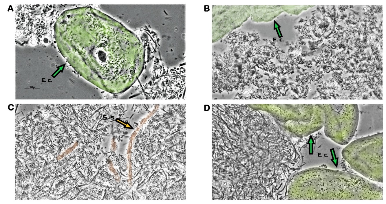
Figure 1: Microscopy images from a healthy patient biofilm. Images were recorded using high power 1000x phase contrast oil immersion microscopy. Epithelial cells are colored in green. (A) Example of normal commensal biofilm, consisting of a static and loosely organized network of cocci and filamentous or lactobacilli form of non-motile bacteria, along with one of numerous epithelial cells in the center. (B) Parts of three epithelial cells at the top of image, present in non-motile bacterial complexes exempt of inflammatory cells. (C) Corn-cob configurations (reminding of Streptococcus sp. adhering to central axis of Corynebacterium sp.) with ends of filamentous rods being surrounded by adherent coccis (orange colored). (D) Mostly filamentous non-motile bacteria and parts of 5 epithelial cells. Green arrows indicate Epithelial cells. Orange arrow indicate Corynebacterium spieces.
Entamoeba gingivalis in periodontal patients
Biofilms of periodontal patients mostly presented continually active motile bacteria, varying degree of leukocytes, and the frequent presence of E. gingivalis [11]. For instance, Fig. 2 shows the presence of many amoebae in one such patient, as revealed using low-power magnification.
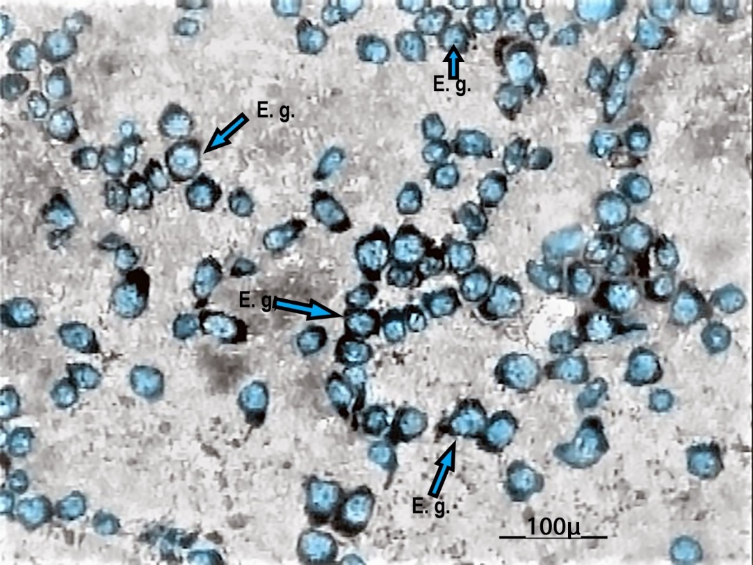
Figure 2: Microscopy observation of amoeba in an infected periodontal patient. Image was taken at low power, phase contrast, pseudo dark field, 10x objective lens, yielding an approximate 100x page magnification. Approximately a hundred E. gingivalis amoebae (blue-colored) are found in this square millimetre image. Blue arrows indicate some E. gingivalis parasites.
In Fig. 3, microscopy is performed at higher resolution, leading to a more precise characterization. Fig. 3A, for instance, shows one large amoeba almost entirely filling the field, with its endoplasm and ectoplasm well differentiated. The nucleus is clearly visible, seen as a circle with a dot offset from the center representing the karyosome. Thickenings of the center ring are chromosomes. The endoplasm contains numerous food vacuoles. In Fig. 3B, the amoeba at the lower center of the image is undergoing binary fission. Asexual cells are normally able to reproduce entirely by themselves, though it has been discovered [20] that in about one-third of Entamoeba invadens dividing cells, contraction of the cleavage furrow may stop before separation is complete. In Fig. 3C, one may observe the progressive organization of the chromosomes until completion of the nucleus as an outer ring and the inner karyosome. Fig. 3D shows a typical adult fed amoeba, presenting with more than fifteen vacuoles; its volume can be up to eight times that of a white cell.
E. gingivalis locomotion
We were able to document amoeba locomotion. As seen in Fig’s 3E and 3F, amoebae movements create an apparent “trail” which may contain remnants of pus and is easily identified under microscopy. This characteristic could explain the presence of “channels” frequently seen in periodontal biofilms [21]. In general, amoebas are found to be elongated in form, with protruding lobopodian and a trailing uroid; less active cells tend to be more spherical. Fig. 3G shows a moving amoeba heading toward the lower right of the image, with many bacteria apparently attached to its uroid while salivary flow is 180° opposite. In Fig. 3H, filopodia formation is apparent at the extremity of an E. gingivalis parasite uroid tail. This capping phenomenon has been suggested as a mechanism for evasion from the host immune response [22]. The uroid or uropod appears as a tail formed by irregular folds of the membrane and filopodia. The fan-shaped uroid appears to be a region of high adhesiveness since debris, bacteria, cell fragments and even red blood cells are attached and trailered to it [22].
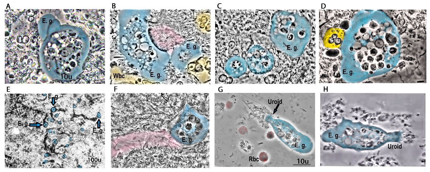
Figure 3: High magnification observations of amoeba biology in the biofilm of periodontal patients. Images were acquired using a phase contrast condenser and 100x Ph objective lens (oil immersion), yielding a page magnification of approximatively x1,000; trophozoite parasites are colored in blue, granular leukocytes in yellow and apparent movement in pink. (A) Observation of a single adult amoeba with 4 microns large nucleus harboring karyosomes in the center with chromatin on the outer ring. (B) Amoeba undergoing fission on the right side leaving a small amoeba with hyalin typical ectoplasm. Three granular leukocytes are also visible. (C) Three amoebae present with the small lower left one completing nucleus formation, whereas the “mature” upper right one presents a circular nucleus and five food vacuoles. (D) A large, fed mature amoeba presenting numerous vacuoles and a clear nucleus; a clear granular leukocyte is present on the left side (yellow). (E) Amoeba locomotion: images were recorded after five minutes, allowing for channel creation seen as a black trail toward amoebae while they move. (F) High power magnification of a moving E. gingivalis amoeba, showing the trail behind it (colored in pink). (G) Another moving amoeba showing protruding lobopodian and a trailing uroid with apparent bacteria attached. (H) Another example of a moving E. gingivalis parasite. Lobopod and uroid are seen on the left and right sides, respectively. Filopodia formation is visible at the extremity of the uroid tail. E. g. indicate Entamoeba gingivalis; Wbc indicate white blood cell; Rbc indicate red blood cell. Black arrow indicate uroid part of amoeba.
E. gingivalis feeding behavior
We were able to observe another biological behavior of amoeba in biofilms from periodontal patients: feeding on human leukocytes. Fig. 4A-D shows time lapse images of a large amoeba feeding on a leukocyte. The amoeba feeds on the white blood cell by “sucking” out the nucleus content. This is helped by peristaltic movement along the pseudopod, bringing the leukocyte’s nuclear material toward digestion in a food vacuole. About fifteen such vacuoles present within the cytoplasm suggest multiple phagocytosis. Note in Fig. 4E-F, the presence of granulated and one degranulated leukocytes around the amoeba. This suggests that the amoeba has the ability to deactivate some of the immune cells, or at least resist their attack, before cell perforation and nucleus ingestion. We term this feeding process “Exonucleophagy”. Feeding duration was 4 minutes in this instance. Interestingly, the presence of two additional vacuoles inside the endoplasm suggests that this amoeba previously ingested two other nuclei.
Fig. 4G-H shows other examples of such exonucleophagy. As reported above, we observed leukocytes in close contact with the amoebae, that were not or only partially degranulated. This suggests the existence of a process by which E. gingivalis secretes a toxic effector to deactivate leukocytes, similar to Entamoeba histolytica [23]. We also observed E. gingivalis phagocytising a leukocyte from two pseudopods simultaneously, as shown in Fig. 4G. Finally, E. gingivalis can feed on up to four nuclei from four different leukocytes, as illustrated in Fig. 4H.
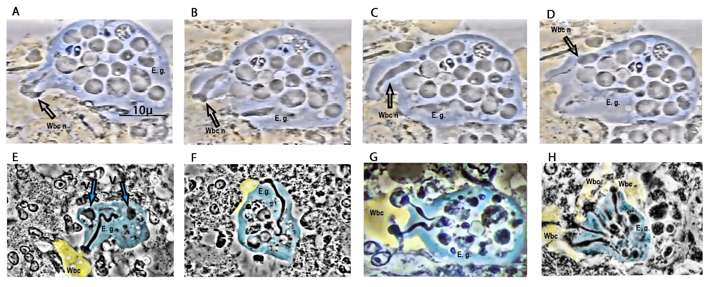
Figure 4: One large amoeba (blue) feeding on a leukocyte (yellow). (A) Remains of the leukocyte nucleus are entering the pseudopod in a form of trogocytosis process, or ingestion of an immune cell fragment. (B) Peristaltic waves are apparent along the pseudopod, moving the leukocyte nuclear material into a food vacuole. (C) Ingestion of the nucleus is about completed. (D) Completion of leukocyte nucleus ingestion, 4 minutes after its initiation. Note the other black vacuoles inside the endoplasm, suggesting that more than fifteen other nuclei were ingested prior to the observed one. A second ingestion phenomena is also visible at the same time lower to the upper one from apparently the same amoeba. (E) Following the exonucleophagy process, one can observe the trilobed nuclear envelope of the remaining leukocyte nucleus, typical of the neutrophil cell. Note that typical neutrophils in the vicinity, contrary to the swallowed one, show granules within their cytoplasm. (F) Two minutes later, the same amoeba (90-degree clockwise displacement) has almost completed the exonucleophagy process, leaving the denucleated cell remnant with an apparent intact cellular membrane. Non-degranulated or partially degranulated leukocytes can be seen on the left and right areas, relative to the amoeba (compare with the fully granulated leukocyte on the upper left). (G) An Entamoeba gingivalis amoeba appears to be feeding on a leukocyte from two pseudopods at the time (top left and bottom left). More than six cells appear to have been phagocytised from this single amoeban cell. Observation of the nucleus present at the center of the ameba endoplasm with chromatin and karyosome clearly confirms the parasitic type of cell. (H) An amoeba feeding on four nuclei from four different leukocytes, simultaneously. Less granulated leukocytes are visible close to those pseudopods. Black arrows named Wbc n indicate White blood cell nucleus, E. g. indicate E. gingivalis parasite; V and blue arrows indicate digesting vacuoles.
Amoeba interactions with human and microbial cells
Our observations led us to document additional behaviors of E. gingivalis. For instance, Fig. 5A shows an amoeba introducing its pseudopod into a leukocyte. The pseudopod end is seen protruding toward the nucleus. Dense “Brownian” granules are again absent from the target cell. This suggests a loss of movement for this monocytic cell, and the granules clump and clear open spaces replacing the dense shimmering within the cell as it degenerates. In Fig. 5B, we see another example of an amoeba interacting with leukocytes that have degranulated and are emptied of their nucleus. The leukocyte granules seem to stop moving upon interaction with the parasite. The amoeba is seen inserting a finger-like projection of cytoplasm through the cell wall of the leukocyte and toward the nucleus. The nuclear membrane is penetrated and its content is aspirated down to the body of the amoeba endoplasm. We call this type of cell a ghost cells to reflect the loss of nuclear content. As seen in Fig. 5C-D, amoebae also seemed to frequently engage into inquilinism, in which one of two species, the inquiline, lives on or in the other (the host), or inside the host's home, obtaining shelter and in some instances hijacking the host's nutriments. In the example shown here, amoebae appear to adhere and coaggregate to a “brush formation” consisting of chains of non-motile rods radiating from a central core and coated with masses of highly motile bacteria that we observed undulating in unison. Amoebae also proliferate in specific areas, thereby forming apparent “nests”. This behavior is similar to what is found in liver abscesses formed by E. histolytica [24]. In deeper periodontal lesions, we frequently found such formations of parasite nests, accompanied by a relative decrease in motile bacteria.
We also observed amoebae feeding on red blood cells, as shown in Fig. 5E-H. The phagocytic process resembled exonucleophagy as described above and lasted about 20 seconds in this instance. A thin undulating black line running from the junction of cells membranes down to the body of the amoeba is observed, ending in a small dense black dot that resembles an ingested bacterium. The images recorded also suggest that the same amoeba fed on other red blood cells prior to the observed phagocytosis event. Therefore, E. gingivalis seemingly has the capacity to feed on both nucleated and non-nucleated human cells.

Figure 5: Interactions of amoebae with leukocytes, Actinomyces-type bacteria, and erythrocytes. (A) An amoeba introducing its pseudopod into a leukocyte cell (left) and reaching the nucleus. Leukocytes here present with a U-shaped nucleus, suggesting monocytes. Note that granules are absent from the target cell. (B) Three amoebae (blue) can be seen on this image, with clearly visible round nuclei. The center one is in contact on the right side with degranulated cells, the centered one colored in yellow. (C) Apparent inquilinism, involving amoebae and Actinomyces sp. appearing to adhere to this “brush formation” consisting of chains of non-motile rods radiating from a central core and coated with masses of highly motile bacteria. (D) Eight amoebae are present in the same area, seemingly forming a nest. On the lower side is one agranular leukocyte (colored yellow). (E-H) Time-lapse images showing E. gingivalis feeding on a red blood cell. Five red blood cells (colored in red) are visible on each image. (E) On the right side of the amoebae, a red blood cell appears to be “pinched” by the pseudopod; a thin line appears that suggests phagocytosis. (F) The pseudopod elongates as feeding continues further. (G) Peristaltic movements are apparent, and apparent torsions of the pseudopod upon the red cell creating a twisted eight figure as a finishing process. (H) Completing the phagocytosis process, the right red blood cell appears pale and of a reduced size. The phagocytosis line is visible from the ectoplasm toward the endoplasm in the typical “wave” suggestive of peristalsis. Note other small dots within the amoeba in the same area, suggesting a repeated behavior. E. g. indicate E. gingivalis; Wbc indicate white blood cell; red arrows with Rbc mention indicate red blood cell. Blue arrows indicate E. gingivalis.
Despite the important contributions of modern genomics and advances in understanding markers of inflammation, optical microscopy can still reveal crucial behaviors and interactions between the different cells of the infected periodontal sulcus [15, 16]. Although identification of E. gingivalis is relatively easy, mishandling the plaque or using mounting liquids other than the patient’s own saliva results in false negatives [25].
E. gingivalis is acquired through direct or indirect contacts. Tap water as well as dental unit tubulure are often positive for many different amoebae [26]. In this study, we were able to analyze in detail the parasitic behavior of this parasite. We documented multiple examples of amoebae feeding on the nuclei of leukocytes, a phenomenon which we call “exonucleophagy”. Following this process, which lasts a few minutes, the amoeba remains viable, whereas the leukocyte visibly degenerates. Individual amoebae were repeatedly seen feeding on as many as four white blood cells simultaneously. Amoebae were seen feeding on up to 15 polynuclear neutrophil nuclei (Fig 4 A-B-C-D). We coined the term “ghost cells” for the denucleated white blood cell left behind following the feeding process. Most likely, ghost cells are unable to achieve either neutrophil extracellular traps (NETS) activity or preprogrammed apoptosis [27, 28]. Ghost cells may promote immune dysregulation and the production of toxic compounds partly responsible for tissue destruction of the supporting bone.
In addition, E. gingivalis was also observed feeding on erythrocytes. The ability of Entamoeba to phagocytose erythrocytes has been proposed as a significant factor differentiating between pathogens and non-pathogens, since the latter do not possess this ability [29]. Thus, our observations of red blood cells attacked by E. gingivalis is most likely highly significant.
Our observations are reminiscent of E. histolytica lytic activity in invasive intestinal amebiasis. E. histolytica-mediated cytolysis is divided into four stages: adhesion, cytolysis following contact, phagocytosis, and intracellular degradation, resulting in tissue destruction [30]. As evidenced in this and other studies, the same stages are present during oral amebiasis periodontal infection. E. gingivalis virulence could also be correlated with the presence of a larger number of parasites as shown in Figure 6 while pocket deepens during active disease.
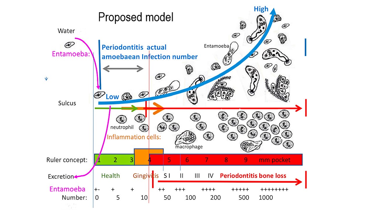
Figure 6: Model proposed for the contribution of E. gingivalis from health to periodontitis assuming possible contaminated water. Infection by amoeba takes advantage of gingivitis inflammation and bleeding, leading to later leukocytes dysregulation and periodontitis. E. gingivalis is present in increasing numbers as periodontal disease progresses. This is explained by the amoebae capacity to feed on both red blood cells and nucleus of leukocytes during immune response and anaerobic deepening of the pocket. Proposed model source: author’s own proposed model and drawing.
Consistent with a role for E. gingivalis in the pathogenesis of periodontitis, inhibiting the growth of this parasite has beneficial effects. Specifically, anti-protozoan treatments, in particular metronidazole, correlate with a more favorable prognosis [31-33].
Pathogenicity of the Entamoeba genus is related to amoeba adherence to mammalian cells, chemotaxis, penetration, invasion, cytolysis, and interaction with symbiotic flora. Our study and others show that E. gingivalis invades tissues and presents with a pathogenic profile [14]. Our microscopic investigations also support previous findings [16, 34, 35], as E. gingivalis is found in the majority of patients suffering from periodontal disease and is relatively absent in healthy or gingivitis patients. Strict topical and systemic pharmaceutical elimination of parasites accompanied by good hygiene habits generally lead to a cessation of bleeding, the elimination of pus and gingival healing, with a 95% pocket depth closure [31]. Clinical improvement is associated with the disappearance of inflammatory cells and reappearance of normal Green complex flora [24, 36].
Some limitation in the methodology might happen as the clinician must review his clinical biology and the basis of general parasitology, as well as go through a learning curve on phase contrast microscopy and finding situations on the slides. Example would be areas with dead neutrophils or pus are also accompanied by dead parasites more difficult to differentiate. Living amoebae are easily found by flying over at 100x magnification on dark field as explained in figures by following the paths of movement in the biofilm related to apparent channels after a 10-minute waiting period.
The microscopic technique can be used without limitations, since it informs the clinician about the state of dysbiosis and the actual inflammatory response on the spot, and in less than 5 minutes operating time. Moreover, it is easy to perform, without any pain for the patient and in my opinion gives a clear diagnostic evaluation of the activity of the periodontal disease.
Professional opinions on the pathogenicity of certain microorganisms evolved with time, as illustrated for Giardia intestinalis and Helicobacter pylori, in which case a radical change has taken place from surgical to pharmacological therapy. E. gingivalis virulence appears similar to that other known pathogenic amoeba like E. histolytica. One distinction between the two is that E. histolytica produces cysts in order to penetrate the intestinal area, which is not needed for oral amebiasis. Some authors have postulated that E. gingivalis is a scavenger of cell debris, but we have never observed this amoeba feeding on apparently already-dead cells.
The current observational study indicate E. gingivalis is highly motile, has phagocytic abilities and exhibits typical pathogen characteristics. The present study reinforces the notion that this amoeba is a causal agent for periodontitis. Specific targeting of this parasite by antimicrobial drugs could represent a very significant advance toward a better management of chronic and aggressive forms of periodontitis. The overwhelming weight of evidence points to E. gingivalis as being an aggressive pathogen.
This study was funded by the authors.
The author Mark Bonner declares to be president and teacher at International Institute of Periodontology, a private dental school dedicated to treatment of periodontal disease.
M. Bonner contributed to most data collection, conception, interpretation, data analysis, drawing and design, drafted the manuscript, and critically revised the manuscript. V. Amard contributed to part of data collection. All authors gave their final approval and agreed to be accountable for all aspect of the work.
M. Bonner ID: https://orcid.org/0000-0002-3379-0120
Dear Editorial Team, Clinical Medical Reviews and Reports. My experience with the journal was highly positive. The peer-review process was rigorous, constructive, and completed in a timely manner. The reviewers provided valuable comments that helped improve the quality and clarity of our manuscript. The editorial office was professional, responsive, and supportive throughout all stages of the publication process. Communication was clear and efficient, and any questions were addressed promptly. Overall, I found the journal to maintain high scientific standards and an excellent publication workflow. I would be pleased to consider submitting future work to this journal. Best wishes from, Elena Popa.

It was my pleasure to submit my testimonial concerning the Reviewer Board of our Scientific Journal “Brain and Neurological Disorders”. The Reviewers focused on some modifications and their contribution was helpful. The ladies of our Editorial Office were also supported my efforts. It was my honor to have such a co-operation and I am looking forward for more collaboration.

Dear Grace Pierce, Editorial Coordinator of Journal of Clinical Research and Reports, Thank you for the speedy and efficient peer review process. I appreciate the fact that your peer reviewers do not take months to respond like with some other journals. I would also like to thank the editorial office for responding quickly to my questions. It is an excellent journal. I plan to submit more manuscripts in the future. Best wishes from, Robert W. McGee

Dear Grace Pierce, Editorial Coordinator of Journal of Clinical Research and Reports, Working with you and your team on our recent publication in JCRR has been a truly wonderful and enjoyable experience. The responses were prompt, and the reviewers were patient, constructive, and highly professional. One reviewer in particular gave me the feeling that a professor was carefully reading and commenting on my coursework, which was deeply touching. The entire process was straightforward and hassle‑free, with no tedious online forms to complete. I highly recommend this journal. Best wishes from, DR Aibing Rao, Head of R&D

I Appreciate the Opportunity to Share my Experience with the Journal of Clinical Research and Reports. The peer review process was timely and constructive, and the feedback provided helped improve the quality of our manuscript. The editorial office was professional, responsive, and supportive throughout the process, ensuring smooth communication and efficient handling of the submission. Overall, it was a positive experience collaborating with your team.

Dear Mercy Grace, Editorial Coordinator of Obstetrics Gynecology and Reproductive Sciences, We would like to express our gratitude for your help at all stages of publishing and editing the article. The editors of the magazine answer all the necessary questions and help at every stage. We will definitely continue to cooperate and publish other works in the Obstetrics Gynecology and Reproductive Sciences! Best wishes from, Alla Konstantinovna Politova,
